
Article Figures & Data
Figures
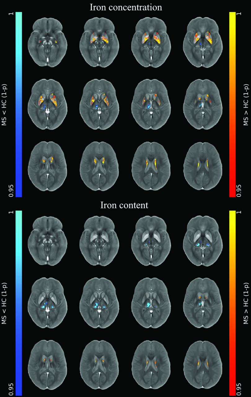
- FIG 1.
Image shows voxelwise analyses of iron maps. Clusters of significant between-group differences regarding unmodulated (top panel) and modulated (lower panel) iron maps for both the MS>HC (red-yellow, according to 1 - P value) and MS<HC (blue-light blue, according to 1 - P value) contrasts are presented, superimposed on the QSM template in the MNI space. Reprinted by permission from Springer Nature Customer Service Center GmbH: Springer Nature, Neuroradiology, European Society of Neuroradiology 2020, Copyright 2020, Springer-Verlag GmbH Germany, part of Springer Nature.
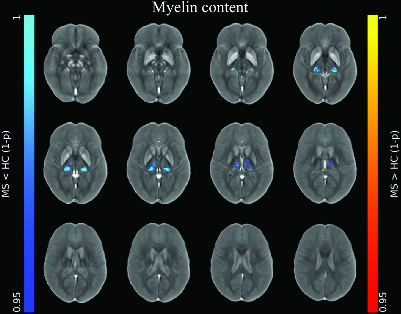
- FIG 2.
Image shows voxelwise analyses of myelin maps. Clusters of significant between-group difference regarding modulated myelin maps for the MS<HC (blue-light blue, according to 1 - P value) contrast are presented, superimposed on the QSM template in the MNI space. No significant differences emerged for the MS>HC contrast or for unmodulated myelin maps. Reprinted by permission from Springer Nature Customer Service Center GmbH: Springer Nature, Neuroradiology, European Society of Neuroradiology 2020, Copyright 2020, Springer-Verlag GmbH Germany, part of Springer Nature.
- FIG 3.
Between-group comparisons of MRI features regarding thalamic subnuclei. Boxplots at the y-axis show z scores (adjusted for the effect of age and sex in HC) of iron concentration and content (upper panel), myelin concentration and content (middle panel), and normalized volume (lower panel) of the corresponding thalamic subregions shown at the x-axis for both the MS and HC groups. Red asterisks mark significant between-group differences.


Tables
- Table 1:
Demographic, clinical, and conventional MRI characteristics of the studied populationa
MS (n = 91) HC (n = 55) P Value (MS vs HC) Age (yr) 38.3 (SD, 11.1) 41.6 (SD, 13.9) .14 Female sexb 54 (59.3) 27 (49.1) .23 Use of DMTb 83 (91.2) – – Progressive courseb 20 (22.0) – – DD (yr) 11.2 (SD, 7.0) – – EDSSc 3.5 (2.5–4.5) – – T2-LL (mL) 11.4 (SD, 13.3) – – Normalized brain volume (mL) 1485.8 (SD, 88.6) 1552.9 (SD, 76.4) <.001 Normalized GM volume (mL) 755.6 (SD, 62.8) 794.7 (SD, 57.5) <.001 Normalized WM volume (mL) 730.2 (SD, 36.5) 758.2 (SD, 31.7) <.001 Note:—– indicates not applicable; DMT, disease-modifying treatment.
↵a Unless otherwise indicated, data are expressed as means (SD). Between-group differences regarding MRI measures are adjusted for age and sex.
↵b Data are the number of subjects, with percentages in parentheses.
↵c Data are medians, with interquartile range in parentheses.
- Table 2:
Results of the ANCOVA analyses for the between-group comparisons regarding DGM structuresa
MS (n = 91) HC (n = 55) Cohen’s D F P Value Normalized volume (mL) Thalamus 19.2 (SD, 2.8) 21.7 (SD, 1.9) 1.14 45.75 <.001 Caudate 8.7 (SD, 1.5) 9.7 (SD, 1.3) 0.88 27.10 <.001 Putamen 12.6 (SD, 1.9) 13.8 (SD, 1.6) 0.82 23.83 <.001 Globus pallidus 4.5 (SD, 0.6) 4.9 (SD, 0.4) 0.76 20.55 <.001 Iron concentration (mg/kg[DW]) Thalamus 5 (SD, 59) 38 (SD, 51) 0.62 13.64 <.001 Caudate 310 (SD, 103) 271 (SD, 76) 0.40 5.55 .02b Putamen 276 (SD, 112) 243 (SD, 94) 0.51 8.94 .03b Globus pallidus 786 (SD, 135) 697 (SD, 128) 0.68 16.51 <.001 Myelin concentration (MVF[DW]) Thalamus 0.24 (SD, 0.06) 0.22 (SD, 0.08) 0.19 1.25 .27 Caudate 0.19 (SD, 0.06) 0.20 (SD, 0.08) 0.06 0.10 .76 Putamen 0.23 (SD, 0.06) 0.22 (SD, 0.08) 0.09 0.29 .59 Globus pallidus 0.21 (SD, 0.06) 0.20 (SD, 0.08) 0.11 0.49 .48 Iron content (µg) Thalamus 0.2 (SD, 1.1) 0.8 (SD, 1.1) 0.63 13.93 <.001 Caudate 2.7 (SD, 1.0) 2.6 (SD, 0.8) 0.03 0.04 .84 Putamen 3.4 (SD, 1.4) 3.3 (SD, 1.2) 0.25 2.13 .15 Globus pallidus 3.5 (SD, 6.8) 3.4 (SD, 6.9) 0.14 0.75 .39 Myelin content (mL) Thalamus 4.6 (SD, 1.4) 4.8 (SD, 1.7) 0.21 1.51 .22 Caudate 1.7 (SD, 0.8) 1.9 (SD, 0.7) 0.29 2.94 .09 Putamen 2.9 (SD, 1.0) 3.1 (SD, 1.0) 0.19 1.32 .25 Globus pallidus 0.9 (SD, 0.3) 1.0 (SD, 0.4) 0.09 0.34 .56 Note:—degrees of freedom (df) , 141.
↵a Descriptive statistics (mean [SD]) for DGM-related MRI features are reported, along with the effect sizes (Cohen’s D), test statistics (F), and exact probability (P value) values regarding between-group (MS versus HC) comparisons.
↵b Not significant after false discovery rate correction.


