Advertisement

Main menu

  • Home
  • Content
    • Current Issue
    • Accepted Manuscripts
    • Article Preview
    • Past Issue Archive
    • Video Articles
    • AJNR Case Collection
    • Case of the Week Archive
    • Case of the Month Archive
    • Classic Case Archive
  • Special Collections
    • AJNR Awards
    • Low-Field MRI
    • Alzheimer Disease
    • ASNR Foundation Special Collection
    • Photon-Counting CT
    • View All
  • Multimedia
    • AJNR Podcasts
    • AJNR SCANtastic
    • Trainee Corner
    • MRI Safety Corner
    • Imaging Protocols
  • For Authors
    • Submit a Manuscript
    • Submit a Video Article
    • Submit an eLetter to the Editor/Response
    • Manuscript Submission Guidelines
    • Statistical Tips
    • Fast Publishing of Accepted Manuscripts
    • Graphical Abstract Preparation
    • Imaging Protocol Submission
    • Author Policies
  • About Us
    • About AJNR
    • Editorial Board
    • Editorial Board Alumni
  • More
    • Become a Reviewer/Academy of Reviewers
    • Subscribers
    • Permissions
    • Alerts
    • Feedback
    • Advertisers
    • ASNR Home

User menu

  • Alerts
  • Log in

Search

  • Advanced search
American Journal of Neuroradiology

American Journal of Neuroradiology

ASHNR American Society of Functional Neuroradiology ASHNR American Society of Pediatric Neuroradiology ASSR
  • Alerts
  • Log in

Advanced Search

  • Home
  • Content
    • Current Issue
    • Accepted Manuscripts
    • Article Preview
    • Past Issue Archive
    • Video Articles
    • AJNR Case Collection
    • Case of the Week Archive
    • Case of the Month Archive
    • Classic Case Archive
  • Special Collections
    • AJNR Awards
    • Low-Field MRI
    • Alzheimer Disease
    • ASNR Foundation Special Collection
    • Photon-Counting CT
    • View All
  • Multimedia
    • AJNR Podcasts
    • AJNR SCANtastic
    • Trainee Corner
    • MRI Safety Corner
    • Imaging Protocols
  • For Authors
    • Submit a Manuscript
    • Submit a Video Article
    • Submit an eLetter to the Editor/Response
    • Manuscript Submission Guidelines
    • Statistical Tips
    • Fast Publishing of Accepted Manuscripts
    • Graphical Abstract Preparation
    • Imaging Protocol Submission
    • Author Policies
  • About Us
    • About AJNR
    • Editorial Board
    • Editorial Board Alumni
  • More
    • Become a Reviewer/Academy of Reviewers
    • Subscribers
    • Permissions
    • Alerts
    • Feedback
    • Advertisers
    • ASNR Home
  • Follow AJNR on Twitter
  • Visit AJNR on Facebook
  • Follow AJNR on Instagram
  • Join AJNR on LinkedIn
  • RSS Feeds

AJNR Awards, New Junior Editors, and more. Read the latest AJNR updates

  • Getting new auth cookie, if you see this message a lot, tell someone!
  • Getting new auth cookie, if you see this message a lot, tell someone!
Research ArticleExtracranial Vascular
Open Access

Symptomatic and Asymptomatic Chronic Carotid Artery Occlusion on High-Resolution MR Vessel Wall Imaging

M. Wan, L. Yan, Z. Xu, Z. Hou, K. Kang, R. Cui, Y. Yu, J. Song, F.K. Hui, Y. Wang, Z. Miao, X. Lou and N. Ma
American Journal of Neuroradiology January 2022, 43 (1) 110-116; DOI: https://doi.org/10.3174/ajnr.A7365
M. Wan
aFrom the Department of Interventional Neuroradiology (M.W., L.Y., Z.H., R.C., Y.Y., J.S., Z.M., N.M.)
cChina National Clinical Research Center for Neurological Diseases (M.W., L.Y., Z.H., K.K., R.C., Y.Y., J.S., Y.W., Z.M., N.M.), Beijing, China
fDepartment of Neurology (M.W.), Shijingshan Teaching Hospital of Capital Medical University, Beijing Shijingshan Hospital, Beijing, China
  • Find this author on Google Scholar
  • Find this author on PubMed
  • Search for this author on this site
  • ORCID record for M. Wan
L. Yan
aFrom the Department of Interventional Neuroradiology (M.W., L.Y., Z.H., R.C., Y.Y., J.S., Z.M., N.M.)
cChina National Clinical Research Center for Neurological Diseases (M.W., L.Y., Z.H., K.K., R.C., Y.Y., J.S., Y.W., Z.M., N.M.), Beijing, China
  • Find this author on Google Scholar
  • Find this author on PubMed
  • Search for this author on this site
  • ORCID record for L. Yan
Z. Xu
dDepartment of Neurology (Z.X.), The First Affiliated Hospital of College of Medicine, Zhejiang University, Hangzhou, China
  • Find this author on Google Scholar
  • Find this author on PubMed
  • Search for this author on this site
  • ORCID record for Z. Xu
Z. Hou
aFrom the Department of Interventional Neuroradiology (M.W., L.Y., Z.H., R.C., Y.Y., J.S., Z.M., N.M.)
cChina National Clinical Research Center for Neurological Diseases (M.W., L.Y., Z.H., K.K., R.C., Y.Y., J.S., Y.W., Z.M., N.M.), Beijing, China
  • Find this author on Google Scholar
  • Find this author on PubMed
  • Search for this author on this site
  • ORCID record for Z. Hou
K. Kang
bNeurology (K.K., Y.W.), Beijing Tiantan Hospital, Capital Medical University, Beijing, China
cChina National Clinical Research Center for Neurological Diseases (M.W., L.Y., Z.H., K.K., R.C., Y.Y., J.S., Y.W., Z.M., N.M.), Beijing, China
  • Find this author on Google Scholar
  • Find this author on PubMed
  • Search for this author on this site
  • ORCID record for K. Kang
R. Cui
aFrom the Department of Interventional Neuroradiology (M.W., L.Y., Z.H., R.C., Y.Y., J.S., Z.M., N.M.)
cChina National Clinical Research Center for Neurological Diseases (M.W., L.Y., Z.H., K.K., R.C., Y.Y., J.S., Y.W., Z.M., N.M.), Beijing, China
  • Find this author on Google Scholar
  • Find this author on PubMed
  • Search for this author on this site
  • ORCID record for R. Cui
Y. Yu
aFrom the Department of Interventional Neuroradiology (M.W., L.Y., Z.H., R.C., Y.Y., J.S., Z.M., N.M.)
cChina National Clinical Research Center for Neurological Diseases (M.W., L.Y., Z.H., K.K., R.C., Y.Y., J.S., Y.W., Z.M., N.M.), Beijing, China
  • Find this author on Google Scholar
  • Find this author on PubMed
  • Search for this author on this site
  • ORCID record for Y. Yu
J. Song
aFrom the Department of Interventional Neuroradiology (M.W., L.Y., Z.H., R.C., Y.Y., J.S., Z.M., N.M.)
cChina National Clinical Research Center for Neurological Diseases (M.W., L.Y., Z.H., K.K., R.C., Y.Y., J.S., Y.W., Z.M., N.M.), Beijing, China
  • Find this author on Google Scholar
  • Find this author on PubMed
  • Search for this author on this site
  • ORCID record for J. Song
F.K. Hui
gDepartment of Radiology and Radiological Sciences (F.K.H.), Johns Hopkins Hospital, Baltimore, Maryland
  • Find this author on Google Scholar
  • Find this author on PubMed
  • Search for this author on this site
  • ORCID record for F.K. Hui
Y. Wang
bNeurology (K.K., Y.W.), Beijing Tiantan Hospital, Capital Medical University, Beijing, China
cChina National Clinical Research Center for Neurological Diseases (M.W., L.Y., Z.H., K.K., R.C., Y.Y., J.S., Y.W., Z.M., N.M.), Beijing, China
  • Find this author on Google Scholar
  • Find this author on PubMed
  • Search for this author on this site
  • ORCID record for Y. Wang
Z. Miao
aFrom the Department of Interventional Neuroradiology (M.W., L.Y., Z.H., R.C., Y.Y., J.S., Z.M., N.M.)
cChina National Clinical Research Center for Neurological Diseases (M.W., L.Y., Z.H., K.K., R.C., Y.Y., J.S., Y.W., Z.M., N.M.), Beijing, China
  • Find this author on Google Scholar
  • Find this author on PubMed
  • Search for this author on this site
  • ORCID record for Z. Miao
X. Lou
eDepartment of Radiology (X.L.), Chinese PLA General Hospital, Beijing, China
  • Find this author on Google Scholar
  • Find this author on PubMed
  • Search for this author on this site
  • ORCID record for X. Lou
N. Ma
aFrom the Department of Interventional Neuroradiology (M.W., L.Y., Z.H., R.C., Y.Y., J.S., Z.M., N.M.)
cChina National Clinical Research Center for Neurological Diseases (M.W., L.Y., Z.H., K.K., R.C., Y.Y., J.S., Y.W., Z.M., N.M.), Beijing, China
  • Find this author on Google Scholar
  • Find this author on PubMed
  • Search for this author on this site
  • ORCID record for N. Ma
  • Article
  • Figures & Data
  • Info & Metrics
  • Responses
  • References
  • PDF
Loading

Article Figures & Data

Figures

  • Tables
  • FIG 1.
    • Download figure
    • Open in new tab
    • Download powerpoint
    FIG 1.

    Stump condition: tapered stump (A), blunt stump (B), no stump (C)
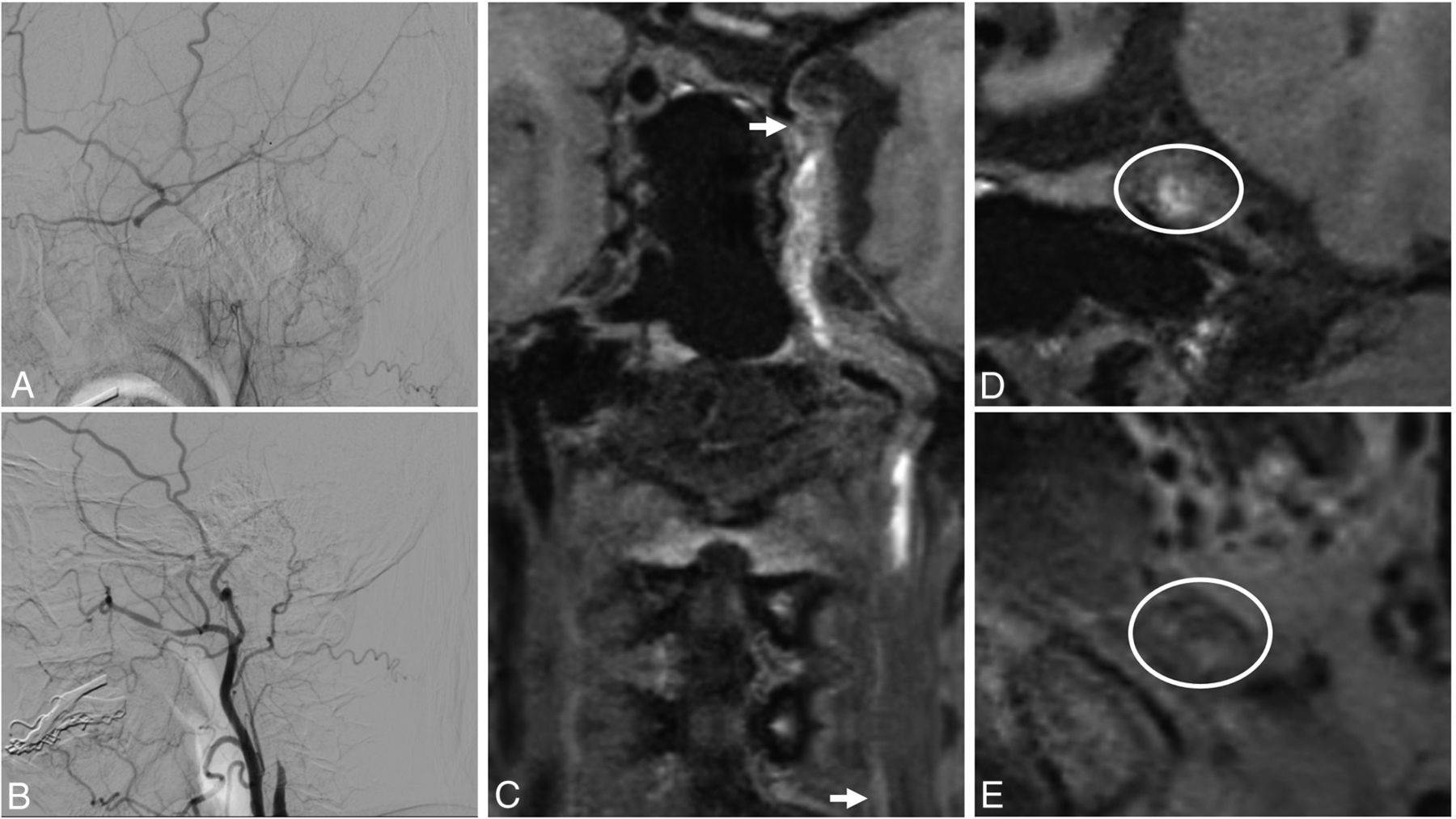
  • FIG 2.
    • Download figure
    • Open in new tab
    • Download powerpoint
    FIG 2.

    An adult man who presented with blurred vision for 1 month and right-limb weakness for 10 days. On DSA, the occluded segment is from C1 (B) to C6 (A). On VWI, 3D T1-weighted turbo spin-echo sequences show that the occluded segment is C1 (arrow) to C6 (arrow) (C). The signal intensity of the distal segment in a elliptical region-of-interest (ROI) (D), and the signal intensity of the origin segment is heterogeneous intensity in a elliptical ROI (E).
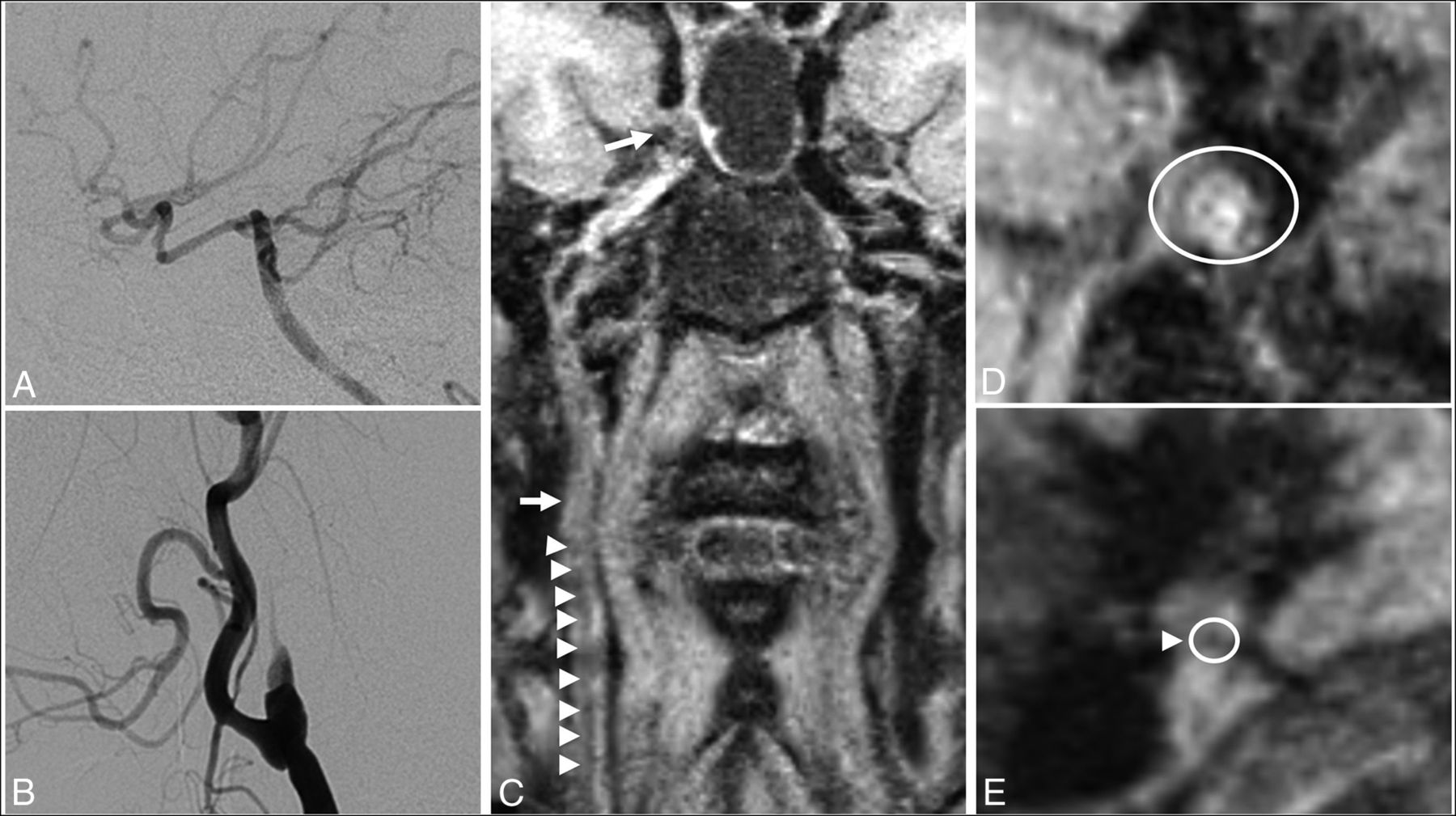
  • FIG 3.
    • Download figure
    • Open in new tab
    • Download powerpoint
    FIG 3.

    An adult woman who presented with dizziness for 2 months. On DSA, the occluded segment is from C1 (B) to C7 (A). On VWI, 3D T1-weighted turbo spin-echo sequences show that the occluded segment was C2 (arrow) to C4 (arrow), and C1 did not have total occlusion (arrowheads, C). The signal intensity of distal segment is isointensity in a elliptical region-of-interest (ROI) (D). The origin segment showed a tiny lumen (arrowhead), in a circular ROI which was considered an occluded segment on DSA (E).

Tables

  • Figures
    • View popup
    Table 1:

    Baseline data of patients with asymptomatic and symptomatic CCAO

    Asymptomatic (n = 9)Symptomatic (n = 35)P Value
    Male (No.) (%)8 (88.9)34 (97.1).37
    Age (mean) (yr)59 (SD, 13.35)60.51 (SD, 11.46).73
    Body mass index (mean)25.30 (SD, 3.49)24.40 (SD, 3.12).46
    Hypertension (No.) (%)7 (77.8)26 (74.3)>.99
    Diabetes mellitus (No.) (%)3 (33.3)12 (34.3)>.99
    Hyperlipidemia (No.) (%)7 (77.8)28 (80.0)>.99
    Coronary artery disease (No.) (%)1 (11.1)3 (8.6)>.99
    Smoker (No.) (%)4 (44.4)15 (42.9)>.99
    Antithrombotic treatment before diagnosis of CCAO (No.) (%)1 (11.1)8 (22.9).75
    Antithrombotic treatment after diagnosis of CCAO (No.) (%)3 (33.3)18 (51.4).55
    History of radiation (No.) (%)0 (0.0)1 (2.9)>.99
    Time intervals between DSA and VWI (median) (IQR) (days)1 (1, 2.5)2 (1, 4).74
    • Note:—IQR indicates interquartile range.

    • View popup
    Table 2:

    Difference in signal intensity at the origin segment of symptomatic CCAO at different stages

    Hyperintensity (n = 0)Isointensity (n = 7)Hypointensity (n = 0)Heterogeneous Intensity (n = 27)P Value
    <7 days (No.) (%)0 (0.0)0 (0.0)0 (0.0)5 (14.7).69
    7–30 days (No.) (%)0 (0.0)4 (57.1)0 (0.0)16 (47.1)
    30–90 days (No.) (%)0 (0.0)2 (28.6)0 (0.0)10 (29.4)
    ≥90 days (No.) (%)0 (0.0)1 (14.3)0 (0.0)3 (8.8)
    • View popup
    Table 3:

    Difference in signal intensity at the distal segment of symptomatic CCAO at different stages

    Hyperintensity(n = 9)Isointensity (n = 14)Hypointensity (n = 0)Heterogeneous Intensity (n = 12)P Value
    <7 days (No.) (%)2 (22.2)1 (7.1)0 (0.0)2 (16.7).29
    7–30 days (No.) (%)2 (22.2)6 (42.9)0 (0.0)8 (67.7)
    30–90 days (No.) (%)4 (44.4)4 (28.6)0 (0.0)2 (16.7)
    ≥90 days (No.) (%)1 (11.1)3 (21.4)0 (0.0)0 (0.0)
PreviousNext
Back to top

In this issue

American Journal of Neuroradiology: 43 (1)
American Journal of Neuroradiology
Vol. 43, Issue 1
1 Jan 2022
  • Table of Contents
  • Index by author
  • Complete Issue (PDF)
Advertisement
Print
Download PDF
Email Article

Thank you for your interest in spreading the word on American Journal of Neuroradiology.

NOTE: We only request your email address so that the person you are recommending the page to knows that you wanted them to see it, and that it is not junk mail. We do not capture any email address.

Enter multiple addresses on separate lines or separate them with commas.
Symptomatic and Asymptomatic Chronic Carotid Artery Occlusion on High-Resolution MR Vessel Wall Imaging
(Your Name) has sent you a message from American Journal of Neuroradiology
(Your Name) thought you would like to see the American Journal of Neuroradiology web site.
CAPTCHA
This question is for testing whether or not you are a human visitor and to prevent automated spam submissions.
Cite this article
M. Wan, L. Yan, Z. Xu, Z. Hou, K. Kang, R. Cui, Y. Yu, J. Song, F.K. Hui, Y. Wang, Z. Miao, X. Lou, N. Ma
Symptomatic and Asymptomatic Chronic Carotid Artery Occlusion on High-Resolution MR Vessel Wall Imaging
American Journal of Neuroradiology Jan 2022, 43 (1) 110-116; DOI: 10.3174/ajnr.A7365

Citation Manager Formats

  • BibTeX
  • Bookends
  • EasyBib
  • EndNote (tagged)
  • EndNote 8 (xml)
  • Medlars
  • Mendeley
  • Papers
  • RefWorks Tagged
  • Ref Manager
  • RIS
  • Zotero
0 Responses
Respond to this article
Share
Bookmark this article
Symptomatic and Asymptomatic Chronic Carotid Artery Occlusion on High-Resolution MR Vessel Wall Imaging
M. Wan, L. Yan, Z. Xu, Z. Hou, K. Kang, R. Cui, Y. Yu, J. Song, F.K. Hui, Y. Wang, Z. Miao, X. Lou, N. Ma
American Journal of Neuroradiology Jan 2022, 43 (1) 110-116; DOI: 10.3174/ajnr.A7365
del.icio.us logo Twitter logo Facebook logo Mendeley logo
  • Tweet Widget
  • Facebook Like
  • Google Plus One
Purchase

Jump to section

  • Article
    • Abstract
    • ABBREVIATIONS:
    • MATERIALS AND METHODS
    • RESULTS
    • DISCUSSION
    • CONCLUSIONS
    • Footnotes
    • References
  • Figures & Data
  • Info & Metrics
  • Responses
  • References
  • PDF

Related Articles

  • No related articles found.
  • PubMed
  • Google Scholar

Cited By...

  • Vessel wall MRI evaluation for the safety of endovascular recanalization of non-acute intracranial anterior circulation artery occlusions
  • Crossref
  • Google Scholar

This article has not yet been cited by articles in journals that are participating in Crossref Cited-by Linking.

More in this TOC Section

  • Proximal Vertebral Artery Variants and Embryology
  • High-Risk Plaque Features in Carotid MRI
  • Nonstenotic Carotid Plaques and Stroke Review
Show more Extracranial Vascular

Similar Articles

Advertisement

Indexed Content

  • Current Issue
  • Accepted Manuscripts
  • Article Preview
  • Past Issues
  • Editorials
  • Editor's Choice
  • Fellows' Journal Club
  • Letters to the Editor
  • Video Articles

Cases

  • Case Collection
  • Archive - Case of the Week
  • Archive - Case of the Month
  • Archive - Classic Case

More from AJNR

  • Trainee Corner
  • Imaging Protocols
  • MRI Safety Corner
  • Book Reviews

Multimedia

  • AJNR Podcasts
  • AJNR Scantastics

Resources

  • Turnaround Time
  • Submit a Manuscript
  • Submit a Video Article
  • Submit an eLetter to the Editor/Response
  • Manuscript Submission Guidelines
  • Statistical Tips
  • Fast Publishing of Accepted Manuscripts
  • Graphical Abstract Preparation
  • Imaging Protocol Submission
  • Evidence-Based Medicine Level Guide
  • Publishing Checklists
  • Author Policies
  • Become a Reviewer/Academy of Reviewers
  • News and Updates

About Us

  • About AJNR
  • Editorial Board
  • Editorial Board Alumni
  • Alerts
  • Permissions
  • Not an AJNR Subscriber? Join Now
  • Advertise with Us
  • Librarian Resources
  • Feedback
  • Terms and Conditions
  • AJNR Editorial Board Alumni

American Society of Neuroradiology

  • Not an ASNR Member? Join Now

© 2025 by the American Society of Neuroradiology All rights, including for text and data mining, AI training, and similar technologies, are reserved.
Print ISSN: 0195-6108 Online ISSN: 1936-959X

Powered by HighWire