
Article Figures & Data
Figures
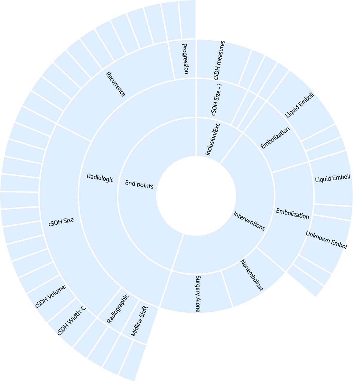
- Fig 1.
Sunburst diagram of data elements in the NK nest for this study. Clicking on each data element outputs a frequency of the tag associated with it, as well as frequently co-occurring tags. See https://nested-knowledge.com/nest/qualitative/461 for an interactive version of this figure.
- Fig 2.
The effect of SDH morphology on volume calculation using the ABC/2. The formula is derived by assuming a crescent shape (the difference between a large ellipsoid and a small ellipsoid, both of which are cut in half). A and B, L = length; W = width, difference between ellipsoids = Wa, Wb; C, Thickness (not shown) (L and C are the same for both ellipsoids). The formula thus reduces to volume of crescent-shaped cSDH = (4/3 π LWaC – 4/3 π LWbC) / 2 = (LWaC – LWbC)/ 2 = LWC/ 2 (= ABC/2). A, The assumed crescent shape allows accurate calculation due to the way the ABC/2 formula is derived (right panel). B, When the SDH is irregular, however, the ABC/2 loses accuracy and can lead to overestimation of the true volume. C, cSDH maximum width perpendicular to the maximum length marked in specific patients with unevenly shaped hematomas. Patient 1: width of the subdural hematoma measured on a section close to vertex (W) is greater than it actually is. Patient 2: inhomogeneous convex- and concave-shaped hematomas. Maximum width (W2) measurement is diagonal and not accurate. W1 would be more accurate in this case. Patient 3: maximum width measured perpendicular to length but slices hematoma diagonally.
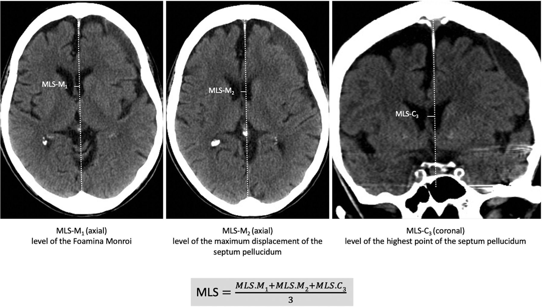
- Fig 3.
Proposed MLS measurement as the mean of maximum at the level and location of the foramen of Monro (MLS-M1), the maximum displacement of the septum pellucidum relative to the midline (MLS-M2), and the maximum MLS of the septum pellucidum at the level of the highest point of the septum on coronal slices (MLS-C3). Overall MLS is determined as the mean of these 3 measurements (or 2 in cases in which coronal reconstructions are missing). MLS-M indicates axial MLS perpendicular to the midline; MLS-C, coronal MLS perpendicular to the midline.


Tables
- Table 1.
Expert suggestions for radiologic measurements of cSDH for volume, width, MLS, and reporting
cSDH Volume/Width MLS Reporting Preferably use computer-assisted volumetric analysis
In case width is measured, report detailed methodology
Measured perpendicular to the midline joining the most anterior and posterior visible points on the falx
Measurements should be conducted on axial and coronal slices
On axial slices measured at the level and location of the foramen of Monro and as the maximum displacement of the septum pellucidum relative to the midline
On coronal slices as the maximum MLS of the septum pellucidum at the level of the highest point of the septum
Overall MLS should then be determined as mean of these 3 (or 2 in case no coronal slices are available) measurements
cSDH width should always be evaluated and reported in context with cSDH volume measurements, MLS, and clinical information
MLS should always be assessed together with other parameters such as clinical information and cSDH volume
Detailed description of the used algorithms when reporting cSDH width, volume, MLS, or other quantitative radiologic measures should be provided
- Table 2.
Advantages and disadvantages of singular and combinations of radiographic measurements to evaluate cSDH progression
Advantage Disadvantage Width Simple, practical, good external applicability, sensitivity easy to adjust (2 vs 5 mm, and so forth) Too dependent on measurement technique and location, sensitivity might be low Volume Intuitively the right choice, presumably most accurate Labor intensive, unlikely to be used in day-to-day practice Volume + width Raises the bar for specificity More challenging to interpret, 2 thresholds to define Less sensitive Volume + width + MLS Raises the bar for specificity even further Even more challenging to interpret, definition issues, 2 df (>2 mm and > 2 mm and > 20 mL) Low sensitivity


Jump to section
Related Articles
Cited By...
- No citing articles found.