
Article Figures & Data
Figures
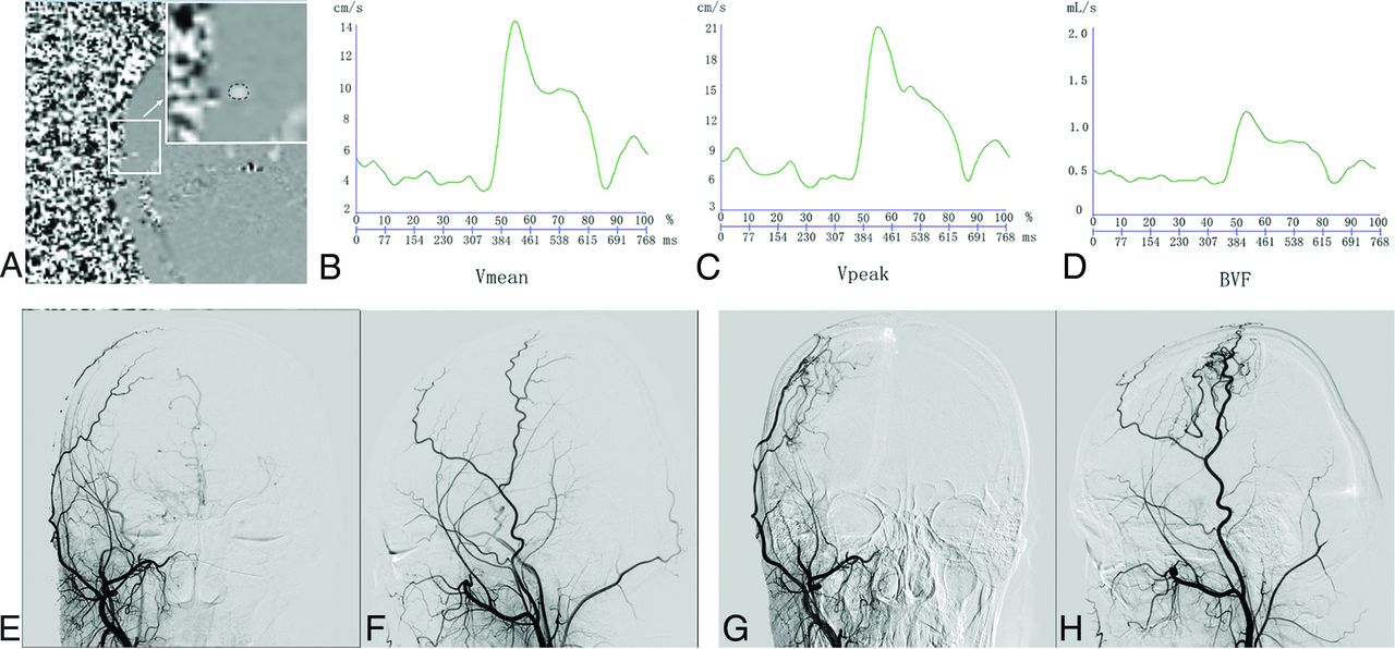
- FIG 1.
PC-MR imaging and DSA examination in a patient with good collateral formation. Images in a female patient diagnosed with MMD who had right EDAS surgery. She underwent PC-MRI and DSA before and 6 months after the operation. A, Phase image shows the ROI of the right STA. A, the arrow represents the enlarged view of the small white square. Modulation of the Vmean (B), Vpeak (C), and BVF (D) of the right STA acquired by PC-MRI. Anterior-posterior and lateral projections of preoperative (E and F) and postoperative (G and H) DSAs of the right external carotid artery show large areas of revascularization (more than one-third but less than two-thirds of MCA territory).
- FIG 2.
PC-MR imaging and DSA examination in a patient with poor collateral formation. Images in a male patient diagnosed with MMD who had right EDAS surgery. He underwent PC-MRI and DSA before and 7 months after the operation. A, Phase image shows the ROI of the right STA. A, the arrow represents the enlarged view of the small white square. Modulation of Vmean (B), Vpeak (C), and BVF (D) of the right STA acquired by PC-MRI. Anterior-posterior and lateral projections of preoperative (E and F) and postoperative (G and H) DSAs of right external carotid artery show small areas of revascularization (less than one-third of the MCA territory).
- FIG 3.
Comparison of PC-MR imaging parameters between patients with good and poor collateral formation (A) and between patients with improved mRS and poor response (B). The asterisk indicates P < .05.


Tables
Patients with MMD, Mean (SD) or No. (%) P Good Collateral Formation (n = 23) Poor Collateral Formation (n = 22) Sex, male 10 (43%) 12 (55%) .458 Age (yr) 30.87 (SD, 18.36) 37.05 (SD, 17.57) .256 Hypertension 8 (35%) 9 (41%) .672 Hyperlipidemia 9 (39%) 11 (50%) .463 CHD 1 (4%) 0 (0%) 1.000 Diabetes 3 (13%) 2 (10%) 1.000 Smoking 0 (0%) 3 (14%) .217 DSA interval (mo) 7.11 (SD, 1.69) 7.73 (SD, 1.91) .256 PCA involvement 4 (17%) 2 (9%) .704 Diameter of MMA (mm) Ipsilateral 2.49 (SD, 0.51) 2.42 (SD, 0.4) .674 Contralateral 2.30 (SD, 0.45) 2.19 (SD, 0.46) .417 Preoperative DSC Ipsilateral rTTP (sec) 3.36 (SD, 2.07) 1.40 (SD, 1.57) .002 Contralateral rTTP (sec) 1.72 (SD, 2.10) 1.07 (SD, 2.18) .353 Ipsilateral CBF (mL/100 g/min) 1.04 (SD, 0.31) 1.31 (SD, 0.44) .021 Contralateral CBF (mL/100 g/min) 1.18 (SD, 0.34) 1.24 (SD, 0.31) .540 Ipsilateral CBV (mL/100 g) 1.77 (SD, 0.62) 2.23 (SD, 0.65) .019 Contralateral CBV (mL/100 g) 1.89 (SD, 0.48) 2.12 (SD, 0.54) .138 Suzuki stage .775 I–II 4 2 III– IV 13 17 V–VI 6 3 Note:—CHD indicates coronary heart disease; DSA interval, interval from surgery to DSA examination; PCA, posterior cerebral artery.
- Table 2:
Comparison of measurements of the STA between participants with good and poor collateral formation
Patients with MMD, Mean (SD) or No. (%) P Good Collateral Formation (n = 23) Poor Collateral Formation (n = 22) Morphologic features of ipsilateral STA Cross-sectional area (mm2) 0.10 (SD, 0.03) 0.09 (SD, 0.02) .258 Straightness 11 (48%) 13 (59%) .449 High bifurcation position 10 (43%) 10 (46%) .894 Morphologic features of contralateral STA Cross-sectional area (mm2) 0.11 (SD, 0.02) 0.12 (SD, 0.04) .095 Straightness 12 (52%) 2 (55%) .873 High bifurcation position 14 (61%) 9 (41%) .181 PC-MR imaging of ipsilateral STA Vmean (cm/s) 6.69 (SD, 2.02) 5.30 (SD, 1.41) .011 Vpeak (cm/s) 22.62 (SD, 5.79) 17.00 (SD, 6.62) .004 BVF (mL/s) 0.70 (SD, 0.32) 0.50 (SD, 0.17) .013 PC-MR imaging of contralateral STA Vmean (cm/s) 7.08 (SD, 2.92) 6.03 (SD, 1.53) .141 Vpeak (cm/s) 23.06 (SD, 6.37) 18.75 (SD, 6.07) .025 BVF (mL/s) 0.74 (SD, 0.35) 0.61 (SD, 0.26) .166 Note:—Ipsilateral STA indicates superficial temporal artery on the operative side; Contralateral STA, superficial temporal artery on the nonoperative side.
- Table 3:
Association of hemodynamic status of the ipsilateral STA and collateral formation using logistic regression analysisa
Univariate Regression Multivariate Regression OR 95% CI P OR 95% CI P Vmean 1.66 1.08–2.53 .020 2.28 1.23–4.25 .009 Vpeak 1.16 1.04–1.30 .009 1.18 1.04–1.34 .010 BVF 1.04 1.01–1.07 .024 1.73 1.02–2.94 .043 ↵a Multivariate regression was adjusted for age, sex, hypertension, hyperlipidemia, diabetes, smoking, interval from the operation to the latest DSA examination, cross-sectional area, straightness and high bifurcation position of the STA, preoperative rCBV, and the diameter of the MMA after EDAS.
- Table 4:
Comparison of PC-MR imaging of STA between participants with improved mRS and those with poor response
Patients with MMD, Mean (SD) or No. (%) P Improved mRS (n = 24) Poor Response mRS (n = 21) PC-MR imaging of ipsilateral STA Vmean (cm/s) 6.43 (SD, 1.91) 5.53 (SD, 1.73) .106 Vpeak (cm/s) 22.13 (SD, 6.59) 17.30 (SD, 6.13) .015 BVF (mL/s) 0.65 (SD, 0.30) 0.55 (SD, 0.24) .268 PC-MR imaging of contralateral STA Vmean (cm/s) 6.92 (SD, 2.37) 6.15 (SD, 2.50) .295 Vpeak (cm/s) 21.97 (SD, 6.60) 19.79 (SD, 6.22) .263 BVF (mL/s) 0.71 (SD, 0.28) 0.64 (SD, 0.34) .453 F P Age 1.829 .186 Sex 1.773 .193 rCBV 1.839 .185 Age × sex 1.781 .192 Age × rCBV 1.832 .186 Sex × rCBV 1.756 .195 Age × sex × rCBV 1.77 .193


Jump to section
Related Articles
Cited By...
- No citing articles found.