
Article Figures & Data
Figures
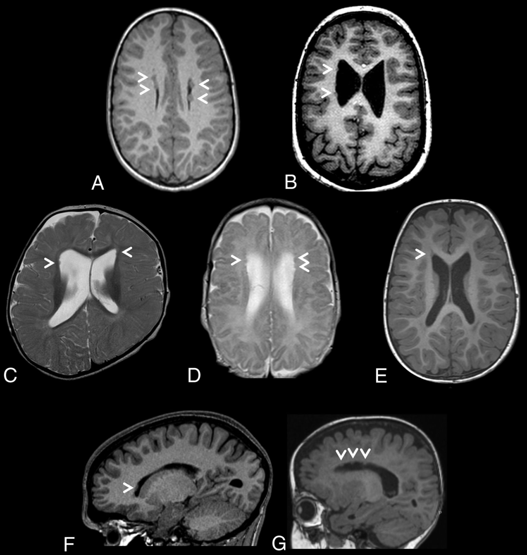
- FIG 1.
Brain MR images depicting periventricular nodular heterotopia distributions in a cohort of 7 unrelated individuals with ZTTK syndrome. A, Axial T1-weighted MRI of the brain of individual 1 at 7 years 7 months of age revealing sparse bilateral frontal heterotopia (white arrowheads). B, Axial T1-weighted MRI of the brain of individual 2 at 11 years and 6 months of age revealing sparse right unilateral frontal and midbody heterotopia (white arrowheads). C, Axial T2-weighted MRI of the brain of individual 3 at 1 year of age revealing sparse bilateral frontal horn heterotopia (white arrowheads). D, Axial T2-weighted MRI of the brain of individual 4 at 1 year 4 months of age revealing sparse bilateral frontal horn heterotopia (white arrowheads). E, Axial T1-weighted MRI of the brain of individual 5 at 1 year 8 months of age revealing a heterotopion of the right frontal horn (white arrowhead). F, Right parasagittal T1-weighted MRI of the brain of individual 6 at 15 years 2 months of age revealing a heterotopion of the right frontal horn (white arrowhead). G, Left parasagittal T1-weighted MRI of the brain of individual 7 at 1 year 2 months of age revealing sparse left frontal horn heterotopia (white arrowheads).
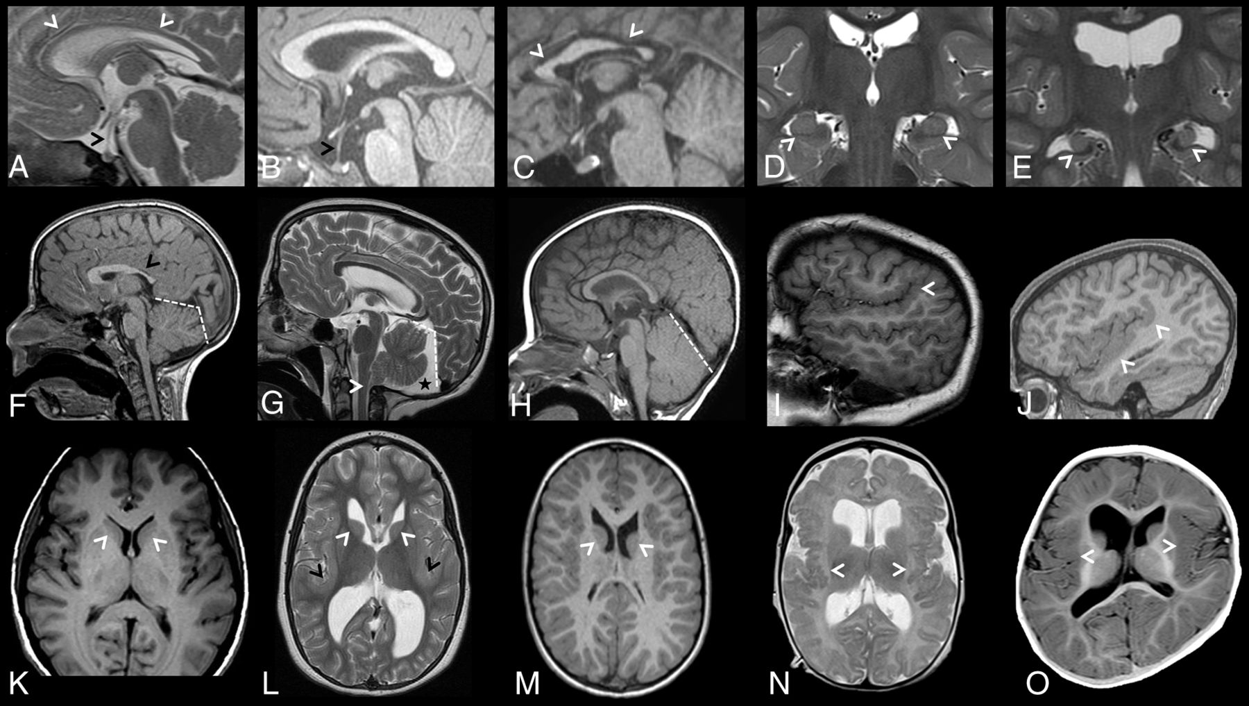
- FIG 2.
Brain MR images depicting additional brain anomalies in a cohort of 7 unrelated individuals with ZTTK syndrome. A, Sagittal midline T2-weighted MRI of the brain of individual 4 at 1 year 4 months of age showing a thin corpus callosum (white arrowheads), an elongated pituitary stalk (black arrowhead), and small pituitary gland. B, Sagittal midline T1-weighted MRI of the brain of individual 5 at 1 year 8 months of age showing an elongated pituitary stalk (black arrowhead) and a small pituitary gland. C, Sagittal midline T1-weighted MRI of the brain of individual 6 at 15 years 2 months of age showing a shortened corpus callosum with a hypoplastic anterior and posterior body (white arrowheads), a normal-sized pituitary stalk, and a normal-sized pituitary gland. D, Coronal T2-weighted MRI of the brain of individual 5 at 1 year 8 months of age revealing normal-sized hippocampi with a lack of internal architecture (white arrowheads). E, Coronal T2-weighted MRI of the brain of individual 2 at 11 years 6 months of age revealing small malrotated hippocampi (white arrowheads). F, Sagittal midline T1-weighted MRI of the brain of individual 1 at 7 years 7 months of age showing a box-shaped posterior fossa (white dashed lines) and a shortened corpus callosum with a hypoplastic posterior body, isthmus, and splenium (black arrow). G, Sagittal midline T2-weighted MRI of the brain of individual 2 at 11 years 6 months of age showing a box-shaped posterior fossa (white dashed lines) with a posterior fossa cyst (black star) and dysmorphic cerebellar tonsils (white arrowhead). H, Sagittal midline T1-weighted MRI of the brain of individual 3 at 1 year of age shows a large cerebellum. I, Right parasagittal T1-weighted MRI of the brain of individual 2 at 11 years 6 months of age revealing perisylvian polymicrogyria with upswept Sylvian fissures (white arrowhead). J, Right parasagittal T1-weighted MRI of the brain of individual 6 at 15 years 2 months of age revealing a thick perisylvian cortex and dysgyria (white arrowheads). K, Axial T1-weighted MRI of the brain of a control individual depicting caudate nuclei with caudate heads (white arrowheads) that are less rounded than in L and M. L, Axial T2-weighted MRI of the brain of individual 2 at 11 years 6 months of age revealing a dysmorphic caudate nucleus with rounded and malrotated caudate heads (white arrowheads), large dysmorphic frontal and occipital horns, and a bilaterally thickened perisylvian cortex (black arrowheads). M, Axial T1-weighted MRI of the brain of individual 1 at 7 years 7 months of age revealing dysmorphic caudate nuclei with rounded and malrotated caudate heads (white arrowheads) and large dysmorphic frontal horns. N, Axial T2-weighted MRI of the brain of individual 4 at 1 year 4 months of age revealing bilateral posterior perisylvian polymicrogyria (white arrowheads). O, Axial T1-weighted MRI of the brain of individual 3 at 1 year of age revealing a thick perisylvian cortex and dysgyria (white arrowheads).


Tables
- Table 1:
Molecular findings identified in a cohort of 7 unrelated individuals with ZTTK syndrome
Individuals 1 2 3 4 5 6 7 cDNA (NM_138927.2) c.5753_5756del c.2357_2358dup c.457del c.1881_1882del c.5753_5756del c.3852_3856del WGD Protein (NP_620305.2) p.Val1918 Glufs*87 p.Ala787* p.Asp153Ilefs*4 p.Val629Alafs*56 p.Val1918Glufs*87 p.Met1284Ilefs*2 WGD Inheritance De novo De novo De novo De novo Unknown Unknown De novo Sequencing method WES WES WES WGS WGS WES CMA Capture platform SureSelect V5 (Agilent) SeqCap EZ V2 (Roche) SureSelect V5+UTRs (Agilent) No capture No capture SureSelect QXT CRE V2 (Agilent) SurePrint G3 CGH ISCA V2 (Agilent) Note:—WGD indicates whole-genome deletion; CMA, chromosomal microarray.
- Table 2:
Neuroradiologic findings identified in a cohort of 7 unrelated individuals with ZTTK syndrome
Individuals 1 2 3 4 5 6 7 PVNH Sparse; bilateral; FH Sparse; bilateral; FH, midbody, peritrigonal Sparse; bilateral; FH, TH Sparse; bilateral; FH, midbody Sparse; bilateral; FH, TH, peritrigonal Single; unilateral; right FH Sparse; unilateral; left FH Vermis height (centile)21 50–97 >97 >97 >97 >97 50 50 Vermis diameter (centile)21 >97 50–97 >97 >97 >97 >97 >97 Cerebellar tonsils Normal Normal Normal Herniated Normal Herniated Normal Posterior fossa Abnormal shape Abnormal shape; cyst Normal Abnormal shape; cyst Abnormal shape Abnormal shape; cyst Normal Corpus callosum diameter (centile)20 <3 50–97 <3 50–97 50–97 <3 50–97 Genu diameter (centile)20 3–50 <3 <3 <3 50–97 <3 <3 Pituitary gland Small Small Small Small Small Normal Normal Pituitary stalk Normal Elongated Elongated Elongated Elongated Normal Elongated Caudate nuclei Dysmorphic Dysmorphic Dysmorphic Dysmorphic Dysmorphic Normal Small Caudate heads Malrotated Malrotated Malrotated Malrotated Malrotated Large Normal Hippocampi Small and malrotated Small and malrotated Small and malrotated Small and malrotated Normal sized; LIA Normal sized; LIA Small; LIA Lentiform nucleus Normal-sized Small globus pallidus Normal-sized Small globus pallidus Small Small globus pallidus Normal-sized Lateral ventricles Dysmorphic; large FH; small OH Dysmorphic; large FH and OH Dysmorphic; large FH Dysmorphic; large FH Dysmorphic; large FH Dysmorphic; large OH Large FH Third ventricle Small Dilated Small Dilated Small Normal Small Fourth ventricle Small Small Small Small Small Small Small Cortex malformations Abnormal temporal lobe sulcation Bilateral PS-PMG; thick PS cortex Bilateral PS-dysgyria; thick PS cortex Bilateral PS-PMG Bilateral PS-PMG Bilateral PS-dysgyria; thick PS cortex Thick PS cortex WM volume Normal Decreased Decreased Decreased Normal Normal Decreased Myelination Normal Normal Delayed Normal Delayed Normal Delayed Note:—FH indicates frontal horns; TH, temporal horns; OH, occipital horns; PS, perisylvian; PMG, polymicrogyria; LIA, lack of internal architecture.


Jump to section
Related Articles
Cited By...
- No citing articles found.