
Article Figures & Data
Figures
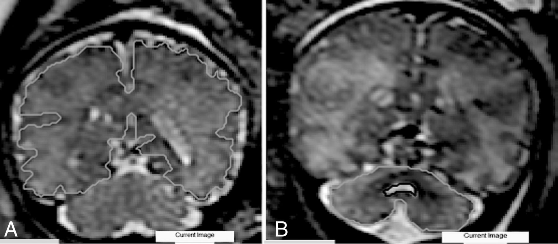
- FIG 1.
Examples of anatomic boundaries. A, ST. B, CER.
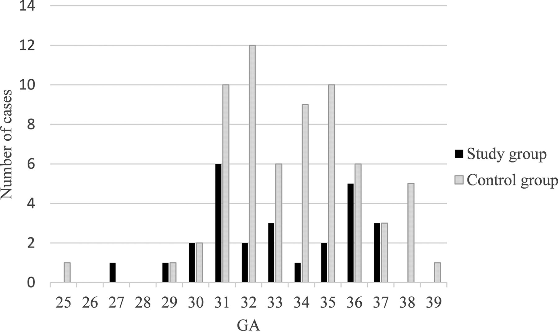
- FIG 2.
Distribution of MR images according to GA in the study and control groups.
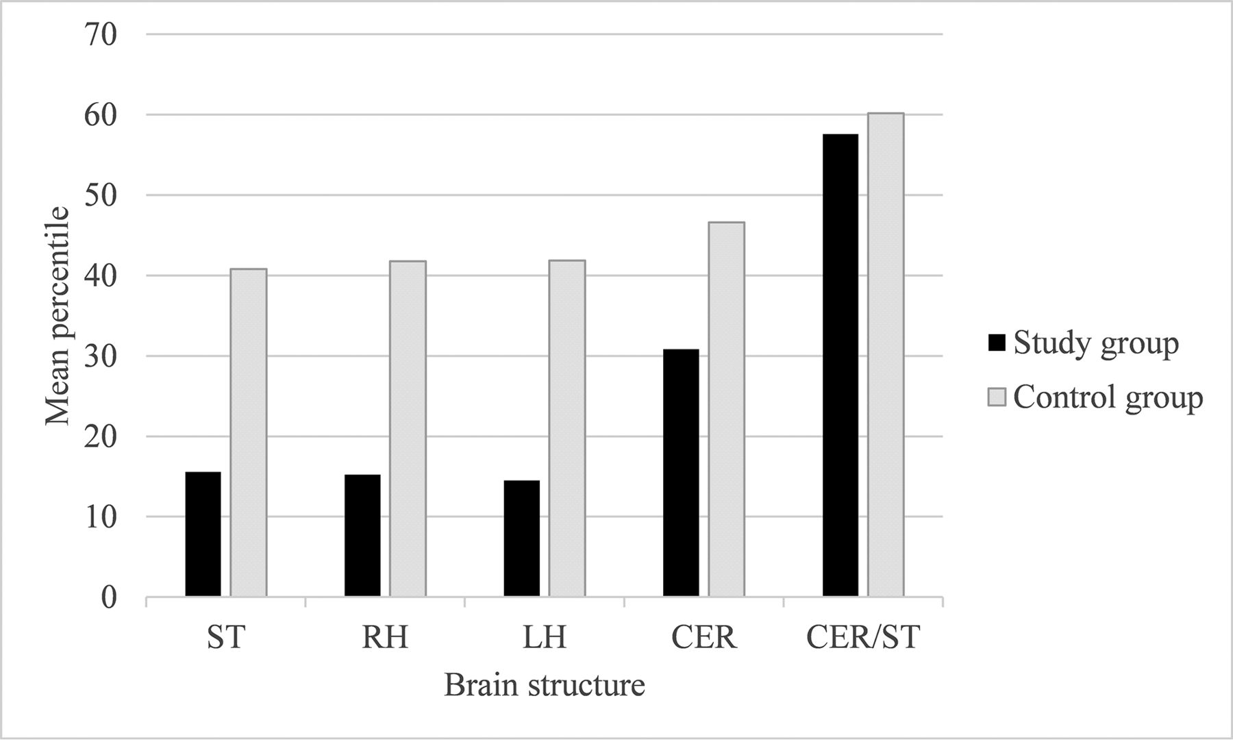
- FIG 3.
Comparison of the mean percentile of brain structures between the 2 groups.
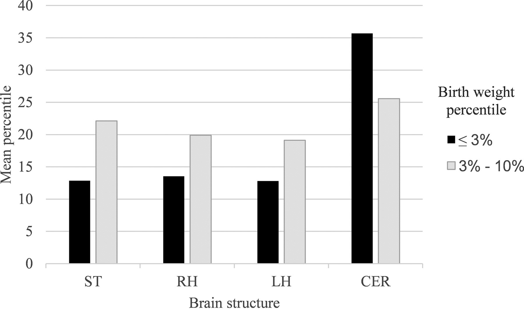
- FIG 4.
Mean percentile of brain structures by birth weight percentiles in the study group.


Tables
- Table 1:
Characteristics of the control population alongside the study population expressed as mean (SD) or frequency and percentage and level of significance
Study Group (n = 26) Control Group (n = 66) P Value Mothers’ characteristics Maternal age at pregnancy (yr) 32.38 (SD, 5.26) 33.03 (SD, 4.66) .57 Thyroid disorders 3/26 (11.5%) 4/66 (6%) .40 Anemia 1/26 (3.8%) 5/66 (7.5%) .67 Blood clotting disorders 1/26 (3.8%) 7/66 (10.6%) .43 Hypertension 1/26 (3.8%) 2/66 (3%) 1 Pregestational diabetes 1/26 (3.8%) 0 .28 Pregnancy characteristics Spontaneous conception 21/26 (80%) 57/65a (88%) .51 Fetal sex Male 15/26 (57%) Male 32/66 (48%) .43 Female 11/26 (42%) Female 34/66 (52%) GA at MR imaging examination 33.3 (SD, 2.79) 34.1 (SD, 2.58) .20 Neonate characteristics Type of birth Vaginal 4/25a (16%) Vaginal 43/64a (67%) <.001 Cesarean 21/25a (84%) Cesarean 21/64a (33%) <.001 Week of birth 35.04 (SD, 2.66) 38.52 (SD, 1.27) <.001 ↵a There are some cases in which the data are not documented in our records.
- Table 2:
Mean volume of brain structures (mL), ratio (CER/ST), and SD in the study group compared with the control group and level of significance
Brain Area Study Group Control Group P Value ST 179.93 (SD, 45.71) 210.66 (SD, 48.99) .006 RH 88.43 (SD, 22.02) 105.34 (SD, 24.59) .003 LH 88.76 (SD, 22.68) 105.22 (SD, 24.82) .004 CER 10.25 (SD, 3.5) 12.16 (SD, 3.27) .015 CER/ST 0.056 (SD, 0.01) 0.058 (SD, 0.01) .39 - Table 3:
Mean percentile of the brain structures or ratio (CER/ST), the SD in the research group compared with the control group, and the level of significance for the difference between them
Brain Area Study Group Control Group P Value ST 15.57 (SD, 23.4) 40.78 (SD, 37.24) <.001 RH 15.25 (SD, 23.03) 41.76 (SD, 37.22) <.001 LH 14.52 (SD, 22.64) 41.86 (SD, 37.7) <.001 CER 30.82 (SD, 30.24) 46.62 (SD, 34.39) .04 CER/ST 57.58 (SD, 34.04) 60.17 (SD, 31.72) .73 - Table 4:
Mean birth weight and birth weight percentile and SD in the study group compared with the control group and level of significance
Study Group (n = 25)a Control Group(n = 63)a P Value Birth weight (g) 1639.16 (SD, 543.70) 3209.4286 (SD, 456.93) <.001 Birth weight percentile 4.04 (SD, 3.29) 54.09 (SD, 26.76) <.001 ↵a There are some cases in which the birth weight and birth weight percentile are not documented in our records.
- Table 5:
Mean percentile of brain structures by birth weight percentiles in the study group and level of significance for the difference between thema
Brain Area <3% (n = 16) 3%–10% (n = 9) P Value ST 12.84 (SD, 25.27) 22.11 (SD, 20.55) .36 RH 13.53 (SD, 24.89) 19.89 (SD, 20.98) .52 LH 12.81 (SD, 25.15) 19.11 (SD, 18.99) .52 CER 35.68 (SD, 32.56) 25.56 (SD, 25.99) .43 ↵a There is 1 case in which the weight percentile is not documented in our records.
- Table 6:
Intraobserver and interobserver reliability of measurements expressed as ICC and 95% CI
Brain Area Intraobserver Interobserver ICC 95% CI ICC 95% CI ST 1 0.999–1 0.998 0.992–1 RH 0.999 0.997–1 0.998 0.993–1 LH 0.997 0.988–0.999 0.996 0.985–0.999 CER 0.999 0.995–1 0.998 0.993–1


