Advertisement

Main menu

  • Home
  • Content
    • Current Issue
    • Accepted Manuscripts
    • Article Preview
    • Past Issue Archive
    • AJNR Case Collection
    • Case of the Week Archive
    • Classic Case Archive
    • Case of the Month Archive
  • Special Collections
    • Spinal CSF Leak Articles (Jan 2020-June 2024)
    • 2024 AJNR Journal Awards
    • Most Impactful AJNR Articles
  • Multimedia
    • AJNR Podcast
    • AJNR Scantastics
    • Video Articles
  • For Authors
    • Submit a Manuscript
    • Author Policies
    • Fast publishing of Accepted Manuscripts
    • Graphical Abstract Preparation
    • Manuscript Submission Guidelines
    • Imaging Protocol Submission
    • Submit a Case for the Case Collection
  • About Us
    • About AJNR
    • Editorial Board
  • More
    • Become a Reviewer/Academy of Reviewers
    • Subscribers
    • Permissions
    • Alerts
    • Feedback
    • Advertisers
    • ASNR Home
  • Other Publications
    • ajnr

User menu

  • Alerts
  • Log in

Search

  • Advanced search
American Journal of Neuroradiology

American Journal of Neuroradiology

ASHNR American Society of Functional Neuroradiology ASHNR American Society of Pediatric Neuroradiology ASSR
  • Alerts
  • Log in

Advanced Search

  • Home
  • Content
    • Current Issue
    • Accepted Manuscripts
    • Article Preview
    • Past Issue Archive
    • AJNR Case Collection
    • Case of the Week Archive
    • Classic Case Archive
    • Case of the Month Archive
  • Special Collections
    • Spinal CSF Leak Articles (Jan 2020-June 2024)
    • 2024 AJNR Journal Awards
    • Most Impactful AJNR Articles
  • Multimedia
    • AJNR Podcast
    • AJNR Scantastics
    • Video Articles
  • For Authors
    • Submit a Manuscript
    • Author Policies
    • Fast publishing of Accepted Manuscripts
    • Graphical Abstract Preparation
    • Manuscript Submission Guidelines
    • Imaging Protocol Submission
    • Submit a Case for the Case Collection
  • About Us
    • About AJNR
    • Editorial Board
  • More
    • Become a Reviewer/Academy of Reviewers
    • Subscribers
    • Permissions
    • Alerts
    • Feedback
    • Advertisers
    • ASNR Home
  • Follow AJNR on Twitter
  • Visit AJNR on Facebook
  • Follow AJNR on Instagram
  • Join AJNR on LinkedIn
  • RSS Feeds

Welcome to the new AJNR, Updated Hall of Fame, and more. Read the full announcements.


AJNR is seeking candidates for the position of Associate Section Editor, AJNR Case Collection. Read the full announcement.


Research ArticleAdult Brain

Perfusion Scotoma: A Potential Core Underestimation in CT Perfusion in the Delayed Time Window in Patients with Acute Ischemic Stroke

K. Abrams and G. Dabus
American Journal of Neuroradiology June 2022, 43 (6) 813-816; DOI: https://doi.org/10.3174/ajnr.A7524
K. Abrams
aFrom the Department of Radiology (K.A.), Baptist Hospital of Miami, Miami Neuroscience Institute, Miami, Florida
  • Find this author on Google Scholar
  • Find this author on PubMed
  • Search for this author on this site
  • ORCID record for K. Abrams
G. Dabus
bDepartment of Neuroendovascular Surgery (G.D.), Miami Cardiac and Vascular Institute, Miami Neuroscience Institute, Miami, Florida
  • Find this author on Google Scholar
  • Find this author on PubMed
  • Search for this author on this site
  • ORCID record for G. Dabus
  • Article
  • Figures & Data
  • Info & Metrics
  • Responses
  • References
  • PDF
Loading

Abstract

SUMMARY: With the growing rise in utilization of CT perfusion for selecting patients for thrombectomy in acute ischemic stroke from large vessel occlusion, some potential pitfalls are becoming more commonly seen particularly when it comes to estimating the core infarct size on CT perfusion. Ghost infarct core has been described to account for overestimating core infarct size in the early time period (<3 hours). Herein, we describe the phenomenon of underestimating core infarct size on CT perfusion in the later time period (>6 hours), which we have termed perfusion scotoma.

As CT perfusion has grown in practice in aiding the selection of patients eligible for thrombectomy in acute ischemic stroke, some potential pitfalls are becoming more commonly seen when it is used to estimate the core infarct size. While DWI remains the criterion standard for visualizing the ischemic stroke core, most patients do not undergo MR imaging at initial presentation. Most institutions that are part of a stroke network use multimodal CT, which includes CTA and CT perfusion. This algorithm works well for most institutions due to the speed and availability of CT imaging.

Following the multiple, positive trials in 2015 for thrombectomy in large-vessel occlusion,1⇓-3 CT perfusion use started to grow exponentially, including for patients in the <6-hour time window. Soon after this growth occurred, reports of “ghost infarct core” were published.4 Ghost infarct core was defined as a core infarct on perfusion (defined as CBF of <30%) that overestimated the final core infarct size. This was seen most commonly in patients presenting in the <3-hour time period. Another study noted this phenomenon in the <6-hour time period.5

Further increasing the demand for this technology were the positive results of the Clinical Mismatch in the Triage of Wake Up and Late Presenting Strokes Undergoing Neurointervention with Trevo (DAWN)6 (2017) and Endovascular Therapy Following Imaging Evaluation for Ischemic Stroke 3 (DEFUSE 3)7 (2018) trials, which used quantitative CT perfusion to stratify which patients would benefit from thrombectomy in the >6-hour time period. Now that the eligible time period was increased in patients last known healthy up to 24 hours (DAWN) or up to 16 hours (DEFUSE 3), widespread CT perfusion has become far more prevalent. With this increase in use, we have seen a new pitfall in estimating the core infarct volume on CT perfusion: underestimating the core infarct volume. This phenomenon was described by Copelan et al,8 who looked at patients in the >6-hour time period who underwent CTA at a spoke hospital before undergoing CT perfusion (RAPID Software; https://www.rapidsoftware.shop/) at the hub hospital. They found that the core infarct was underestimated in this scenario and hypothesized that recent contrast administration rendered the core infarct estimation inaccurate. However, even more recently, Voleti et al9 published an article correlating the ASPECTS with the CT perfusion core in large-vessel occlusion. In general, as the ASPECTS number decreases, one would expect core infarct volume to increase. The scatterplot from that article demonstrated outliers in the correlation of the core infarct volume in patients with a lower ASPECTS who also showed low core infarct volumes on perfusion. We have termed this phenomenon of a small core infarct on CT perfusion but a low ASPECTS “perfusion scotoma.”

We have seen this phenomenon in our clinical practice almost exclusively in the >6-hour time period, when ASPECTS abnormalities are apparent. These findings appear to be independent of the vendor used for CT perfusion, judging by what has appeared in the literature as well as our own clinical experience.

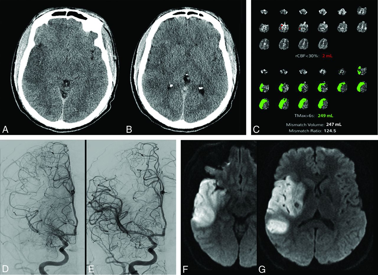
Figure 1 is an example of the perfusion scotoma from our clinical practice where a patient presented to one of our spoke hospitals last known healthy more than 8 hours before arrival. Only noncontrast CT of the brain was performed before transfer to our Comprehensive Stroke Center. The initial brain CT demonstrated a hyperdense acute clot in the M1 segment of the right MCA, with early acute ischemic changes and an ASPECTS of 6. On arrival at our comprehensive stroke center, CTA and CT perfusion were performed and demonstrated an occlusive or near-occlusive clot in the M1 segment of the right MCA, with a large area of acute ischemia (time-to-maximum, >6 seconds of 249 mL), but a minimal core infarct (CBF of <30% of 2 mL). The patient underwent angiography and successful thrombectomy of a nearly totally occlusive clot of the M1 segment of the right MCA. Follow-up MR imaging demonstrated areas of restricted diffusion corresponding to the areas of ASPECTS abnormality on the original CT scan, the “perfusion scotoma.”

FIG 1.
  • Download figure
  • Open in new tab
  • Download powerpoint
FIG 1.

A patient presented with right MCA syndrome to an outside hospital, last known healthy >8 hours before arrival. The initial brain CT at the outside institution demonstrated acute ischemic changes in the right temporal lobe and insula with an ASPECTS of 6 (A and B). CT perfusion performed at our hospital reconstructed using both manual (Advantage Workstation; GE Healthcare, not shown) and automated (Viz.ai Intelligent Care Coordination; https://www.viz.ai; C) perfusion software demonstrates a large area of acute ischemia (time-to maximum, >6 seconds of 249 mL), but a minimal core infarct (CBF of <30% of 2 mL). Cerebral angiography prethrombectomy demonstrates partial recanalization of the right M1 segment with some antegrade flow (D). The patient underwent successful thrombectomy of the M1 segment (E). Follow-up MR imaging demonstrates areas of restricted diffusion corresponding to the areas of ASPECTS abnormality on the original CT scan, the “perfusion scotoma” (F and G). rCBF indicates relative CBF; Tmax, time-to-maximum.

The likely etiology for this case and many cases of this perfusion scotoma is early reperfusion from partial recanalization as well as luxury perfusion. Although we tend to think of luxury perfusion in the 48- to 72-hour timeframe, it can also be seen within 24 hours and may contribute to this phenomenon.10 We can think of the infarcted tissue on perfusion as a continuum from reduced CBF in the <6-hour timeframe, slowly increasing with time to when it may become equivalent to normal tissue in the 6- to 24-hour timeframe and elevated in the >24-hour timeframe.

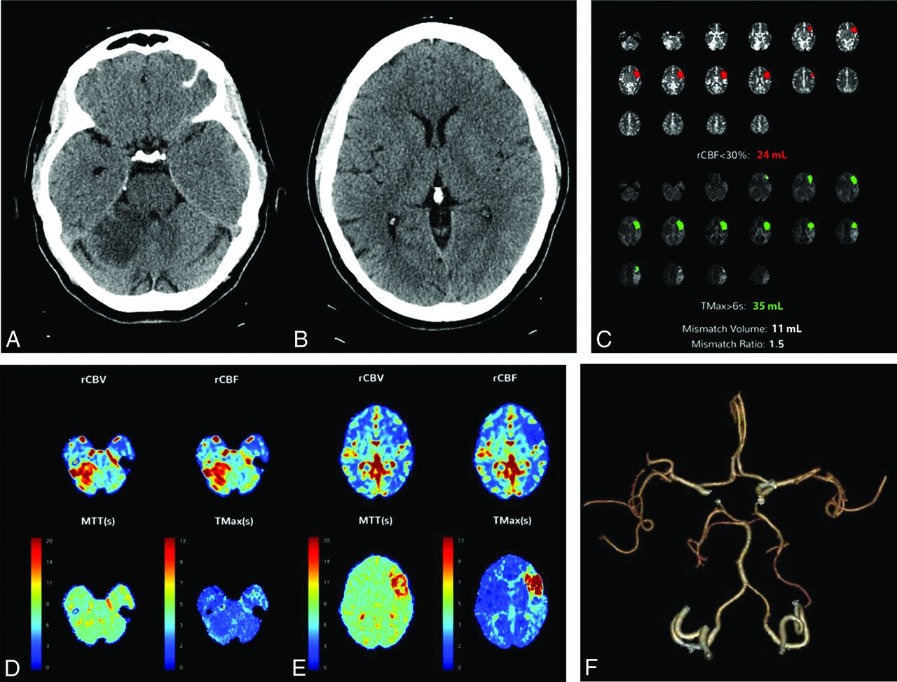
Figure 2 is an example of a patient with a subacute right superior cerebellar infarct >24 hours old but who presented with an acute left MCA syndrome. Note on the quantitative perfusion maps, not only the acute left MCA ischemia but also the absence of core infarct in the right superior cerebellar hemisphere. However, elevated CBF and CBV were noted in the cerebellar infarct on the qualitative color maps, consistent with reperfusion. The CTA also demonstrated that the right superior cerebellar artery was patent and the M2 segment of the left middle cerebral artery was occluded.

FIG 2.
  • Download figure
  • Open in new tab
  • Download powerpoint
FIG 2.

A patient presenting with an acute left MCA syndrome. CT also demonstrates an acute/subacute right superior cerebellar infarct (A and B). CT perfusion was performed and reconstructed using both manual (Advantage Workstation, not shown) and automated (C–E) perfusion software and demonstrates similar results. The quantitative perfusion maps demonstrate acute left MCA ischemia but no evidence of core infarct in the right superior cerebellar hemisphere (C). D and E, Qualitative color maps demonstrate elevated CBF and CBV in the right superior cerebellar infarct, consistent with reperfusion. CTA also demonstrates the right superior cerebellar artery to be patent and the M2 segment of the left MCA to be occluded (F). rCBF indicates relative CBF; rCBV, relative CBV; Tmax, time-to-maximum.

Why does the perfusion scotoma matter? When communicating the quantitative CT perfusion findings, it is imperative for the radiologist to also incorporate the ASPECTS to accurately assess the true ischemic penumbra. For example, in a patient with large-vessel occlusion and a large time-to-maximum of >6 seconds (eg, 150 mL) as the area of acute ischemia, the CBF of <30% (core infarct) being very low (eg, 5 mL), with a high ASPECTS (eg, >6), would imply that the patient is a thrombectomy candidate. However, if in that same patient, the ASPECTS is low (eg, 3 as seen in the scatterplot), this patient would, most likely, not be a thrombectomy candidate, even though the mismatch may be listed as 145 mL in this example; furthermore, thrombectomy may even be deleterious. It also allows the neurointerventionalist to give realistic expectations to the family regarding the procedure. Although we have not correlated the CT perfusion images to DWI in the early phase (our patients had MRIs at later time periods), we believe that this finding can be extrapolated, and it may represent a future area for research.

In summary, like the importance of understanding the concept of overestimating the core infarct in the <3-hour time period (ghost infarct core), physicians involved in acute stroke care should understand the concept of underestimating the infarct core in the >6-hour time period (perfusion scotoma).

Footnotes

  • Disclosure forms provided by the authors are available with the full text and PDF of this article at www.ajnr.org.

References

  1. 1.↵
    1. Goyal M,
    2. Demchuk AM,
    3. Menon BK, et al
    ; ESCAPE Trial Investigators. Randomized assessment of rapid endovascular treatment of ischemic stroke. N Engl J Med 2015;372:1019–30 doi:10.1056/NEJMoa1414905 pmid:25671798
    CrossRefPubMed
  2. 2.↵
    1. Saver JL,
    2. Goyal M,
    3. Bonafe A, et al
    ; SWIFT PRIME Investigators. Stent-retriever thrombectomy after intravenous t-PA vs. t-PA alone in stroke. N Engl J Med 2015;372:2285–95 doi:10.1056/NEJMoa1415061 pmid:25882376
    CrossRefPubMed
  3. 3.↵
    1. Campbell BC,
    2. Mitchell PJ,
    3. Kleinig TJ, et al
    ; EXTEND-IA Investigators. Endovascular therapy for ischemic stroke with perfusion-imaging selection. N Engl J Med 2015;372:1009–18 doi:10.1056/NEJMoa1414792 pmid:25671797
    CrossRefPubMed
  4. 4.↵
    1. Martins N,
    2. Aires A,
    3. Mendez B, et al
    . Ghost infarct core and admission computed tomography perfusion: redefining the role of neuroimaging in acute ischemic stroke. Interv Neurol 2018;7:513–21 doi:10.1159/000490117 pmid:30410531
    CrossRefPubMed
  5. 5.↵
    1. Rodrigues GM,
    2. Mohammaden MH,
    3. Haussen DC, et al
    . Ghost infarct core following endovascular reperfusion: a risk for computed tomography perfusion misguided selection in stroke. Int J Stroke 2021 Nov 19. [Epub ahead of print] doi:10.1177/17474930211056228 pmid:34796765
    CrossRefPubMed
  6. 6.↵
    1. Nogueira RG,
    2. Jadhav AP,
    3. Haussen DC, et al
    ; DAWN Trial Investigators. Thrombectomy 6 to 24 hours after stroke with a mismatch between deficit and infarct. N Engl J Med 2018;378:11–21 doi:10.1056/NEJMoa1706442 pmid:29129157
    CrossRefPubMed
  7. 7.↵
    1. Albers GW,
    2. Marks MP,
    3. Kemp S, et al
    ; DEFUSE 3 Investigators. Thrombectomy for stroke at 6 to 16 hours with selection by perfusion imaging. N Engl J Med 2018;378:708–18 doi:10.1056/NEJMoa1713973 pmid:29364767
    CrossRefPubMed
  8. 8.↵
    1. Copelan AZ,
    2. Smith ER,
    3. Drocton GT, et al
    . Recent administration of iodinated contrast renders core infarct estimation inaccurate using RAPID software. AJNR Am J Neuroradiol 2020;41:2235–42 doi:10.3174/ajnr.A6908 pmid:33214184
    Abstract/FREE Full Text
  9. 9.↵
    1. Voleti S,
    2. Vidovich J,
    3. Corcoran B, et al
    . Correlation of Alberta Stroke Program Early Computed Tomography Score with computed tomography perfusion core in large vessel occlusion in delayed time windows. Stroke 2021;52:498–504 doi:10.1161/STROKEAHA.120.030353 pmid:33406866
    CrossRefPubMed
  10. 10.↵
    1. Nagar VA,
    2. McKinney AM,
    3. Karagulle AT, et al
    . Reperfusion phenomenon masking acute and subacute infarcts at dynamic perfusion CT: confirmation by fusion of CT and diffusion-weighted MR images. AJNR Am J Neuroradiol 2009;193:1629–38 doi:10.2214/AJR.09.2664 pmid:19933658
    CrossRefPubMed
  • Received February 15, 2022.
  • Accepted after revision April 7, 2022.
  • © 2022 by American Journal of Neuroradiology
PreviousNext
Back to top

In this issue

American Journal of Neuroradiology: 43 (6)
American Journal of Neuroradiology
Vol. 43, Issue 6
1 Jun 2022
  • Table of Contents
  • Index by author
  • Complete Issue (PDF)
Advertisement
Print
Download PDF
Email Article

Thank you for your interest in spreading the word on American Journal of Neuroradiology.

NOTE: We only request your email address so that the person you are recommending the page to knows that you wanted them to see it, and that it is not junk mail. We do not capture any email address.

Enter multiple addresses on separate lines or separate them with commas.
Perfusion Scotoma: A Potential Core Underestimation in CT Perfusion in the Delayed Time Window in Patients with Acute Ischemic Stroke
(Your Name) has sent you a message from American Journal of Neuroradiology
(Your Name) thought you would like to see the American Journal of Neuroradiology web site.
CAPTCHA
This question is for testing whether or not you are a human visitor and to prevent automated spam submissions.
Cite this article
K. Abrams, G. Dabus
Perfusion Scotoma: A Potential Core Underestimation in CT Perfusion in the Delayed Time Window in Patients with Acute Ischemic Stroke
American Journal of Neuroradiology Jun 2022, 43 (6) 813-816; DOI: 10.3174/ajnr.A7524

Citation Manager Formats

  • BibTeX
  • Bookends
  • EasyBib
  • EndNote (tagged)
  • EndNote 8 (xml)
  • Medlars
  • Mendeley
  • Papers
  • RefWorks Tagged
  • Ref Manager
  • RIS
  • Zotero
0 Responses
Respond to this article
Share
Bookmark this article
CT Perfusion Scotoma in Acute Stroke
K. Abrams, G. Dabus
American Journal of Neuroradiology Jun 2022, 43 (6) 813-816; DOI: 10.3174/ajnr.A7524
del.icio.us logo Twitter logo Facebook logo Mendeley logo
  • Tweet Widget
  • Facebook Like
  • Google Plus One
Purchase

Jump to section

  • Article
    • Abstract
    • Footnotes
    • References
  • Figures & Data
  • Info & Metrics
  • Responses
  • References
  • PDF

Related Articles

  • No related articles found.
  • PubMed
  • Google Scholar

Cited By...

  • No citing articles found.
  • Crossref (11)
  • Google Scholar

This article has been cited by the following articles in journals that are participating in Crossref Cited-by Linking.

  • Comparison of CT Perfusion Software Packages for Thrombectomy Eligibility
    Leonardo Pisani, Diogo C. Haussen, Mahmoud Mohammaden, Catarina Perry da Camara, Dinesh V. Jillella, Gabriel M. Rodrigues, Mehdi Bouslama, Alhamza Al‐Bayati, Adam Prater, Bernardo Liberato, Michael R. Frankel, Raul G. Nogueira
    Annals of Neurology 2023 94 5
  • Cerebral Infarct Growth: Pathophysiology, Pragmatic Assessment, and Clinical Implications
    Umberto Pensato, Andrew M. Demchuk, Bijoy K. Menon, Thanh N. Nguyen, Gabriel Broocks, Bruce C.V. Campbell, Diego A. Gutierrez Vasquez, Peter J. Mitchell, Michael D. Hill, Mayank Goyal, Johanna M. Ospel
    Stroke 2025 56 1
  • Automated advanced imaging in acute ischemic stroke. Certainties and uncertainties
    Enrico Fainardi, Giorgio Busto, Andrea Morotti
    European Journal of Radiology Open 2023 11
  • Effect of Hemoglobin and Blood Glucose Levels on CT Perfusion Ischemic Core Estimation
    Umberto Pensato, Salome Bosshart, Alexander Stebner, Axel Rohr, Timothy J. Kleinig, Rishi Gupta, Götz Thomalla, Ji Hoe Heo, Mayank Goyal, Andrew M. Demchuk, Michael D. Hill, Johanna M. Ospel
    Neurology 2024 103 10
  • Acute Ischemic Stroke in the Clinic and the Laboratory: Targets for Translational Research
    Bart Franx, Rick M. Dijkhuizen, Diederik W.J. Dippel
    Neuroscience 2024 550
  • Optimal Cerebral Blood Flow Thresholds for Ischemic Core Estimation Using Computed Tomography Perfusion and Diffusion‐Weighted Imaging
    Nakhoon Kim, Wi‐Sun Ryu, Sue Young Ha, Jun Yup Kim, Jihoon Kang, Sung Hyun Baik, Cheolkyu Jung, Moon‐Ku Han, Hee‐Joon Bae, Longting Lin, Mark Parsons, Beom Joon Kim
    Annals of Neurology 2025 97 5
  • Artificial intelligence in neuroradiology: brain CT perfusion imaging for acute ischemic stroke management
    Umberto ROZZANIGO, Giulia CASAGRANDA, Marianna MOCHEN, Mauro FERRARI
    Journal of Radiological Review 2023 10 3
  • Association of CT perfusion parameters with outcomes in patients with medium vessel occlusion undergoing endovascular thrombectomy
    Yoel Schwartzmann, Hamza Joubran, Tamer Jubeh, Issa Metanis, Aviva Alpernas, Tali Jonas-Kimchi, Udi Sadeh, John M. Gomori, Jose E. Cohen, Hen Hallevi, Jeremy Molad, Ronen R. Leker
    Therapeutic Advances in Neurological Disorders 2025 18
  • Computed tomography perfusion ischemic core underestimation: a perfusion scotoma case
    Giorgio Busto, Elisa Scola, Enrico Fainardi
    Neurological Sciences 2024 45 2
  • Deep transfer learning radiomics for distinguishing sinonasal malignancies: a preliminary MRI study
    Naier Lin, Yiqian Shi, Min Ye, Yiyin Zhang, Xianhao Jia
    Future Oncology 2025 21 8

More in this TOC Section

  • Diagnostic Neuroradiology of Monoclonal Antibodies
  • ML for Glioma Molecular Subtype Prediction
  • Segmentation of Brain Metastases with BLAST
Show more Adult Brain

Similar Articles

Advertisement

Indexed Content

  • Current Issue
  • Accepted Manuscripts
  • Article Preview
  • Past Issues
  • Editorials
  • Editors Choice
  • Fellow Journal Club
  • Letters to the Editor

Cases

  • Case Collection
  • Archive - Case of the Week
  • Archive - Case of the Month
  • Archive - Classic Case

Special Collections

  • Special Collections

Resources

  • News and Updates
  • Turn around Times
  • Submit a Manuscript
  • Author Policies
  • Manuscript Submission Guidelines
  • Evidence-Based Medicine Level Guide
  • Publishing Checklists
  • Graphical Abstract Preparation
  • Imaging Protocol Submission
  • Submit a Case
  • Become a Reviewer/Academy of Reviewers
  • Get Peer Review Credit from Publons

Multimedia

  • AJNR Podcast
  • AJNR SCANtastic
  • Video Articles

About Us

  • About AJNR
  • Editorial Board
  • Not an AJNR Subscriber? Join Now
  • Alerts
  • Feedback
  • Advertise with us
  • Librarian Resources
  • Permissions
  • Terms and Conditions

American Society of Neuroradiology

  • Not an ASNR Member? Join Now

© 2025 by the American Society of Neuroradiology All rights, including for text and data mining, AI training, and similar technologies, are reserved.
Print ISSN: 0195-6108 Online ISSN: 1936-959X

Powered by HighWire