
Article Figures & Data
Figures
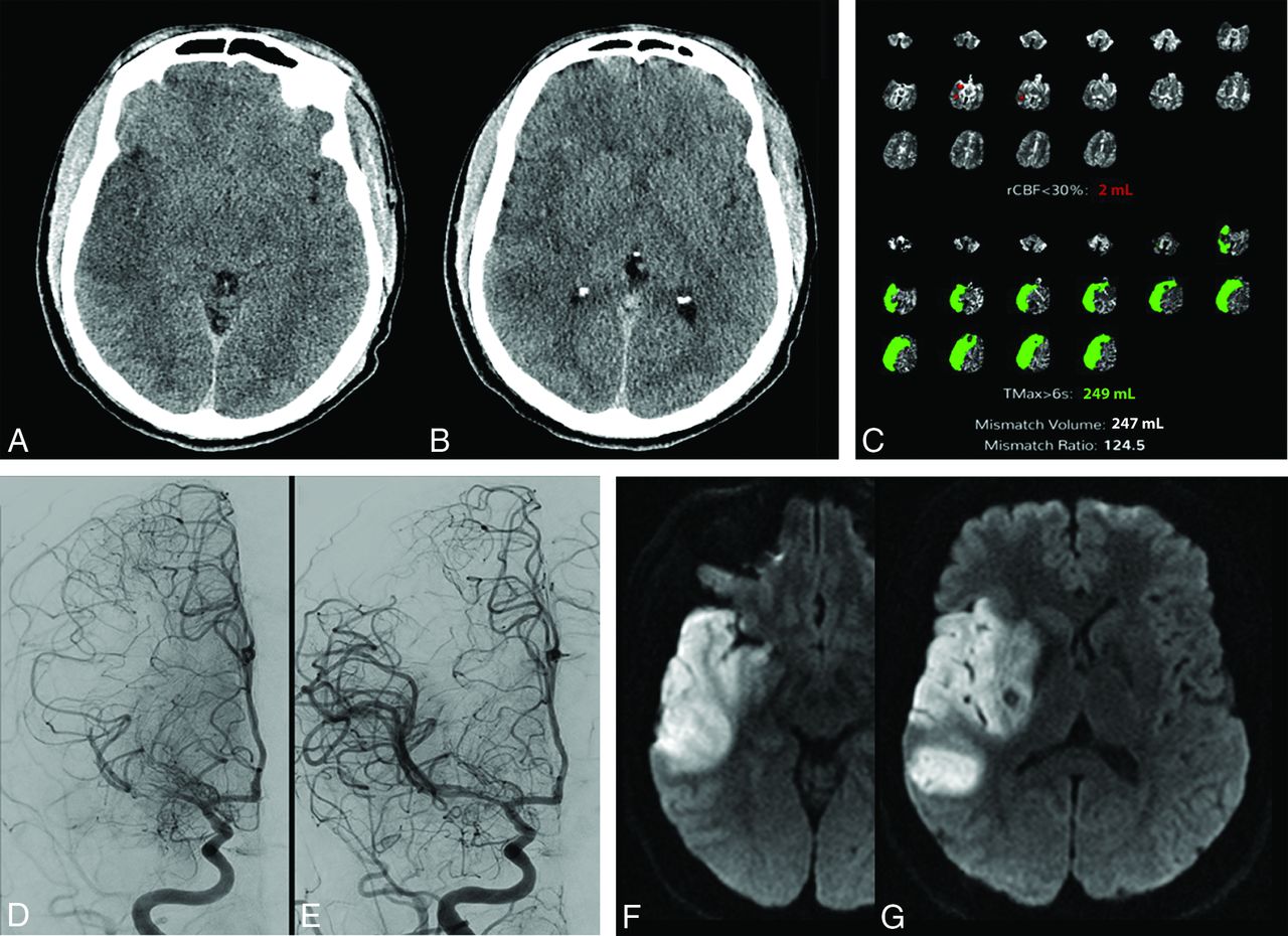
- FIG 1.
A patient presented with right MCA syndrome to an outside hospital, last known healthy >8 hours before arrival. The initial brain CT at the outside institution demonstrated acute ischemic changes in the right temporal lobe and insula with an ASPECTS of 6 (A and B). CT perfusion performed at our hospital reconstructed using both manual (Advantage Workstation; GE Healthcare, not shown) and automated (Viz.ai Intelligent Care Coordination; https://www.viz.ai; C) perfusion software demonstrates a large area of acute ischemia (time-to maximum, >6 seconds of 249 mL), but a minimal core infarct (CBF of <30% of 2 mL). Cerebral angiography prethrombectomy demonstrates partial recanalization of the right M1 segment with some antegrade flow (D). The patient underwent successful thrombectomy of the M1 segment (E). Follow-up MR imaging demonstrates areas of restricted diffusion corresponding to the areas of ASPECTS abnormality on the original CT scan, the “perfusion scotoma” (F and G). rCBF indicates relative CBF; Tmax, time-to-maximum.
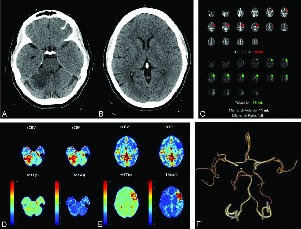
- FIG 2.
A patient presenting with an acute left MCA syndrome. CT also demonstrates an acute/subacute right superior cerebellar infarct (A and B). CT perfusion was performed and reconstructed using both manual (Advantage Workstation, not shown) and automated (C–E) perfusion software and demonstrates similar results. The quantitative perfusion maps demonstrate acute left MCA ischemia but no evidence of core infarct in the right superior cerebellar hemisphere (C). D and E, Qualitative color maps demonstrate elevated CBF and CBV in the right superior cerebellar infarct, consistent with reperfusion. CTA also demonstrates the right superior cerebellar artery to be patent and the M2 segment of the left MCA to be occluded (F). rCBF indicates relative CBF; rCBV, relative CBV; Tmax, time-to-maximum.


Jump to section
Related Articles
Cited By...
- No citing articles found.