Advertisement

Main menu

  • Home
  • Content
    • Current Issue
    • Accepted Manuscripts
    • Article Preview
    • Past Issue Archive
    • AJNR Case Collection
    • Case of the Week Archive
    • Classic Case Archive
    • Case of the Month Archive
  • Special Collections
    • Spinal CSF Leak Articles (Jan 2020-June 2024)
    • 2024 AJNR Journal Awards
    • Most Impactful AJNR Articles
  • Multimedia
    • AJNR Podcast
    • AJNR Scantastics
    • Video Articles
  • For Authors
    • Submit a Manuscript
    • Author Policies
    • Fast publishing of Accepted Manuscripts
    • Graphical Abstract Preparation
    • Manuscript Submission Guidelines
    • Imaging Protocol Submission
    • Submit a Case for the Case Collection
  • About Us
    • About AJNR
    • Editorial Board
  • More
    • Become a Reviewer/Academy of Reviewers
    • Subscribers
    • Permissions
    • Alerts
    • Feedback
    • Advertisers
    • ASNR Home
  • Other Publications
    • ajnr

User menu

  • Alerts
  • Log in

Search

  • Advanced search
American Journal of Neuroradiology

American Journal of Neuroradiology

ASHNR American Society of Functional Neuroradiology ASHNR American Society of Pediatric Neuroradiology ASSR
  • Alerts
  • Log in

Advanced Search

  • Home
  • Content
    • Current Issue
    • Accepted Manuscripts
    • Article Preview
    • Past Issue Archive
    • AJNR Case Collection
    • Case of the Week Archive
    • Classic Case Archive
    • Case of the Month Archive
  • Special Collections
    • Spinal CSF Leak Articles (Jan 2020-June 2024)
    • 2024 AJNR Journal Awards
    • Most Impactful AJNR Articles
  • Multimedia
    • AJNR Podcast
    • AJNR Scantastics
    • Video Articles
  • For Authors
    • Submit a Manuscript
    • Author Policies
    • Fast publishing of Accepted Manuscripts
    • Graphical Abstract Preparation
    • Manuscript Submission Guidelines
    • Imaging Protocol Submission
    • Submit a Case for the Case Collection
  • About Us
    • About AJNR
    • Editorial Board
  • More
    • Become a Reviewer/Academy of Reviewers
    • Subscribers
    • Permissions
    • Alerts
    • Feedback
    • Advertisers
    • ASNR Home
  • Follow AJNR on Twitter
  • Visit AJNR on Facebook
  • Follow AJNR on Instagram
  • Join AJNR on LinkedIn
  • RSS Feeds

Welcome to the new AJNR, Updated Hall of Fame, and more. Read the full announcements.


AJNR is seeking candidates for the position of Associate Section Editor, AJNR Case Collection. Read the full announcement.


Review ArticleMolecular Neuroimaging/Nuclear Medicine
Open Access

PET/MRI in Pediatric Neuroimaging: Primer for Clinical Practice

C. Pedersen, M. Aboian, J.E. McConathy, H. Daldrup-Link and A.M. Franceschi
American Journal of Neuroradiology July 2022, 43 (7) 938-943; DOI: https://doi.org/10.3174/ajnr.A7464
C. Pedersen
aFrom the Department of Radiology (C.P., M.A.), Yale School of Medicine, New Haven, Connecticut
  • Find this author on Google Scholar
  • Find this author on PubMed
  • Search for this author on this site
M. Aboian
aFrom the Department of Radiology (C.P., M.A.), Yale School of Medicine, New Haven, Connecticut
  • Find this author on Google Scholar
  • Find this author on PubMed
  • Search for this author on this site
  • ORCID record for M. Aboian
J.E. McConathy
bDivision of Molecular Imaging and Therapeutics (J.E.M.), Department of Radiology, University of Alabama at Birmingham, Birmingham, Alabama
  • Find this author on Google Scholar
  • Find this author on PubMed
  • Search for this author on this site
  • ORCID record for J.E. McConathy
H. Daldrup-Link
cDepartment of Radiology and Pediatrics (H.D.-L.), Stanford University School of Medicine, Palo Alto, California
  • Find this author on Google Scholar
  • Find this author on PubMed
  • Search for this author on this site
A.M. Franceschi
dNeuroradiology Division (A.M.F.), Department of Radiology, Northwell Health/Donald and Barbara Zucker School of Medicine, Lenox Hill Hospital, New York, New York
  • Find this author on Google Scholar
  • Find this author on PubMed
  • Search for this author on this site
  • ORCID record for A.M. Franceschi
  • Article
  • Figures & Data
  • Supplemental
  • Info & Metrics
  • Responses
  • References
  • PDF
Loading

Abstract

SUMMARY: Modern pediatric imaging seeks to provide not only exceptional anatomic detail but also physiologic and metabolic information of the pathology in question with as little radiation penalty as possible. Hybrid PET/MR imaging combines exquisite soft-tissue information obtained by MR imaging with functional information provided by PET, including metabolic markers, receptor binding, perfusion, and neurotransmitter release data. In pediatric neuro-oncology, PET/MR imaging is, in many ways, ideal for follow-up compared with PET/CT, given the superiority of MR imaging in neuroimaging compared with CT and the lower radiation dose, which is relevant in serial imaging and long-term follow-up of pediatric patients. In addition, although MR imaging is the main imaging technique for the evaluation of spinal pathology, PET/MR imaging may provide useful information in several clinical scenarios, including tumor staging and follow-up, treatment response assessment of spinal malignancies, and vertebral osteomyelitis. This review article covers neuropediatric applications of PET/MR imaging in addition to considerations regarding radiopharmaceuticals, imaging protocols, and current challenges to clinical implementation.

ABBREVIATIONS:

DOPA
dioxyphenylalanine
DOTATATE
[tetrazetan-D-Phe1,Tyr3]-octreotate
FET
fluoroethyltyrosine
mFBG
meta-fluorobenzylguanidine
MIBG
metaiodobenzylguanidine
LCH
Langerhans cell histiocytosis
max
maximum
MET
methionine
SUV
standard uptake value

Serial imaging and radiation dose reduction should remain balanced in pediatric imaging. Repeat PET/CTs, especially in pediatric neuro-oncology, result in a considerable cumulative radiation dose and may increase the risk of secondary cancer.1⇓-3 The risk of radiation-induced malignancy is increased at exposures of >50–100 mSv.2 A retrospective review of 78 pediatric patients found that the average cumulative dose from PET/CT during a 5-year period amounted to 78.9 mSV.3 Meanwhile, reduction in the cumulative dose by PET/MR imaging has been reported to be as high as 50%–70% in pediatric lymphoma.4⇓-6 Further dose reduction may be achieved by lowering radiopharmaceutical doses with artificial intelligence–based algorithms, which is an area of active research and product development.7,8

Hybrid PET/MR imaging is particularly promising in pediatric neuroimaging because it allows functional and soft-tissue characterization with a low radiation dose and comparable agreement with PET/CT reported in several recent studies.5,6,9 Unlike PET applications in body imaging, the CT component of brain PET/CT typically provides little clinically useful information beyond attenuation correction, but it does contribute to the radiation dose. PET can be used to differentiate high-grade from low-grade tumors at the initial work-up, provide prognosis for patient progression-free survival and overall survival, identify the site for optimal biopsy, and determine the extent of tumor to optimize resection and radiation therapy.10 PET is also useful to evaluate tumor recurrence posttreatment in the setting of equivocal MR imaging findings and to detect transformation of tumor to a higher-grade malignancy.10 MR imaging allows DWI and FLAIR sequences, which are valuable in brain tumor assessment, and also whole-body evaluation of metastatic disease.11⇓-13 MR imaging–based attenuation correction methods for PET are more complex than for CT but are possible and typically involve tissue-segmentation techniques.14

PET/MR imaging can be performed with a sequential or synchronous system. In a synchronous system, the solid-state PET detectors are located between the body and gradient coils in the 3T MR imaging gantry, which allows truly synchronous data acquisition. In a sequential system, PET and MR imaging are performed separately with transportation of the patient between scanners. A sequential system is technically easier to achieve because most US hospitals currently own separate MR imaging and PET machines, but 1 major drawback is the asynchronous nature of the scans, potentially giving rise to misregistration artifacts, longer scan times, and longer sedation requirements.15,16 Therefore, a synchronous PET/MR imaging system is generally preferable in pediatric neuroimaging.4

Reduction in sedation need and sedation time is best achieved by decreasing the overall MR imaging scan time, eg, by specifically tailored protocols and sequence design.17,18 In 1 study, the use of a rapid brain-tailored protocol in 1308 pediatric emergency department encounters decreased head CT use by >20% without a missed diagnosis on follow-up imaging.19 Sequence design may be optimized with SyntheticMR (https://syntheticmr.com/company/), which allows reconstruction of multiple sequences for qualitative and quantitative analysis from a single acquisition20 and has been applied to multiple disease processes including cancer, neurodegenerative disorders, and stroke.21,22

Combining PET and MR imaging results in imaging time reduction, decreases sedation time and need, and, overall, has the potential to increase throughput of scans.4 Cost analysis of savings from increased throughput of patients remains an area of future investigation.

PET/MR Imaging in Pediatric Neuro-Oncology

CNS cancer is the leading cause of death in children and adolescents.23 Standard-of-care treatment for pediatric brain tumors is maximum safe resection, targeted radiation therapy, and chemotherapy. PET may help differentiate high-grade from low-grade tumors, prognosticate progression-free and overall survival, identify an optimal biopsy site, and determine tumor extent to optimize resection and radiation therapy.10 Postoperative MR imaging may be equivocal or miss small residual lesions. In this setting, PET can be used to evaluate tumor recurrence and detect transformation to a higher tumor grade.

[18F] FDG-PET.

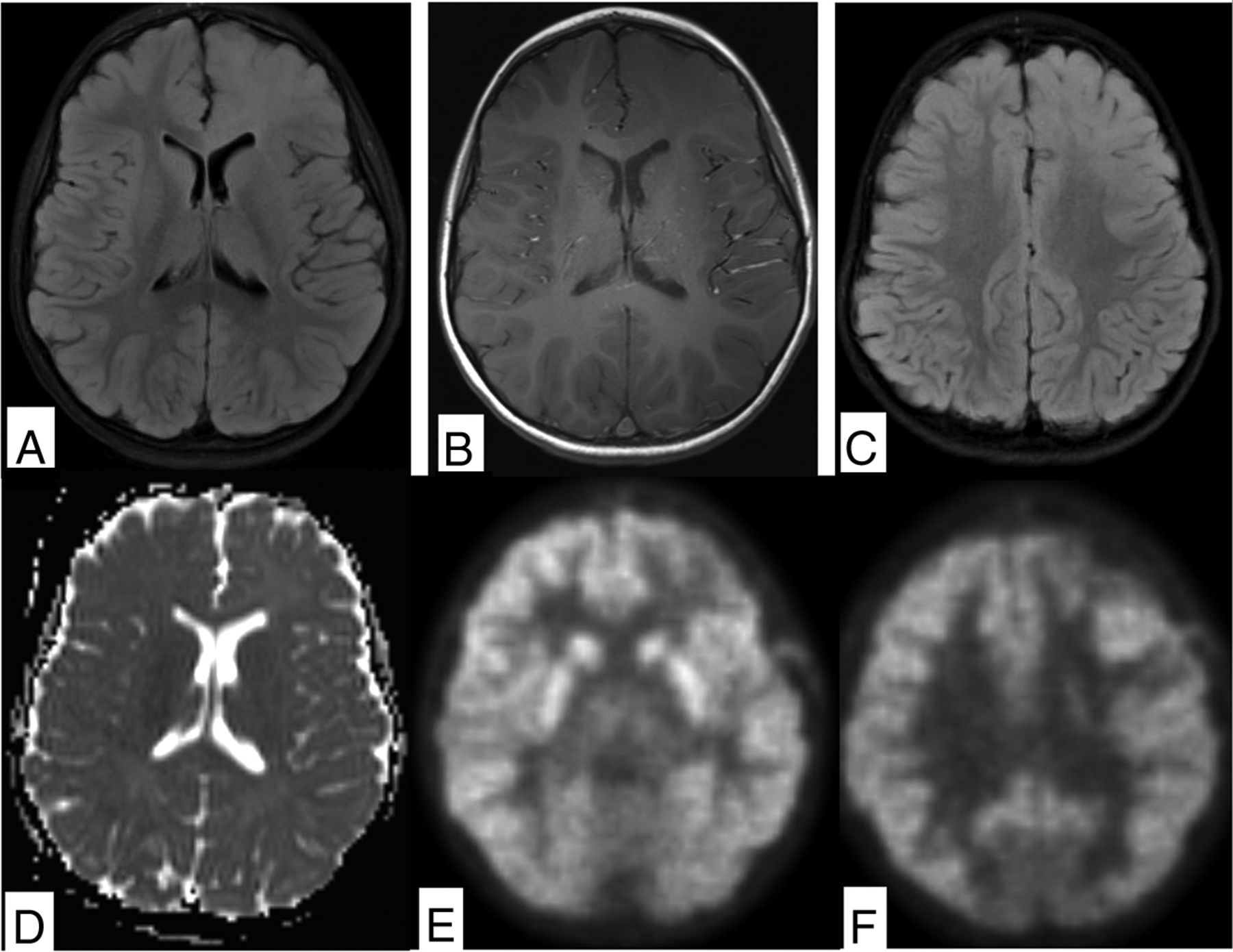
FDG is a positron-emitting analog of glucose that uses glucose transporters to transport labeled FDG into the cells. Once the FDG is inside the cell, it gets phosphorylated by hexokinase, resulting in intracellular retention.24 This method allows imaging of cellular glucose uptake and thus allows for assessment of cellular glucose metabolism. A higher level of FDG uptake has been shown to correlate with a higher tumor grade and lead to survival prediction in primary brain tumors such as gliomas.10 Disadvantages of FDG-PET include high normal brain parenchymal uptake, which may lead to poor visualization of low-grade tumors, and limitations in tumor-margin assessment (Figure). Prominent FDG uptake can also be seen in inflammatory lesions, so careful multimodal lesion evaluation is recommended.25 FDG-PET is particularly valuable when differentiating posttreatment changes from tumor recurrence in the clinical setting because the former is not expected to be FDG-avid (Online Supplemental Data).24

FIGURE.
  • Download figure
  • Open in new tab
  • Download powerpoint
FIGURE.

Diffuse astrocytoma in 4-year-old child demonstrates a nonenhancing FLAIR hyperintense mass involving the gray and white matter of the left frontal lobe (A and B). There is no evidence of reduced diffusion or hypointense signal on ADC (C and D). The inferior aspect of the lesion demonstrates a subtle decrease in FDG uptake compared with the contralateral side (E), while the superior portion demonstrates relative hypermetabolism on FDG-PET (F).

Amino Acid PET.

Amino acids are critical substrates in cellular metabolic pathways for synthesis of proteins and nucleotides and generation of adenosine triphosphate, all essential for cell function and growth. Up-regulation of amino acid transporters is an early step in carcinogenesis.26,27 The main advantage of amino acid–based tracer imaging is very low background uptake by normal brain parenchyma, allowing better lesion detection and improved tumor-border visualization. Natural and modified amino acids have been used to study amino acid metabolism of tumors, with the most commonly used tracers including 11C-methionine (MET), [18F] dioxyphenylalanine (DOPA), [18F] fluoroethyltyrosine ([18F] FET), and [18F] fluciclovine (Online Supplemental Data).26,28,29 11C-MET is a natural amino acid labeled with a carbon-11 radionuclide that has a very short half-life of 20 minutes and is, thus, limited to centers with an on-site cyclotron. The main benefit of 11C-MET in pediatrics is the low radiation dose that is administered, but the inconvenience of a short half-life outweighs the benefits. [18F] FET and [18F] DOPA are modified amino acids that use the same pathway as natural amino acids but are labeled with [18F], which has led to more widespread use due to a longer half-life (110 minutes). Both tracers can be analyzed on static imaging, but interpretation of dynamic PET is more established for [18F] FET.30,31 One of the main applications of [18F] FET-PET in pediatric neuro-oncology is the definition of residual tumor after resection. In a study evaluating residual brain tumors in 27 pediatric patients, [18F] FET-PET found at least 1 residual tumor not clearly identified on MR imaging, which significantly altered management.32 There are also promising results for the differentiation of radiation necrosis from tumor recurrence and the definition of tumor hypermetabolic regions, though most of the studies are performed in adults.33

[18F] FDG- and 11C-MET-PET were compared in 27 children with newly diagnosed brain tumors with diverse histology.34 Twenty-two of 23 patients had increased uptake on MET-PET. High-grade tumors demonstrated a significantly elevated mean standard uptake value (SUVmean) and maximum SUV (SUVmax) compared with low-grade tumors. When the same patients were imaged with FDG-PET, 52% of tumors were hypermetabolic; 38%, eumetabolic; and 10%, hypometabolic. Because FDG uptake was higher in high-grade tumors, it was proposed that FDG-PET may provide information on tumor grade, while MET-PET provides superior information on tumor location and border delineation.

An emerging amino acid PET tracer in pediatrics [18F] fluciclovine PET, also known as Axumin PET, is an FDA-approved tracer for imaging metastatic prostate cancer, but it is also showing promising results in neuro-oncology.29,35⇓⇓-38 It has lower background uptake compared with 11C-MET and is transported by both L‐type amino acid transporter 1 and system alanine‐serine‐cysteine amino acid transporter‐2 into the cell. Upcoming trials in pediatric brain tumors will elucidate the role and applications of this tracer in clinical neuro-oncology.

Head and Neck Tumors

Orbital Malignancies.

Hybrid PET/MR imaging in pediatric orbital disease is still under evaluation. Orbital involvement by lymphoma and other lymphoproliferative malignancies can be evaluated using [18F] FDG-PET, while malignancies of the optic nerves can be evaluated by both [18F] FDG and amino acid tracers.39 Uveal melanoma has been described as FDG-avid, while intraorbital retinoblastoma demonstrates heterogeneous FDG avidity.39,40 Meningiomas may originate along the optic nerve sheath or extend into the orbit through the sphenoid bone or foramen rotundum and can be evaluated using somatostatin receptor analogs such as gallium-68 [tetrazetan-D-Phe1,Tyr3]-octreotate (DOTATATE).41

Rhabdomyosarcoma in the Head and Neck.

Rhabdomyosarcoma in the head and neck region requires multimodal evaluation. The primary tumor is best evaluated with high-resolution MR imaging including noncontrast and non-fat-suppressed T1 and postcontrast T1 while [18F] FDG-PET can help delineate metastases and detect tumor recurrence (Online Supplemental Data). Recent literature indicates that FDG-PET is superior to conventional imaging in characterization of metastatic lesions and prediction of treatment response and patient outcomes.42,43 Notably, chest CT is currently still required to evaluate pulmonary metastases in these patients. While MR imaging and PET are making progress, the sensitivity of CT for pulmonary metastases remains superior.

Neuroendocrine Malignancies.

Neuroblastoma originates in primitive neural crest cells of the sympathetic nervous system and is the most common solid extracranial tumor of childhood. Most cases are diagnosed before 5 years of age and up to 50% of patients present with metastatic disease, commonly involving lymph nodes, bone, liver, and skin (Online Supplemental Data).44

Iodine-123-metaiodobenzylguanidine (123I-MIBG) imaging is based on iodine accumulation in tumor cells and is the primary imaging technique for staging and treatment-response assessment and provides the foundation for targeted therapy with 131I-MIBG.45,46 23I-MIBG SPECT and SPECT/CT can be acquired advantageously to assist with tracer-uptake identification. In 1 meta-analysis, 123I-MIBG had lower per-lesion accuracy but was more specific compared with [18F] FDG-PET. MR imaging is a valuable for initial local staging and treatment response and lends itself well to the multivariate presentations of neuroblastoma. The sensitivity of MR imaging was superior to that of MIBG in 1 study, though less specific.47 Whole-body MR imaging demonstrated good sensitivity for lymph node metastases, though with lower specificity compared with [18F] FDG-PET, partly due to difficulty in distinguishing treated and viable disease.48

More recently, 124I-MIBG-PET has demonstrated a favorable dosimetry profile and allows high-resolution images with increased accuracy for the detection of metastatic lesions in the head, neck, and spine compared with conventional 123I-MIBG SPECT in several studies.49,50

[18F] FDG-PET is generally preferred in non-MIBG-avid neuroblastomas, in aggressive and dedifferentiated tumors with loss of iodine uptake, and when high background activity complicates the evaluation of spinal involvement.51,52 FDG-PET has good accuracy in metastatic lesion detection compared with 123I and 131I-MIBG scans.11,43,44 Novel PET tracers such as [18F] DOPA have shown high accuracy and good agreement with MIBG in patients with relapse.53⇓-55 [18F] meta-fluorobenzylguanidine ([18F] mFBG) has been developed as a radiotracer that can provide MIBG-equivalent whole-body staging on PET/MR imaging.56,57 Thus, [18F] mFBG PET/MR imaging may combine local and whole-body staging in 1 session, which is particularly helpful in pediatric patients who typically require anesthesia for medical imaging.

Additional imaging agents for neuroblastoma include 68Ga-DOTATATE, which is a somatostatin receptor analog approved for the detection of neuroendocrine tumors expressing somatostatin receptors such as neuroblastoma.58 Somatostatin receptor analogs may be combined with peptide receptor radionuclide therapy for refractory neuroblastoma, eg, 177Lu-DOTATATE, because the tumor expresses somatostatin receptors, which allow select targeting.59

Thyroid Cancer.

Thyroid nodules are less common in children than in adults but carry a higher risk of malignancy.60 [18F] FDG-PET is not used routinely for thyroid nodules but may be useful in staging non-radioiodine-avid metastatic disease. MR imaging of the neck in this setting helps evaluate the thyroid resection bed for local recurrence and depicts anatomically complex regions such as the skull base and spine.60

PET/MR Imaging in Pediatric Spine Imaging

[18F] FDG-PET may provide useful information in treatment-response assessment of spinal malignancies, tumor staging, and follow-up. The role of FDG-PET in neuroblastomas was covered above (Online Supplemental Data). Nononcologic spinal imaging applications of PET/MR imaging include Langerhans cell histiocytosis (LCH) and spinal infection. Furthermore, [18F] DOPA may detect hormonally active beta cells in patients with congenital hyperinsulinism.

Pediatric Spinal Malignancy.

Astrocytomas are the most common primary intramedullary tumor in children and young adults. Most are slow-growing, low-grade tumors, while about 10%–15% are high-grade and may demonstrate elevated tracer uptake on [18F]-FDG- and 11C-MET-PET. The classic imaging presentation is an eccentrically located infiltrating tumor with fusiform spinal cord expansion, variable enhancement, and often an associated cyst or syrinx. Ependymomas arise from ependymal cells of the central canal and typically present as an enhancing mass with surrounding edema, associated cysts, and hemorrhage. Due to their central location, even a small ependymoma may cause partial obstruction of the central canal and lead to formation of a syrinx.

Ewing sarcoma is the second most common primary malignant osseous tumor and typically occurs between 10 and 20 years of age. Primary vertebral Ewing sarcoma may present as either a lytic, sclerotic, or mixed lytic and sclerotic mass and may involve any part of the spine. Paravertebral Ewing sarcoma may extend directly through the neuroforamina, and spinal invasion is common.61 Necrosis, cystic change, hemorrhage, and robust enhancement are common imaging findings. These tumors demonstrate avid tracer uptake on all 3 phases of technetium Tc99m methylene diphosphonate bone scans. [18F] FDG-PET may be used for staging purposes and to evaluate residual disease following treatment (Online Supplemental Data).

Sacrococcygeal teratomas are the most common congenital tumor in the fetus and neonate and can be classified into 4 types depending on their location within and outside the pelvis. They are often large tumors composed of different tissues with a variable appearance on T1, T2, and postcontrast images. About a third are immature or malignant. [18F] FDG-PET may aid in staging and posttreatment follow-up of malignant sacrococcygeal tumors.

Lymphoma

Lymphoma is one of the most common indications for PET imaging in pediatric oncology (Online Supplemental Data). The Deauville or Lugano criteria are endorsed by the Children’s Oncology Group and rely on semiquantitative measurements of glucose metabolism, which is helpful to avoid radiation therapy of non-FDG-avid residual soft-tissue masses.4 Current clinical practice includes PET/CT before, during, and after initiation of therapy, with a resultant high cumulative radiation dose. Meanwhile, PET/MR imaging provides excellent soft-tissue contrast and is either equivalent or superior for malignant lymph node detection.4,62,63 There is a high correlation between FDG-PET/MR imaging tumor SUV compared with PET/CT, though 1 study reported systematically lower SUVs on PET/MR imaging compared with PET/CT.4,63,64 The greatest benefit of PET/MR imaging in lymphoma is radiation-dose reduction.6,64 PET/MR imaging for lymphoma in children is typically acquired as a whole-body scan from head to toe.13

Langerhans Cell Histiocytosis

LCH is a proliferative process of histiocytes in children and young adults with a predilection for the vertebral bodies, which may result in vertebral body collapse (vertebra plana). The prognosis depends on disease extension, and [18F] FDG-PET is used to evaluate metastatic disease and residual tumor following resection (Online Supplemental Data).65

Vertebral Osteomyelitis

Clinical manifestations of osteomyelitis are diverse and depend on location, causative microorganism, immune status, comorbidities of the host, and route of contamination. Conventional radiographs are nonspecific and only show late findings of osteomyelitis. CT provides excellent resolution and good osseous evaluation but is limited in the evaluation of soft tissues, which are commonly involved in osteomyelitis. Three-phase bone scintigraphy is based on hydroxyapatite deposition and is sensitive for detection of osteomyelitis (83%), though not specific (45%).66 A leukocyte (white blood cell) scan is based on leukocyte recruitment and is more specific (88%) with reasonable sensitivity (73%) but requires cumbersome labeling of white blood cells that might not be routinely available. MR imaging and PET/CT have excellent sensitivity for vertebral osteomyelitis.67⇓–69 MR imaging provides excellent evaluation of surrounding soft tissues and is more sensitive than PET/CT for the evaluation of small epidural abscesses. FDG-PET excels in the detection of distant sites of infection. Thus, MR imaging is used as the primary imaging tool to evaluate uncomplicated unifocal cases, while FDG-PET may be considered for possible multifocal disease. PET/MR imaging fared better than PET/CT in a small study,70 though larger prospective studies are yet to confirm these results.

Congenital Hyperinsulinism

Congenital hyperinsulinism is characterized by persistent hypoglycemia in infancy due to abnormal insulin secretion. Genetic analysis and [18F] DOPA-PET help differentiate focal and diffuse histologic subtypes, which, in medically refractory cases, may undergo focal pancreatic resection or total pancreatectomy, respectively.

[18F] DOPA-PET is based on L-3,4-DOPA uptake in pancreatic islet cells by amino acid transporters, where it is converted to dopamine by DOPA decarboxylase.71 DOPA decarboxylase activity is high in focal and diffuse forms of congenital hyperinsulinism. Thus, [18F] DOPA can be used as an indirect marker of aromatic amino acid decarboxylase activity due to the increased detection of [18F] DOPA in B-cells with a high rate of insulin synthesis and secretion.72 In focal congenital hyperinsulinism, [18F] DOPA uptake is greater in the focally abnormal part of the pancreas, while diffuse forms of congenital hyperinsulinism show higher uptake in the pancreatic head compared with other parts of the pancreas. SUV suggests focal disease with >1.5-fold localized [18F] DOPA uptake compared with background pancreatic uptake.73 Euglycemia must be maintained during scanning, and glucagon therapy should be stopped 2 days before scanning due to potential interference with B-cell activity.74

CONCLUSIONS

Hybrid PET/MR imaging is a promising technique in pediatric neuroimaging and provides functional and anatomic information in combination with a reduction in the radiation dose, sedation time, and sedation events. Availability and technical implementation are still limited, but the improved diagnostic capabilities are quite attractive and applicable to a wide range of oncologic and nononcologic pediatric pathologies.

Acknowledgments

The authors would like to thank the St. Baldrick’s Foundation for a research grant that supported acquisition of the FET-PET/MR imaging images.

Footnotes

  • Disclosure forms provided by the authors are available with the full text and PDF of this article at www.ajnr.org.

Indicates open access to non-subscribers at www.ajnr.org

References

  1. 1.↵
    1. Mathews JD,
    2. Forsythe AV,
    3. Brady Z, et al
    . Cancer risk in 680,000 people exposed to computed tomography scans in childhood or adolescence: data linkage study of 11 million Australians. BMJ 2013;346:f2360 doi:10.1136/bmj.f2360 pmid:23694687
    Abstract/FREE Full Text
  2. 2.↵
    1. Pearce MS,
    2. Salotti JA,
    3. Little MP, et al
    . Radiation exposure from CT scans in childhood and subsequent risk of leukaemia and brain tumours: a retrospective cohort study. Lancet 2012;380:499–505 doi:10.1016/S0140-6736(12)60815-0 pmid:22681860
    CrossRefPubMed
  3. 3.↵
    1. Chawla SC,
    2. Federman N,
    3. Zhang D, et al
    . Estimated cumulative radiation dose from PET/CT in children with malignancies: a 5-year retrospective review. Pediatr Radiol 2010;40:681–86 doi:10.1007/s00247-009-1434-z pmid:19967534
    CrossRefPubMed
  4. 4.↵
    1. Daldrup-Link H
    . How PET/MR can add value for children with cancer. Curr Radiol Rep 2017;5:15 doi:10.1007/s40134-017-0207-y pmid:28695063
    CrossRefPubMed
  5. 5.↵
    1. Schafer JF, et al
    . Simultaneous whole-body PET/MR imaging in comparison to PET/CT in pediatric oncology: initial results. Radiology 2014;273:220–31 doi:10.1148/radiol.14131732 pmid:24877983
    CrossRefPubMed
  6. 6.↵
    1. Ponisio MR,
    2. McConathy J,
    3. Laforest R, et al
    . Evaluation of diagnostic performance of whole-body simultaneous PET/MRI in pediatric lymphoma. Pediatr Radiol 2016;46:1258–68 doi:10.1007/s00247-016-3601-3 pmid:27003132
    CrossRefPubMed
  7. 7.↵
    1. Schmall JP,
    2. Surti S,
    3. Otero HJ, et al
    . Investigating low-dose image quality in whole-body pediatric (18)F-FDG scans using TOF-PET/MRI. J Nucl Med 2021;62:123–30 doi:10.2967/jnumed.119.240127 pmid:32482791
    Abstract/FREE Full Text
  8. 8.↵
    1. Kaplan S,
    2. Zhu YM
    . Full-dose PET image estimation from low-dose PET image using deep learning: a pilot study. J Digit Imaging 2019;32:773–78 doi:10.1007/s10278-018-0150-3 pmid:30402670
    CrossRefPubMed
  9. 9.↵
    1. Klenk C,
    2. Gawande R,
    3. Uslu L, et al
    . Ionising radiation-free whole-body MRI versus F-18-fluorodeoxyglucose PET/CT scans for children and young adults with cancer: a prospective, non-randomised, single-centre study. Lancet Oncol 2014;15:275–85 doi:10.1016/S1470-2045(14)70021-X pmid:24559803
    CrossRefPubMed
  10. 10.↵
    1. Padma MV,
    2. Said S,
    3. Jacobs M, et al
    . Prediction of pathology and survival by FDG PET in gliomas. J Neurooncol 2003;64:227–37 doi:10.1023/A:1025665820001 pmid:14558598
    CrossRefPubMed
  11. 11.↵
    1. Ishiguchi H,
    2. Ito S,
    3. Kato K, et al
    . Diagnostic performance of (18)F-FDG PET/CT and whole-body diffusion-weighted imaging with background body suppression (DWIBS) in detection of lymph node and bone metastases from pediatric neuroblastoma. Ann Nucl Med 2018;32:348–62 doi:10.1007/s12149-018-1254-z pmid:29667143
    CrossRefPubMed
  12. 12.↵
    1. Maggialetti N,
    2. Ferrari C,
    3. Minoia C, et al
    . Role of WB-MR/DWIBS compared to 18F-FDG PET/CT in the therapy response assessment of lymphoma. Radiol Med 2016;121:132–43 doi:10.1007/s11547-015-0581-6 pmid:26349573
    CrossRefPubMed
  13. 13.↵
    1. Pareek A,
    2. Muehe AM,
    3. Theruvath
    AJ, et al. Whole-body PET/MRI of pediatric patients: the details that matter. J Vis Exp 2017;57128 doi:10.3791/57128 pmid:9286486
    CrossRefPubMed
  14. 14.↵
    1. Tudisca C,
    2. Nasoodi A,
    3. Fraioli F
    . PET-MRI: clinical application of the new hybrid technology. Nucl Med Commun 2015;36:666–78 doi:10.1097/MNM.0000000000000312 pmid:25830717
    CrossRefPubMed
  15. 15.↵
    1. Brendle CB,
    2. Schmidt H,
    3. Fleischer S, et al
    . Simultaneously acquired MR/PET images compared with sequential MR/PET and PET/CT: alignment quality. Radiology 2013;268:190–99 doi:10.1148/radiol.13121838 pmid:23657890
    CrossRefPubMed
  16. 16.↵
    1. Broski SM,
    2. Goenka AH,
    3. Kemp BJ, et al
    . Clinical PET/MRI: 2018 Update. AJR Am J Roentgenol 2018;211:295–313 doi:10.2214/AJR.18.20001 pmid:29949413
    CrossRefPubMed
  17. 17.↵
    1. Barkovich MJ,
    2. Xu D,
    3. Desikan RS, et al
    . Pediatric neuro MRI: tricks to minimize sedation. Pediatr Radiol 2018;48:50–55 doi:10.1007/s00247-017-3785-1 pmid:28432404
    CrossRefPubMed
  18. 18.↵
    1. Patel DM,
    2. Tubbs RS,
    3. Pate G, et al
    . Fast-sequence MRI studies for surveillance imaging in pediatric hydrocephalus. J Neurosurg Pediatr. 2014;13:440–47 doi:10.3171/2014.1.PEDS13447 pmid:24559278
    CrossRefPubMed
  19. 19.↵
    1. Ramgopal S,
    2. Karim SA,
    3. Subramanian S, et al
    . Rapid brain MRI protocols reduce head computerized tomography use in the pediatric emergency department. BMC Pediatr 2020;20:14 doi:10.1186/s12887-020-1919-3 pmid:31931764
    CrossRefPubMed
  20. 20.↵
    1. Andica C,
    2. Hagiwara A,
    3. Hori M, et al
    . Review of synthetic MRI in pediatric brains: Basic principle of MR quantification, its features, clinical applications, and limitations. J Neuroradiol 2019;46:268–75 doi:10.1016/j.neurad.2019.02.005 pmid:30853545
    CrossRefPubMed
  21. 21.↵
    1. Haacke EM,
    2. Chen Y,
    3. Utriainen D, et al
    . STrategically Acquired Gradient Echo (STAGE) imaging, part III: technical advances and clinical applications of a rapid multi-contrast multi-parametric brain imaging method. Magn Reson Imaging 2020;65:15–26 doi:10.1016/j.mri.2019.09.006 pmid:31629075
    CrossRefPubMed
  22. 22.↵
    1. Tanenbaum LN,
    2. Tsiouris AJ,
    3. Johnson AN, et al
    . Synthetic MRI for clinical neuroimaging: results of the Magnetic Resonance Image Compilation (MAGiC) prospective, multicenter, multireader trial. AJNR Am J Neuroradiol 2017;38:1103–10 doi:10.3174/ajnr.A5227 pmid:28450439
    Abstract/FREE Full Text
  23. 23.↵
    1. Siegel RL,
    2. Miller KD,
    3. Jemal A
    . Cancer statistics, 2020. CA Cancer J Clin 2020;70:7–30 doi:10.3322/caac.21590 pmid:31912902
    CrossRefPubMed
  24. 24.↵
    1. Langleben DD,
    2. Segall GM
    . PET in differentiation of recurrent brain tumor from radiation injury. J Nucl Med 2000;41:1861–67 pmid:11079496
    Abstract/FREE Full Text
  25. 25.↵
    1. Love C,
    2. Tomas MB,
    3. Tronco GG, et al
    . FDG PET of infection and inflammation. Radiographics 2005;25:1357–68 doi:10.1148/rg.255045122 pmid:16160116
    CrossRefPubMed
  26. 26.↵
    1. Youland RS,
    2. Kitange GJ,
    3. Peterson TE, et al
    . The role of LAT1 in (18)F-DOPA uptake in malignant gliomas. J Neurooncol 2013;111:11–88 doi:10.1007/s11060-012-0986-1 pmid:23086431
    CrossRefPubMed
  27. 27.↵
    1. Lu X
    . The role of large neutral amino acid transporter (LAT1) in cancer. Curr Cancer Drug Targets 2019;19:863–76 doi:10.2174/1568009619666190802135714 pmid:31376820
    CrossRefPubMed
  28. 28.↵
    1. Dunet V,
    2. Rossier C,
    3. Buck A, et al
    . Performance of 18F-fluoro-ethyl-tyrosine (18F-FET) PET for the differential diagnosis of primary brain tumor: a systematic review and meta-analysis. J Nucl Med 2012;53:207–14 doi:10.2967/jnumed.111.096859 pmid:22302961
    Abstract/FREE Full Text
  29. 29.↵
    1. Albano D,
    2. Tomasini D,
    3. Bonù M, et al
    . (18)F-fluciclovine ((18)F-FACBC) PET/CT or PET/MRI in gliomas/glioblastomas. Ann Nucl Med 2020;34:81–86 doi:10.1007/s12149-019-01426-w pmid:31773466
    CrossRefPubMed
  30. 30.↵
    1. Röhrich M,
    2. Huang K,
    3. Schrimpf D, et al
    . Integrated analysis of dynamic FET PET/CT parameters, histology, and methylation profiling of 44 gliomas. Eur J Nucl Med Mol Imaging 2018;45:1573–84 doi:10.1007/s00259-018-4009-0 pmid:29732524
    CrossRefPubMed
  31. 31.↵
    1. Zaragori T,
    2. Ginet M,
    3. Marie P-Y, et al
    . Use of static and dynamic [(18)F]-F-DOPA PET parameters for detecting patients with glioma recurrence or progression. EJNMMI Res 2020;10:56 doi:10.1186/s13550-020-00645-x pmid:32472232
    CrossRefPubMed
  32. 32.↵
    1. Marner L,
    2. Nysom K,
    3. Sehested A, et al
    . Early postoperative (18)F-FET PET/MRI for pediatric brain and spinal cord tumors. J Nucl Med 2019;60:1053–58 doi:10.2967/jnumed.118.220293 pmid:30683767
    Abstract/FREE Full Text
  33. 33.↵
    1. Chiaravalloti A,
    2. Filippi L,
    3. Ricci M, et al
    . Molecular imaging in pediatric brain tumors. Cancers (Basel) 2019;11:1853 doi:10.3390/cancers11121853 pmid:31771237
    CrossRefPubMed
  34. 34.↵
    1. Utriainen M,
    2. Metsähonkala L,
    3. Salmi TT, et al
    . Metabolic characterization of childhood brain tumors: comparison of 18F-fluorodeoxyglucose and 11C-methionine positron emission tomography. Cancer 2002;95:1376–86 doi:10.1002/cncr.10798 pmid:12216107
    CrossRefPubMed
  35. 35.↵
    1. Bogsrud TV,
    2. Londalen A,
    3. Brandal P, et al
    . 18F-fluciclovine PET/CT in suspected residual or recurrent high-grade glioma. Clin Nucl Med 2019;44:605–11 doi:10.1097/RLU.0000000000002641 pmid:31274605
    CrossRefPubMed
  36. 36.↵
    1. Henderson F,
    2. Brem S,
    3. O’Rourke DM Jr., et al
    . (18)F-fluciclovine PET to distinguish treatment-related effects from disease progression in recurrent glioblastoma: PET fusion with MRI guides neurosurgical sampling. Neurooncol Pract 2020;7:152–57 doi:10.1093/nop/npz068 pmid:32206320
    CrossRefPubMed
  37. 37.↵
    1. Kondo A,
    2. Ishii H,
    3. Aoki S, et al
    . Phase IIa clinical study of [(18)F]fluciclovine: efficacy and safety of a new PET tracer for brain tumors. Ann Nucl Med 2016;30:608–18 doi:10.1007/s12149-016-1102-y pmid:27418267
    CrossRefPubMed
  38. 38.↵
    1. Michaud L,
    2. Beattie BJ,
    3. Akhurst T, et al
    . 18 F-Fluciclovine (18 F-FACBC) PET imaging of recurrent brain tumors. Eur J Nucl Med Mol Imaging 2020;47:1353–67 doi:10.1007/s00259-019-04433-1 pmid:31418054
    CrossRefPubMed
  39. 39.↵
    1. Kalemaki MS,
    2. Karantanas AH,
    3. Exarchos D, et al
    . PET/CT and PET/MRI in ophthalmic oncology (Review). Int J Oncol 2020;56:417–29 doi:10.3892/ijo.2020.4955 pmid:31939615
    CrossRefPubMed
  40. 40.↵
    1. Moll AC,
    2. Hoekstra OS,
    3. Imhof SM, et al
    . Fluorine-18 fluorodeoxyglucose positron emission tomography (PET) to detect vital retinoblastoma in the eye: preliminary experience. Ophthalmic Genet 2004;25:31–35 doi:10.1076/opge.25.1.31.29001 pmid:15255112
    CrossRefPubMed
  41. 41.↵
    1. Pelak MJ,
    2. d’Amico A,
    3. Pecka KM
    . The prognostic usability of Ga68-DOTA-TATE PET-CT in irradiated meningioma. Neuro-Oncology 2016;18(Suppl 5):iv14–15 doi:10.1093/neuonc/now188.047
    CrossRef
  42. 42.↵
    1. Dong Y,
    2. Zhang X,
    3. Wang S, et al
    . 18F-FDG PET/CT is useful in initial staging, restaging for pediatric rhabdomyosarcoma. Q J Nucl Med Mol Imaging 2017;61:438–46 doi:10.23736/S1824-4785.17.02792-3 pmid:25996974
    CrossRefPubMed
  43. 43.↵
    1. Eugene T,
    2. Corradini N,
    3. Carlier T, et al
    . 18F-FDG-PET/CT in initial staging and assessment of early response to chemotherapy of pediatric rhabdomyosarcomas. Nucl Med Commun 2012;33:1089–95 doi:10.1097/MNM.0b013e328356741f pmid:22929116
    CrossRefPubMed
  44. 44.↵
    1. Berthold F,
    2. Spix C,
    3. Kaatsch P, et al
    . Incidence, survival, and treatment of localized and metastatic neuroblastoma in Germany 1979-2015. Paediatr Drugs 2017;19:577–93 doi:10.1007/s40272-017-0251-3 pmid:28786082
    CrossRefPubMed
  45. 45.↵
    1. Bar-Sever Z,
    2. Biassoni L,
    3. Shulkin B, et al
    . Guidelines on nuclear medicine imaging in neuroblastoma. Eur J Nucl Med Mol Imaging 2018;45:2009–24 doi:10.1007/s00259-018-4070-8 pmid:29938300
    CrossRefPubMed
  46. 46.↵
    1. Mueller WP,
    2. Coppenrath E,
    3. Pfluger T
    . Nuclear medicine and multimodality imaging of pediatric neuroblastoma. Pediatr Radiol 2013;43:418–27 doi:10.1007/s00247-012-2512-1 pmid:23151727
    CrossRefPubMed
  47. 47.↵
    1. Pfluger T,
    2. Schmied C,
    3. Porn U, et al
    . Integrated imaging using MRI and 123I metaiodobenzylguanidine scintigraphy to improve sensitivity and specificity in the diagnosis of pediatric neuroblastoma. AJR Am J Roentgenol 2003;181:1115–24 doi:10.2214/ajr.181.4.1811115 pmid:14500242
    CrossRefPubMed
  48. 48.↵
    1. Goo HW
    . Whole-body MRI of neuroblastoma. Eur J Radiol 2010;75:306–14 doi:10.1016/j.ejrad.2009.09.014 pmid:19781884
    CrossRefPubMed
  49. 49.↵
    1. Aboian MS,
    2. Huang Sy,
    3. Hernandez-Pampaloni M, et al
    . I-MIBG PET-CT to monitor metastatic disease in children with relapsed neuroblastoma. J Nucl Med 2021;62:43–47 doi:10.2967/jnumed.120.243139 pmid:32414950
    Abstract/FREE Full Text
  50. 50.↵
    1. Huang Sy,
    2. Bolch WE,
    3. Lee C, et al
    . Patient-specific dosimetry using pretherapy [(1)(2)(4)I]m-iodobenzylguanidine ([(1)(2)(4)I]mIBG) dynamic PET/CT imaging before [(1)(3)(1)I]mIBG targeted radionuclide therapy for neuroblastoma. Mol Imaging Biol 2015;17:284–94 doi:10.1007/s11307-014-0783-7 pmid:25145966
    CrossRefPubMed
  51. 51.↵
    1. Papathanasiou ND,
    2. Gaze MN,
    3. Sullivan K, et al
    . 18F-FDG PET/CT and 123I-metaiodobenzylguanidine imaging in high-risk neuroblastoma: diagnostic comparison and survival analysis. J Nucl Med 2011;52:519–25 doi:10.2967/jnumed.110.083303 pmid:21421719
    Abstract/FREE Full Text
  52. 52.↵
    1. Dhull VS,
    2. Sharma P,
    3. Patel C, et al
    . Diagnostic value of 18F-FDG PET/CT in paediatric neuroblastoma: comparison with 131I-MIBG scintigraphy. Nucl Med Commun 2015;36:1007–13 doi:10.1097/MNM.0000000000000347 pmid:26049371
    CrossRefPubMed
  53. 53.↵
    1. Lopci E,
    2. Piccardo A,
    3. Nanni C, et al
    . 18F-DOPA PET/CT in neuroblastoma: comparison of conventional imaging with CT/MR. Clin Nucl Med 2012;37:e73–78 doi:10.1097/RLU.0b013e3182485172 pmid:22391727
    CrossRefPubMed
  54. 54.↵
    1. Piccardo A,
    2. Lopci E,
    3. Conte M, et al
    . Comparison of 18F-DOPA PET/CT and 123I-MIBG scintigraphy in stage 3 and 4 neuroblastoma: a pilot study. Eur J Nucl Med Mol Imaging 2012;39:57–71 doi:10.1007/s00259-011-1938-2 pmid:21932116
    CrossRefPubMed
  55. 55.↵
    1. Piccardo A,
    2. Puntoni M,
    3. Lopci E, et al
    . Prognostic value of (1)(8)F-DOPA PET/CT at the time of recurrence in patients affected by neuroblastoma. Eur J Nucl Med Mol Imaging 2014;41:1046–56 doi:10.1007/s00259-014-2691-0 pmid:24562643
    CrossRefPubMed
  56. 56.↵
    1. Pandit-Taskar N,
    2. Zanzonico P,
    3. Staton KD, et al
    . Biodistribution and dosimetry of (18)F-meta-fluorobenzylguanidine: a first-in-human PET/CT imaging study of patients with neuroendocrine malignancies. J Nucl Med 2018;59:147–53 doi:10.2967/jnumed.117.193169 pmid:28705916
    Abstract/FREE Full Text
  57. 57.↵
    1. Zhang H,
    2. Huang R,
    3. Cheung NK, et al
    . Imaging the norepinephrine transporter in neuroblastoma: a comparison of [18F]-MFBG and 123I-MIBG. Clin Cancer Res 2014;20:2182–91 doi:10.1158/1078-0432.CCR-13-1153 pmid:24573553
    Abstract/FREE Full Text
  58. 58.↵
    1. Gains JE,
    2. Aldridge MD,
    3. Mattoli MV, et al
    . 68Ga-DOTATATE and 123I-mIBG as imaging biomarkers of disease localisation in metastatic neuroblastoma: implications for molecular radiotherapy. Nucl Med Commun 2020;41:1169–77 doi:10.1097/MNM.0000000000001265 pmid:32796449
    CrossRefPubMed
  59. 59.↵
    1. Gains JE,
    2. Bomanji JB,
    3. Fersht NL, et al
    . 177Lu-DOTATATE molecular radiotherapy for childhood neuroblastoma. J Nucl Med 2011;52:1041–47 doi:10.2967/jnumed.110.085100 pmid:21680680
    Abstract/FREE Full Text
  60. 60.↵
    1. Iakovou I,
    2. Giannoula E,
    3. Sachpekidis C
    . Imaging and imaging-based management of pediatric thyroid nodules. J Clin Med 2020;9:384 doi:10.3390/jcm9020384] pmid:32024056
    CrossRefPubMed
  61. 61.↵
    1. Ilaslan H,
    2. Sundaram M,
    3. Unni KK, et al
    . Primary Ewing’s sarcoma of the vertebral column. Skeletal Radiol 2004;33:506–13 doi:10.1007/s00256-004-0810-x pmid:15232658
    CrossRefPubMed
  62. 62.↵
    1. Lyons K,
    2. Seghers V,
    3. Sorensen JIL, et al
    . Comparison of standardized uptake values in normal structures between PET/CT and PET/MRI in a tertiary pediatric hospital: a prospective study. AJR Am J Roentgenol 2015;205:1094–101 doi:10.2214/AJR.15.14304 pmid:26496558
    CrossRefPubMed
  63. 63.↵
    1. Sher AC,
    2. Seghers V,
    3. Paldino MJ, et al
    . Assessment of sequential PET/MRI in comparison with PET/CT of pediatric lymphoma: a prospective study. AJR Am J Roentgenol 2016;206:623–31 doi:10.2214/AJR.15.15083 pmid:26901021
    CrossRefPubMed
  64. 64.↵
    1. Hirsch FW,
    2. Sattler B,
    3. Sorge I, et al
    . PET/MR in children. Initial clinical experience in paediatric oncology using an integrated PET/MR scanner. Pediatr Radiol 2013;43:860–75 doi:10.1007/s00247-012-2570-4 pmid:23306377
    CrossRefPubMed
  65. 65.↵
    1. Jessop S,
    2. Crudgington D,
    3. London K, et al
    . FDG PET-CT in pediatric Langerhans cell histiocytosis. Pediatr Blood Cancer 2020;67:e28034 doi:10.1002/pbc.28034 pmid:31599488
    CrossRefPubMed
  66. 66.↵
    1. Wang GL,
    2. Zhao K,
    3. Liu ZF, et al
    . A meta-analysis of fluorodeoxyglucose-positron emission tomography versus scintigraphy in the evaluation of suspected osteomyelitis. Nucl Med Commun 2011;32:1134–42 doi:10.1097/MNM.0b013e32834b455c pmid:21934545
    CrossRefPubMed
  67. 67.↵
    1. Kouijzer IJE,
    2. Scheper H,
    3. de Rooy JWJ, et al
    . The diagnostic value of (18)F-FDG-PET/CT and MRI in suspected vertebral osteomyelitis: a prospective study. Eur J Nucl Med Mol Imaging 2018;45:798–805 doi:10.1007/s00259-017-3912-0 pmid:29256136
    CrossRefPubMed
  68. 68.↵
    1. Termaat MF,
    2. Raijmakers PG,
    3. Scholten HJ, et al
    . The accuracy of diagnostic imaging for the assessment of chronic osteomyelitis: a systematic review and meta-analysis. J Bone Joint Surg Am 2005;87:2464–71 doi:10.2106/JBJS.D.02691 pmid:16264122
    Abstract/FREE Full Text
  69. 69.↵
    1. Demirev A,
    2. Weijers R,
    3. Geurts J, et al
    . Comparison of [18 F]FDG PET/CT and MRI in the diagnosis of active osteomyelitis. Skeletal Radiol 2014;43:665–72 doi:10.1007/s00256-014-1844-3 pmid:24609810
    CrossRefPubMed
  70. 70.↵
    1. Hulsen DJ,
    2. Geurts J,
    3. Arts JJ, et al
    . Hybrid FDG-PET/MR imaging of chronic osteomyelitis: a prospective case series. Eur J Hybrid Imaging 2019;3:7 doi:10.1186/s41824-019-0055-5 pmid:34191175
    CrossRefPubMed
  71. 71.↵
    1. Jager PL,
    2. Chirakal R,
    3. Marriott CJ, et al
    . 6-L-18F-fluorodihydroxyphenylalanine PET in neuroendocrine tumors: basic aspects and emerging clinical applications. J Nucl Med 2008;49:573–86 doi:10.2967/jnumed.107.045708 pmid:18344441
    Abstract/FREE Full Text
  72. 72.↵
    1. de Lonlay P,
    2. Simon-Carre A,
    3. Ribeiro MJ, et al
    . Congenital hyperinsulinism: pancreatic [18F]fluoro-L-dihydroxyphenylalanine (DOPA) positron emission tomography and immunohistochemistry study of DOPA decarboxylase and insulin secretion. J Clin Endocrinol Metab 2006;91:933–40 doi:10.1210/jc.2005-1713 pmid:16403819
    CrossRefPubMed
  73. 73.↵
    1. Mohnike K,
    2. Blankenstein O,
    3. Minn H, et al
    . [18F]-DOPA positron emission tomography for preoperative localization in congenital hyperinsulinism. Horm Res 2008;70:65–72 doi:10.1159/000137655 pmid:18547951
    CrossRefPubMed
  74. 74.↵
    1. Ribeiro MJ,
    2. Boddaert N,
    3. Bellanné-Chantelot C, et al
    . The added value of [18F]fluoro-L-DOPA PET in the diagnosis of hyperinsulinism of infancy: a retrospective study involving 49 children. Eur J Nucl Med Mol Imaging 2007;34:2120–28 doi:10.1007/s00259-007-0498-y pmid:17661030
    CrossRefPubMed
  • Received November 2, 2021.
  • Accepted after revision December 13, 2021.
  • © 2022 by American Journal of Neuroradiology
Back to top
Advertisement
Print
Download PDF
Email Article

Thank you for your interest in spreading the word on American Journal of Neuroradiology.

NOTE: We only request your email address so that the person you are recommending the page to knows that you wanted them to see it, and that it is not junk mail. We do not capture any email address.

Enter multiple addresses on separate lines or separate them with commas.
PET/MRI in Pediatric Neuroimaging: Primer for Clinical Practice
(Your Name) has sent you a message from American Journal of Neuroradiology
(Your Name) thought you would like to see the American Journal of Neuroradiology web site.
CAPTCHA
This question is for testing whether or not you are a human visitor and to prevent automated spam submissions.
Cite this article
C. Pedersen, M. Aboian, J.E. McConathy, H. Daldrup-Link, A.M. Franceschi
PET/MRI in Pediatric Neuroimaging: Primer for Clinical Practice
American Journal of Neuroradiology Jul 2022, 43 (7) 938-943; DOI: 10.3174/ajnr.A7464

Citation Manager Formats

  • BibTeX
  • Bookends
  • EasyBib
  • EndNote (tagged)
  • EndNote 8 (xml)
  • Medlars
  • Mendeley
  • Papers
  • RefWorks Tagged
  • Ref Manager
  • RIS
  • Zotero
0 Responses
Respond to this article
Share
Bookmark this article
PET/MRI in PEDS: Clinical Primer
C. Pedersen, M. Aboian, J.E. McConathy, H. Daldrup-Link, A.M. Franceschi
American Journal of Neuroradiology Jul 2022, 43 (7) 938-943; DOI: 10.3174/ajnr.A7464
del.icio.us logo Twitter logo Facebook logo Mendeley logo
  • Tweet Widget
  • Facebook Like
  • Google Plus One
Purchase

Jump to section

  • Article
    • Abstract
    • ABBREVIATIONS:
    • CONCLUSIONS
    • Acknowledgments
    • Footnotes
    • References
  • Figures & Data
  • Supplemental
  • Info & Metrics
  • Responses
  • References
  • PDF

Related Articles

  • PubMed
  • Google Scholar

Cited By...

  • No citing articles found.
  • Crossref (6)
  • Google Scholar

This article has been cited by the following articles in journals that are participating in Crossref Cited-by Linking.

  • Advances in Neuro-Oncological Imaging: An Update on Diagnostic Approach to Brain Tumors
    Paniz Sabeghi, Paniz Zarand, Sina Zargham, Batis Golestany, Arya Shariat, Myles Chang, Evan Yang, Priya Rajagopalan, Daniel Phung, Ali Gholamrezanezhad
    Cancers 2024 16 3
  • First Results of the 4D-PET Brain System
    Andrea Gonzalez-Montoro, Santiago Jiménez-Serrano, Jorge Álamo, Julio Barberá, Alejandro Lucero, Neus Cucarella, Karel Díaz, Marta Freire, Antonio J. Gonzalez, Laura Moliner, Álvaro Mondejar, Constantino Morera-Ballester, John Prior, David Sánchez, Jose M. Benlloch
    IEEE Transactions on Radiation and Plasma Medical Sciences 2024 8 7
  • Combined Metabolic and Functional Tumor Volumes on [18F]FDG-PET/MRI in Neuroblastoma Using Voxel-Wise Analysis
    Maryanna Chaika, Simon Männlin, Sebastian Gassenmaier, Ilias Tsiflikas, Helmut Dittmann, Tim Flaadt, Steven Warmann, Brigitte Gückel, Jürgen Frank Schäfer
    Journal of Clinical Medicine 2023 12 18
  • Diagnostic performance of simultaneous PET-MR versus PET-CT in oncology with an overview on clinical utility and referral pattern of PET-MR: a single institutional study
    Shanmuga Sundaram Palaniswamy, Padma Subramanyam
    Nuclear Medicine Communications 2024 45 12
  • Emerging technologies in pediatric radiology: current developments and future prospects
    Bethlehem T. Kibrom, Tsegahun Manyazewal, Biruk D. Demma, Tesfahunegn H. Feleke, Abisiniya S. Kabtimer, Nitsuh D. Ayele, Eyasu W. Korsa, Samuel S. Hailu
    Pediatric Radiology 2024 54 9
  • Will PET/MR Imaging Replace PET/CT for Pediatric Applications?
    Gabriele Masselli, Chiara Di Bella
    Diagnostics 2025 15 9

More in this TOC Section

  • PET/MR AC Accuracy in Amyloid Imaging
  • DOTATATE PET/MR Distinguishes Meningioma Types
  • Optimization of [18F]-FDOPA Brain PET Acquisition Times for Assessment of Parkinsonism in the Clinical Setting
Show more Molecular Neuroimaging/Nuclear Medicine

Similar Articles

Advertisement

Indexed Content

  • Current Issue
  • Accepted Manuscripts
  • Article Preview
  • Past Issues
  • Editorials
  • Editors Choice
  • Fellow Journal Club
  • Letters to the Editor

Cases

  • Case Collection
  • Archive - Case of the Week
  • Archive - Case of the Month
  • Archive - Classic Case

Special Collections

  • Special Collections

Resources

  • News and Updates
  • Turn around Times
  • Submit a Manuscript
  • Author Policies
  • Manuscript Submission Guidelines
  • Evidence-Based Medicine Level Guide
  • Publishing Checklists
  • Graphical Abstract Preparation
  • Imaging Protocol Submission
  • Submit a Case
  • Become a Reviewer/Academy of Reviewers
  • Get Peer Review Credit from Publons

Multimedia

  • AJNR Podcast
  • AJNR SCANtastic
  • Video Articles

About Us

  • About AJNR
  • Editorial Board
  • Not an AJNR Subscriber? Join Now
  • Alerts
  • Feedback
  • Advertise with us
  • Librarian Resources
  • Permissions
  • Terms and Conditions

American Society of Neuroradiology

  • Not an ASNR Member? Join Now

© 2025 by the American Society of Neuroradiology All rights, including for text and data mining, AI training, and similar technologies, are reserved.
Print ISSN: 0195-6108 Online ISSN: 1936-959X

Powered by HighWire