
Article Figures & Data
Figures
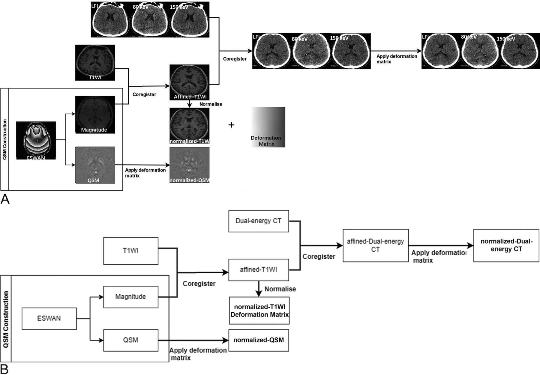
- FIG 1.
Main flow chart. A, The specific process of image registration. B, The process description: First, the QSM image was reconstructed and the magnitude map was normalized to the MNI standard space using the T1 structure image to obtain the T1-MNI, and the transformation matrix was recorded. DECT images were aligned with T1-MNI; and finally, QSM was transformed to the standard space using the same transformation matrix. After we matched the normalized MR imaging, QSM, and CT images with the AALv3 template, we extracted the MSV and CT values (average values) of the corresponding 3D ROIs using enhanced T2*-weighted angiography (ESWAN). LF1 indicates linear fusion image.
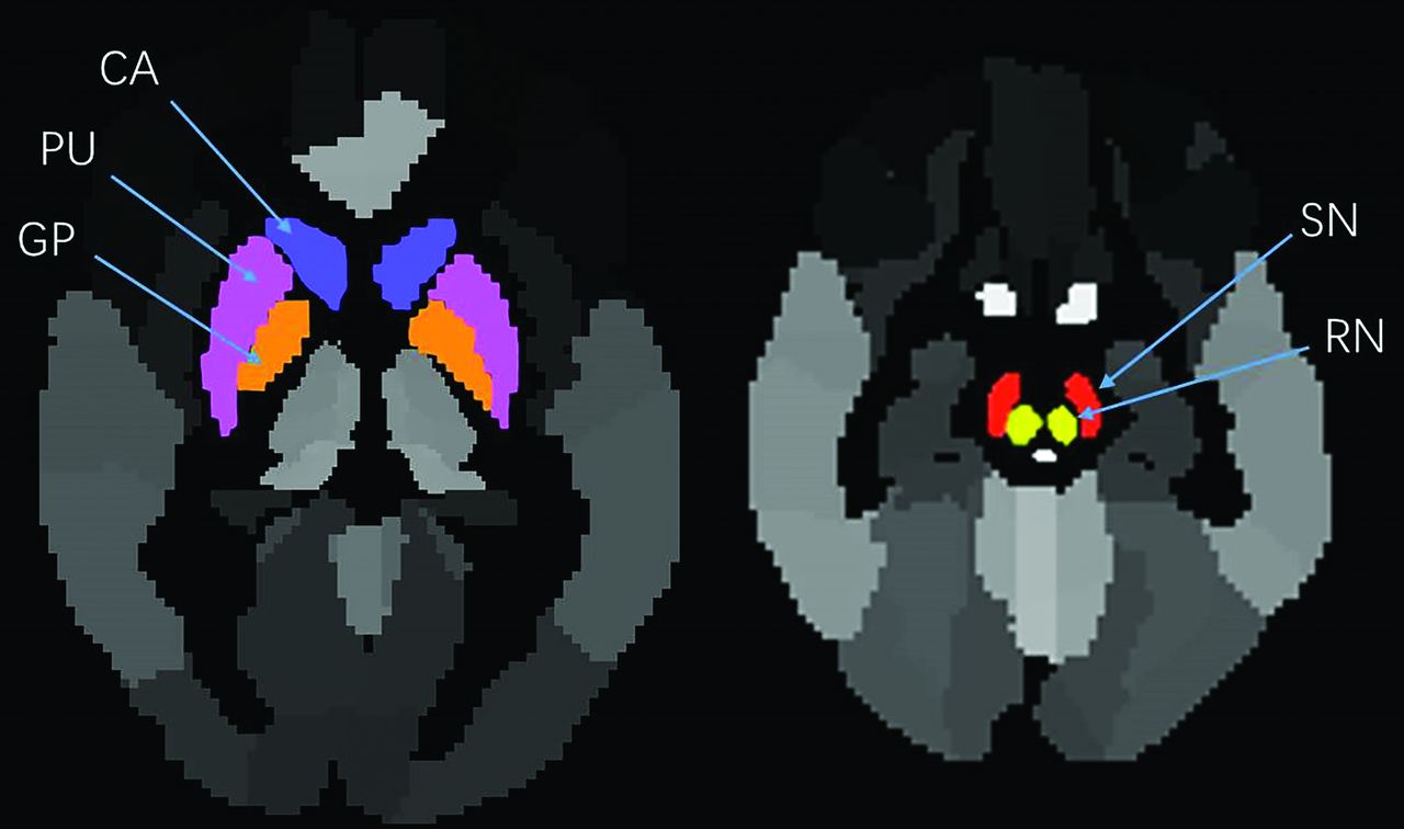
- FIG 2.
ROI settings. The ROIs used to extract MSV and CT values (average values) were set in the bilateral CA (blue), PU (purple), GP (orange), SN (red), and RN (yellow).
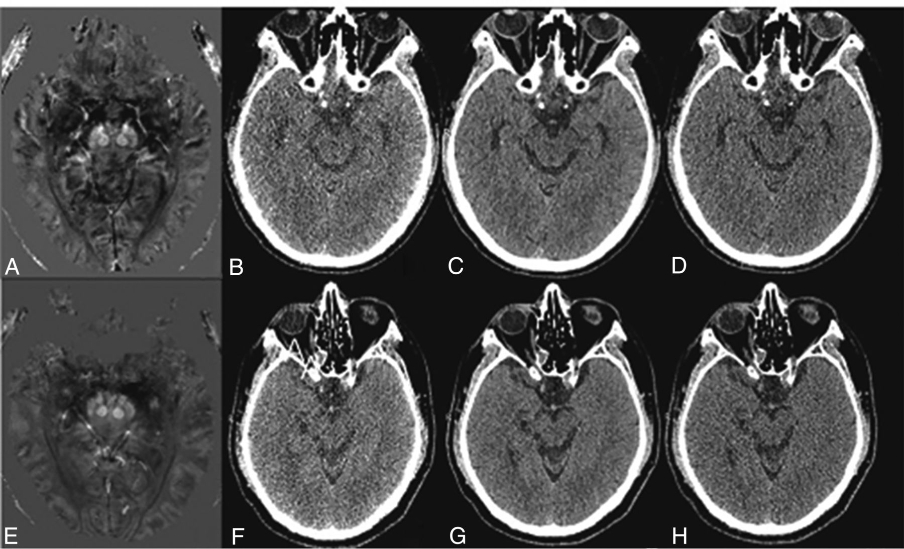
- FIG 3.
Comparison of QSM and CT images of patients with PD and HC groups. A–D, A 67-year-old female patient with PD. A, QSM image of a patient with PD showing the increased signal in the bilateral SN, suggesting increased iron deposition; B–D, CT images of a patient with PD at 80 kV(p), linear fusion, and Sn150 kV(p) levels, respectively. E–H, A 68-year-old healthy woman. E, QSM image of a healthy person showing no obvious abnormal signal changes in the bilateral SN. F–H, CT images of a healthy person at the level of 80 kV(p), linear fusion, and Sn150 kV(p), respectively.
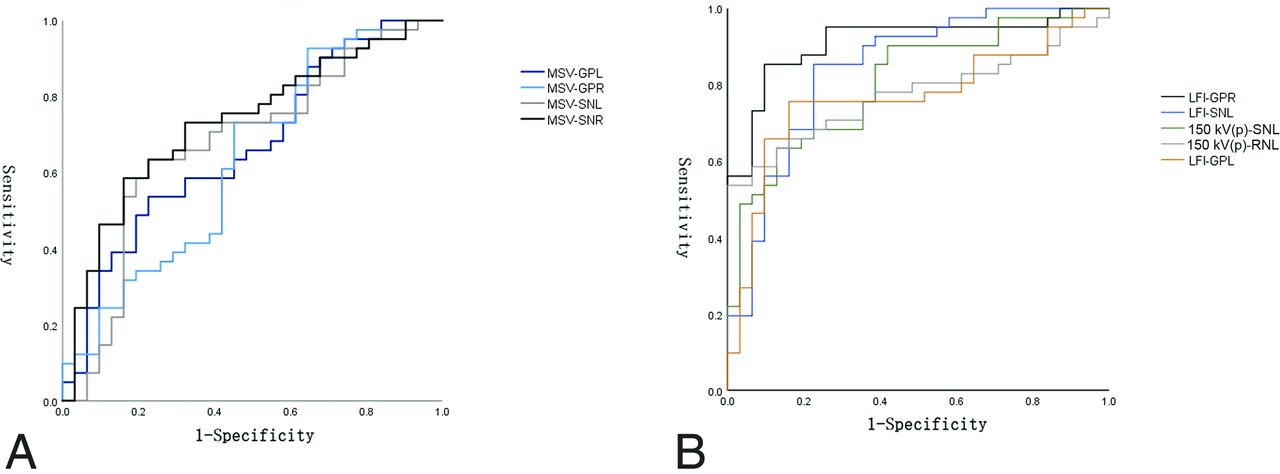
- FIG 4.
ROC curves of MSV and CT values. A, The MSV in the left GP has the highest diagnostic performance (black). B, The LFI-based CT value in GPR had the highest diagnostic performance (black). GPL indicates globus pallidus left; GPR, globus pallidus right; SNL, substantia nigra left; SNR, substantia nigra right; LFI, linear fusion image; RNL, red nucleus left.
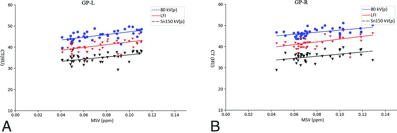
- FIG 5.
Correlations between MSV and CT values of the bilateral GP of patients with PD. MSV of the left (A) and right GPs (B) in the PD group were positively correlated with CT values at the 80 kV(p) level, linear fusion images, and Sn150 kV(p) values. GP-L indicates globus pallidus left; LFI, linear fusion image; GP-R, globus pallidus right.


Tables
Demographics and clinical status of the study participants
No. (Female/Male) Age (mean) (yr) Disease Duration (mean) H-Y (Stage) Patients with PD 41 (18:23) 65.0 (SD, 7.5) 4.5 (SD, 2.7) 1.9 (SD, 1.1) HCs 31 (20:11) 62.4 (SD, 7.5) Note:—H-Y indicates Hoehn and Yahr stage.


Jump to section
Related Articles
Cited By...
- No citing articles found.