
Article Figures & Data
Figures
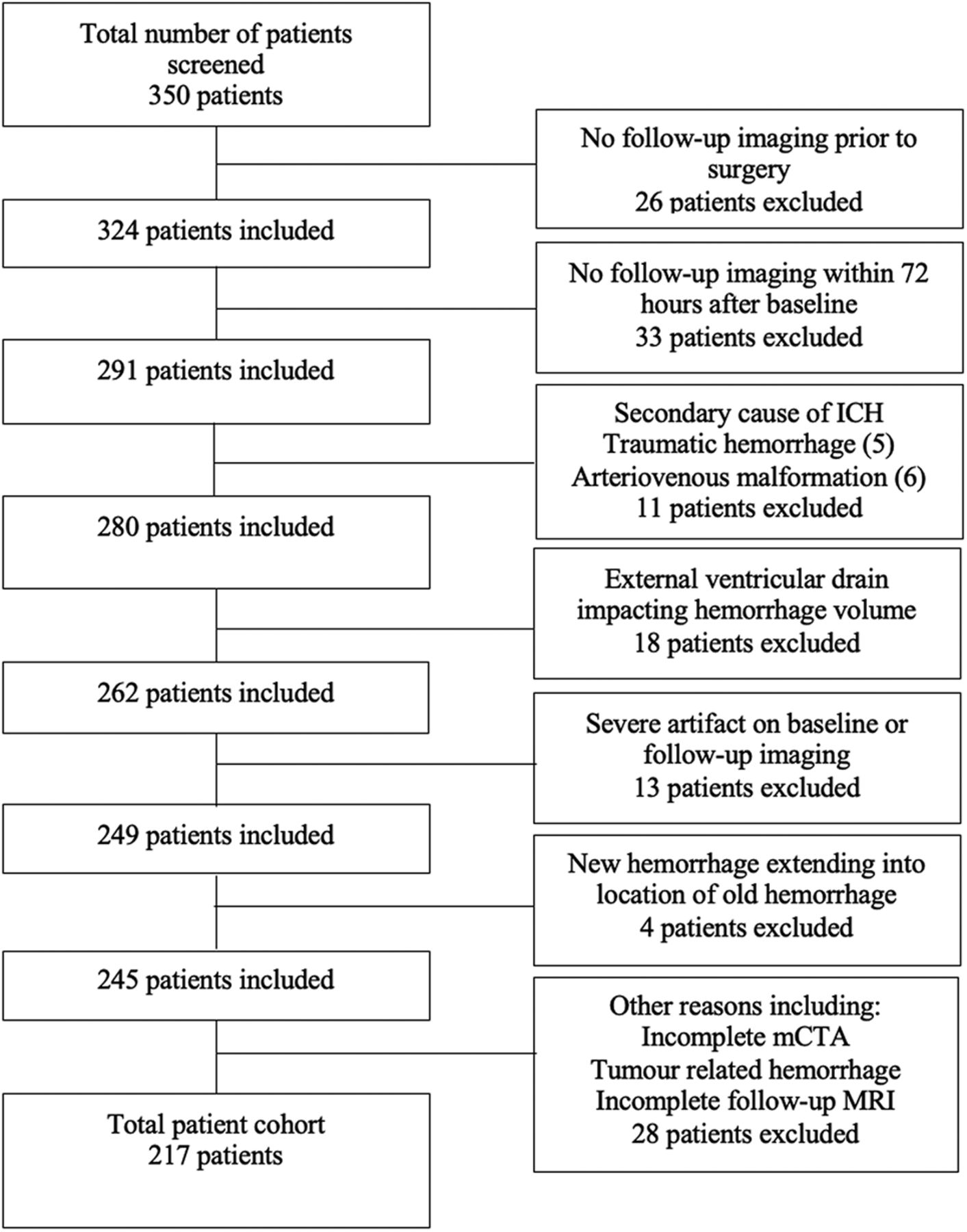
- FIG 1.
Patient inclusion/exclusion flow chart. Patients were screened according to the inclusion/exclusion criteria of the study. Of the 350 patients screened, 217 patients were included in the study.
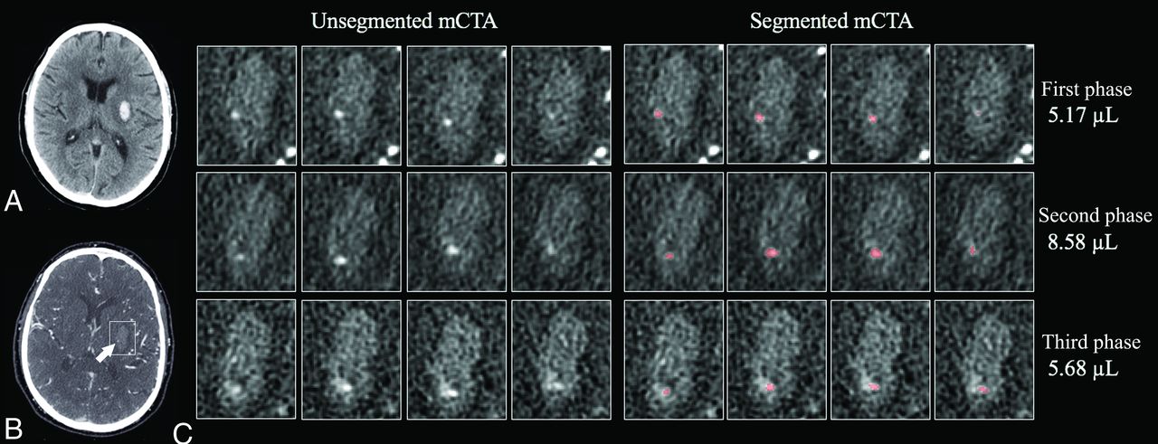
- FIG 2.
Representative slices of the spot sign in each phase of mCTA. A, Noncontrast CT shows a hematoma in the left putamen. B, mCTA shows a spot sign in the hematoma in the first phase (arrow). C, Segmentation of the spot sign in each phase. The volume of the spot sign in the first phase increases in the second phase. In the third phase, the contrast seems to partially disperse into the hematoma, and the volume of the spot sign decreases.
- FIG 3.
Predicted spot sign volume by mCTA phase time stratified by hematoma growth category. A mixed-effects regression model estimated a change in spot sign volume across time stratified by different ICH growth category. The x-axis measures time in seconds under the assumption that phase 1 of the mCTA is acquired at time = 0 seconds, and the relationship between the predicted spot sign volume change and time are shown in patients without hematoma growth and hematoma growth of ≤6 mL and >6 mL. The area shows 95% CIs.
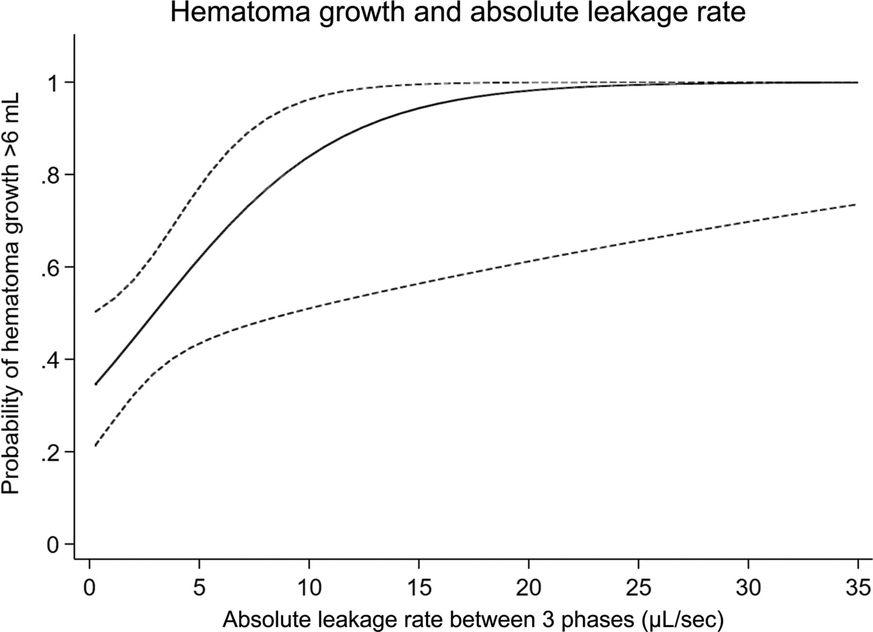
- FIG 4.
Hematoma growth probability as predicted by absolute spot sign leakage rate. The probability of hematoma growth of >6 mL is predicted by the absolute spot sign leakage rate ([|leakage rate between first and second phases| + |leakage rate between second and third phases|]/2). The area shows 95% CIs.


Tables
Variable Total (n = 217) Hematoma Growth P Value Yes (n = 44) No (n = 173) Age (yr) 70 (59.5–80) 75 (61–83) 69 (59–79) .209 Sex (male) 131 (60.4) 27 (61.4) 104 (60.1) .880 History of stroke 21 (9.7) 9 (20.5) 12 (6.9) .018 Ischemic 16 (7.4) 8 (18.2) 8 (4.6) Hemorrhagic 5 (2.3) 1 (2.3) 4 (2.3) Hypertension 182 (83.9) 39 (88.6) 143 (82.7) .491 Diabetes mellitus 41 (18.9) 10 (22.7) 31 (17.9) .467 Current smoking 23 (10.6) 3 (6.8) 20 (11.6) .583 Alcohol consumption 9 (4.2) 1 (2.3) 8 (4.6) .690 Prior antithrombotic medication 59 (27.2) 17 (38.6) 42 (24.3) .056 Antiplatelets 26 (12.0) 8 (18.2) 18 (10.4) .192 Anticoagulants 37 (17.1) 11 (25.0) 26 (15.0) .116 SBP (mm Hg) 190 (171–205) 200 (174–210) 190 (168–200) .160 Hemoglobin (g/L) 141 (131–153) 141 (132–148) 142 (131–154) .432 Serum glucose (mmol/L) 6.9 (5.7–8.2) 6.9 (5.7–8.2) 6.7 (4.9–8.1) .751 International normalized ratio 1 (1.0–1.1) 1 (1.0–2.4) 1 (1.0–1.1) .146 NIHSS score on admission 10 (5–20) 18.5 (9–23) 8 (3.25–17) <.001 Onset-to-imaging time (min) 225 (109–392) 151 (102–248) 243 (134–422) .008 Baseline ICH volume (mL) 18.9 (5.4–34.4) 28.7 (13.7–58.8) 12.8 (4.2–30.9) <.001 Hematoma location .150 Deep cerebral 115 (53.0) 26 (59.1) 89 (51.5) Lobar 80 (36.9) 17 (38.6) 63 (36.4) Infratentorial 22 (10.1) 1 (2.3) 21 (12.1) Timing of spot sign appearance <.001 No spot sign 148 (68.2) 10 (22.7) 138 (79.8) First appearing in the first phase 47 (21.7) 26 (59.1) 21 (12.1) First appearing in the second phase 20 (9.2) 8 (18.2) 12 (6.9) First appearing in the third phase 2 (0.9) 0 (0) 2 (1.2) Note:—SBP indicates systolic blood pressure.
↵a Hematoma growth is defined as a >6-mL increase in volume of the hematoma from baseline. Data are presented as No. (%) or median (interquartile range).
Spot Sign Volume Parameter Totala Spot Sign First Appearing in the First Phase Spot Sign First Appearing in the Second Phase Hematoma Growth P Value Hematoma Growth P Value Yes (n = 26) No (n = 21) Yes (n = 8) No (n = 12) Volume in the first phase (μL) 19.7 (5.7–64.3) 32.7 (12.9–74.3) 6.7 (4.4–59.0) .033 Leakage rate between first and second phases (μL/sec) 2.6 (0.6–7.7) 3.8 (1.1–9.7) 1.2 (0.4–6.5) .034 Volume in the second phase (μL) 31.4 (7.4–108.8) 95.9 (44.1–172.0) 19.7 (8.2–133.0) .017 17.0 (5.8–40.3) 6.3 (4.8–14.4) .216 Leakage rate between second and third phases (μL/sec) 0.1 (−0.2–1.3) 0.4 (−0.1–2.5) 0.0 (−2.1–2.1) .153 0.6 (−0.1–2.1) 0.1 (0.1–0.5) .396 Volume in the third phase (μL) 34.8 (10.0–129.0) 58.0 (32.9–172.6) 34.8 (7.3–156.7) .121 31.5 (1.9–79.1) 10.8 (6.2–18.5) .440 Absolute leakage rate (μL/sec) 1.02 (0.5–4.7) 3.5 (1.0–8.1) 0.9 (0.5–3.8) .017 ↵a The number of patients was 47 for volume in the first phase and leakage rate between the first and second phases, 67 for volume in the second phase and leakage rate between second and third phases, and 69 for volume in the third phase. Data are presented as median (interquartile range).
OR (95% CI) P value C-Statistic BIC AIC Model 1 0.735 190.2 183.5 No spot sign in the first phase (reference) 1.0 Spot sign in the first phase 10.5 (5.0–22.7) <.001 Model 2 0.798 182.8 172.7 No spot sign (reference) 1.0 Spot sign first appearing in the first phase 17.1 (7.2–40.4) <.001 Spot sign first appearing in the second or third phase 7.9 (2.7–23.2) <.001 Model 3 0.807 185.6 172.3 No spot sign (reference) 1.0 Spot sign first appearing in the first phase with volume <19.7 μL 10.6 (3.7–30.9) <.001 Spot sign first appearing in the first phase with volume ≥19.7 μL 27.6 (9.9–84.6) <.001 Spot sign first appearing in the second or third phase 7.9 (2.7–23.2) <.001 Model 4 0.800 182.4 172.2 No spot sign (reference) 1.0 Spot sign with positive leakage rate between phases 10.1 (4.2–24.1) <.001 Spot sign with negative leakage rate between any phases 23.0 (8.1–65.5) <.001 Model 5a 0.713 95.4 99.9 Absolute leakage rate (per 1-μL/sec increase) 1.26 (1.04–1.52) .019 Note:—AIC indicates Akaike information criterion; BIC, Bayesian information criterion.
↵a Model 5 includes only patients with the spot sign (n = 69).


