Advertisement

Main menu

  • Home
  • Content
    • Current Issue
    • Accepted Manuscripts
    • Article Preview
    • Past Issue Archive
    • AJNR Case Collection
    • Case of the Week Archive
    • Classic Case Archive
    • Case of the Month Archive
  • Special Collections
    • Spinal CSF Leak Articles (Jan 2020-June 2024)
    • 2024 AJNR Journal Awards
    • Most Impactful AJNR Articles
  • Multimedia
    • AJNR Podcast
    • AJNR Scantastics
    • Video Articles
  • For Authors
    • Submit a Manuscript
    • Author Policies
    • Fast publishing of Accepted Manuscripts
    • Graphical Abstract Preparation
    • Manuscript Submission Guidelines
    • Imaging Protocol Submission
    • Submit a Case for the Case Collection
  • About Us
    • About AJNR
    • Editorial Board
  • More
    • Become a Reviewer/Academy of Reviewers
    • Subscribers
    • Permissions
    • Alerts
    • Feedback
    • Advertisers
    • ASNR Home
  • Other Publications
    • ajnr

User menu

  • Alerts
  • Log in

Search

  • Advanced search
American Journal of Neuroradiology

American Journal of Neuroradiology

ASHNR American Society of Functional Neuroradiology ASHNR American Society of Pediatric Neuroradiology ASSR
  • Alerts
  • Log in

Advanced Search

  • Home
  • Content
    • Current Issue
    • Accepted Manuscripts
    • Article Preview
    • Past Issue Archive
    • AJNR Case Collection
    • Case of the Week Archive
    • Classic Case Archive
    • Case of the Month Archive
  • Special Collections
    • Spinal CSF Leak Articles (Jan 2020-June 2024)
    • 2024 AJNR Journal Awards
    • Most Impactful AJNR Articles
  • Multimedia
    • AJNR Podcast
    • AJNR Scantastics
    • Video Articles
  • For Authors
    • Submit a Manuscript
    • Author Policies
    • Fast publishing of Accepted Manuscripts
    • Graphical Abstract Preparation
    • Manuscript Submission Guidelines
    • Imaging Protocol Submission
    • Submit a Case for the Case Collection
  • About Us
    • About AJNR
    • Editorial Board
  • More
    • Become a Reviewer/Academy of Reviewers
    • Subscribers
    • Permissions
    • Alerts
    • Feedback
    • Advertisers
    • ASNR Home
  • Follow AJNR on Twitter
  • Visit AJNR on Facebook
  • Follow AJNR on Instagram
  • Join AJNR on LinkedIn
  • RSS Feeds

Welcome to the new AJNR, Updated Hall of Fame, and more. Read the full announcements.


AJNR is seeking candidates for the position of Associate Section Editor, AJNR Case Collection. Read the full announcement.


  • Getting new auth cookie, if you see this message a lot, tell someone!
  • Getting new auth cookie, if you see this message a lot, tell someone!
Research ArticlePediatric Neuroimaging

The Clinical Value of Cranial CT Venography for Predicting Fusobacterium necrophorum as the Causative Agent in Children with Complicated Acute Mastoiditis

Shelly I. Shiran, Li-tal Pratt, Ari DeRowe, Sophie Matot, Narin Carmel Neiderman and Oshri Wasserzug
American Journal of Neuroradiology June 2024, 45 (6) 761-768; DOI: https://doi.org/10.3174/ajnr.A8217
Shelly I. Shiran
aFrom the Department of Radiology (S.I.S., L.-t.P.), Tel Aviv Sourasky Medical Center, Tel Aviv, Israel
  • Find this author on Google Scholar
  • Find this author on PubMed
  • Search for this author on this site
  • ORCID record for Shelly I. Shiran
Li-tal Pratt
aFrom the Department of Radiology (S.I.S., L.-t.P.), Tel Aviv Sourasky Medical Center, Tel Aviv, Israel
  • Find this author on Google Scholar
  • Find this author on PubMed
  • Search for this author on this site
Ari DeRowe
bPediatric Otorhinolaryngology Unit, “Dana” Children’s Hospital (A.D., O.W.), Tel Aviv Sourasky Medical Center, Tel Aviv, Israel
  • Find this author on Google Scholar
  • Find this author on PubMed
  • Search for this author on this site
  • ORCID record for Ari DeRowe
Sophie Matot
cDepartment of Otolaryngology, Head & Neck and Maxillofacial Surgery (S.M., N.C.N.), Tel Aviv Sourasky Medical Center, Faculty of Medicine, Tel Aviv University, Tel Aviv, Israel.
  • Find this author on Google Scholar
  • Find this author on PubMed
  • Search for this author on this site
Narin Carmel Neiderman
cDepartment of Otolaryngology, Head & Neck and Maxillofacial Surgery (S.M., N.C.N.), Tel Aviv Sourasky Medical Center, Faculty of Medicine, Tel Aviv University, Tel Aviv, Israel.
  • Find this author on Google Scholar
  • Find this author on PubMed
  • Search for this author on this site
Oshri Wasserzug
bPediatric Otorhinolaryngology Unit, “Dana” Children’s Hospital (A.D., O.W.), Tel Aviv Sourasky Medical Center, Tel Aviv, Israel
  • Find this author on Google Scholar
  • Find this author on PubMed
  • Search for this author on this site
  • Article
  • Figures & Data
  • Info & Metrics
  • Responses
  • References
  • PDF
Loading

Article Figures & Data

Figures

  • Tables
  • FIG 1.
    • Download figure
    • Open in new tab
    • Download powerpoint
    FIG 1.

    Perisigmoid abscess classification in 4 different cases of complicated mastoiditis: class I (A), normal dura with no thickening; class II (B), linear smooth halo of thickened dura (arrows); class III (C), focal nodular dural thickening ≤4 mm thick (arrow); class IV (D), large nodular halo >4 mm thick (arrow). Classes III and IV comprise the patients considered positive for perisigmoid abscess on imaging. Note that there is an extracranial subperiosteal abscess present in B and C (arrowhead).

  • FIG 2.
    • Download figure
    • Open in new tab
    • Download powerpoint
    FIG 2.

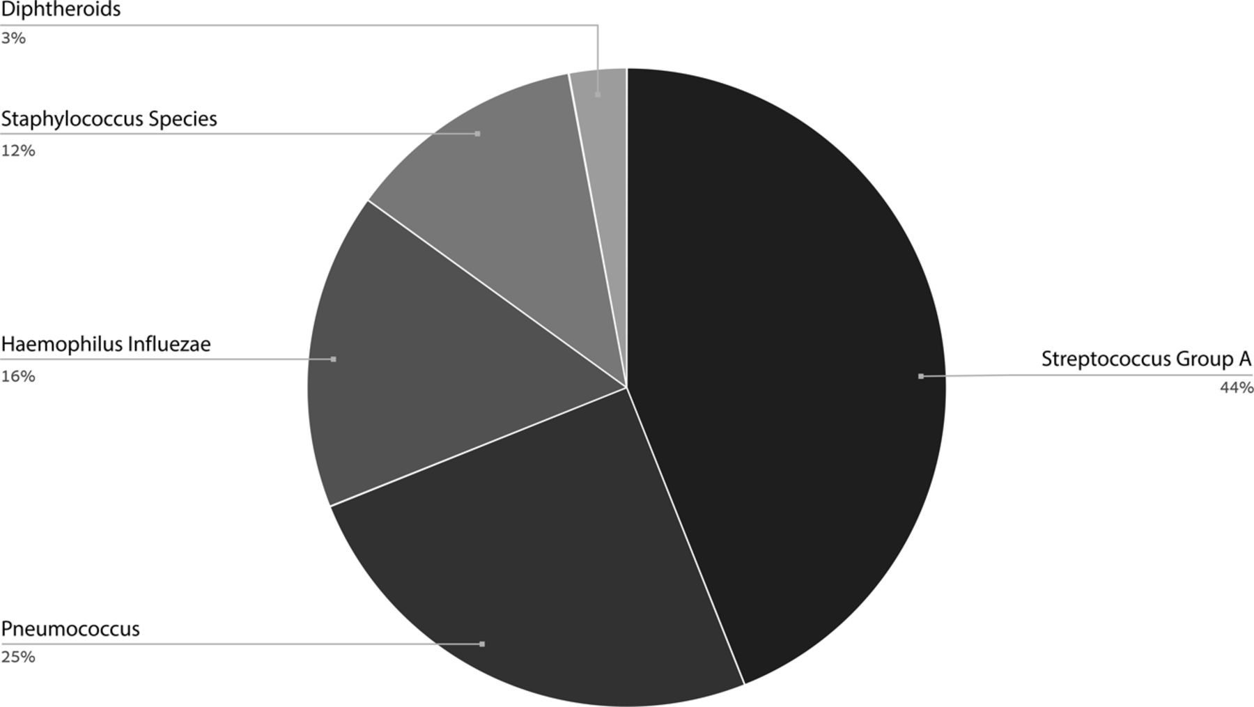
    The prevalence of bacteria species in the non-F necrophorum–related disease group.

  • FIG 3.
    • Download figure
    • Open in new tab
    • Download powerpoint
    FIG 3.

    A 9-month-old old boy with F necrophorum–related left-sided CAM causing otogenic Lemierre syndrome variant. Axial CTV image (A) at the level of the sigmoid sinus demonstrates an obstructing filling defect in the left sigmoid sinus consistent with SVT (white arrows), as apposed to normal contrast filling in the right sigmoid sinus (black arrow). No contrast is seen in the left IJV (white arrowhead) with normal contrast filling in the right IJV (black arrowhead). Also note extensive retroauricular soft-tissue phlegmon (asterisk). Sagittal-oblique MPR centered on the left jugular bulb (B) demonstrates extension of thrombus with complete obstruction of the jugular bulb and proximal IJV, with abrupt transition (arrow) where the thrombus ends. Coronal reformat of the sella region (C) demonstrates asymmetric contrast enhancement of the cavernous sinuses with a filling defect on the left (arrow), consistent with thrombosis. Coronal reformat of the orbits (D) demonstrates thrombophlebitis of the right superior ophthalmic vein with enhancement and fat-stranding along the obstructed vein (arrow).

  • FIG 4.
    • Download figure
    • Open in new tab
    • Download powerpoint
    FIG 4.

    Extramastoid osteomyelitis–related bone changes in 3 different children with F necrophorum–related CAM. A 3-year-old girl (A) with left mastoid CAM. The mastoid air cells are opacified bilaterally, but only on the left is there demineralization of the mastoid sinus plate as well as extension of focal cortical lytic changes posteriorly along the left sigmoid plate, consistent with subtle extramastoid osteomyelitis (arrows). A 2-year-old boy (B) with right-sided CAM with more extensive destructive bone changes posterior and anterior to the mastoid (arrows). A 4-year-old boy (C) with left-sided CAM has abnormal air deposits in the nonpneumatized sphenoid bone, consistent with emphysematous osteomyelitis (circle). Note also the presence of SVT in the left jugular bulb (arrow).

  • FIG 5.
    • Download figure
    • Open in new tab
    • Download powerpoint
    FIG 5.

    An 11-month-old girl with F necrophorum–related right-sided CAM with TMJ abscess. Coronal reformat (A) and axial (B) CTV images demonstrate a large subperiosteal abscess (arrows) extending to the zygomatic arch and into the glenoid fossa of the right TMJ.
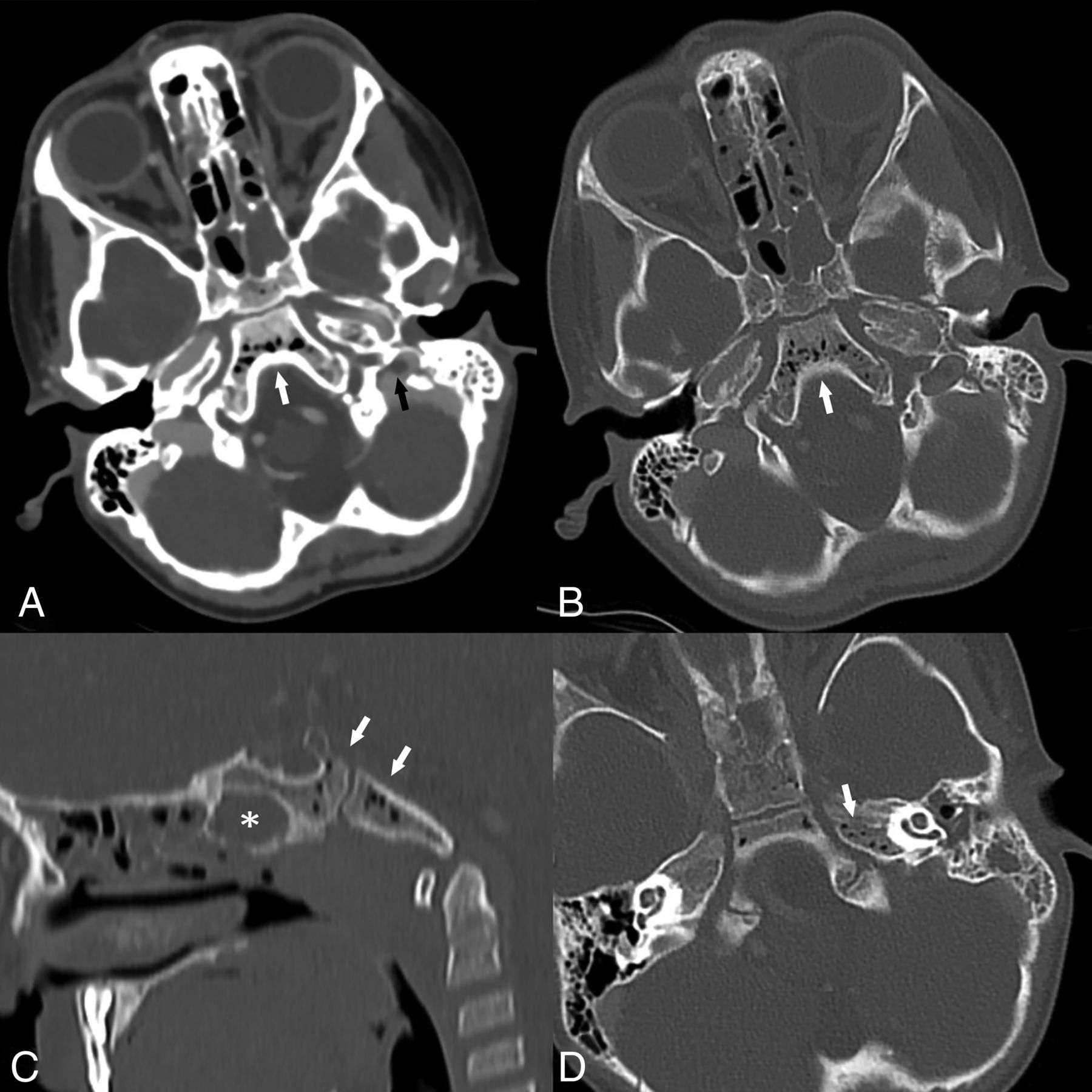
  • FIG 6.
    • Download figure
    • Open in new tab
    • Download powerpoint
    FIG 6.

    A 4-year-old boy with left-sided F necrophorum–related CAM complicated with a pumice bone pattern extramastoid osteomyelitis. The hallmark of this pattern is abnormal air deposits in a nonpneumatized bone without extensive cortical destruction, as seen in this child. Axial CTV image in a soft-tissue window (A) and in the bone algorithm (B) demonstrates abnormal air deposits in the nonpneumatized clivus (white arrows). A sagittal reformat (C) shows that both sphenoidal and basilar parts of the clivus are involved (arrows), distinguished from the sphenoid sinus that has mucosal thickening (asterisk). Similar changes are seen in the left petrous apex (arrow) (D) compared with the normal bone in the right petrous apex. Note also the presence of SVT in the left jugular bulb (back arrow) (A).

Tables

  • Figures
    • View popup
    Table 1:

    Results

    FindingFusobacterium (n = 37)Other (n = 39)P Value
    Male/female ratio19:18 (1.01)23:16 (1.4).967
    Site of ear infection RT:LT ratio19:18 (1.01)22:17 (1.3).658
    Chronic ear changes6 (16%)5 (13%).674
    Intracranial perisigmoid epidural abscess27 (73%)16 (41%).036
    Intracranial middle cranial fossa epidural abscess10 (27%)4 (10%).199
    Subdural empyema1 (2.7%)1 (2.6%)1
    SVT24 (64%)4 (10%)< .001
    Extramastoid bone changes consistent with osteomyelitis20 (54%)4 (10%)< .001
    Emphysematous osteomyelitis8 (22%)0.015
    Subperiosteal abscess30 (81%)30 (77%)1
    Inflammatory phlegmon (mild and extensive)34 (92%)38 (97%).961
    TMJ abscess7 (19%)1 (2.6%).099
    Ipsilateral neck lymphadenopathy32 (87%)33 (85%)1
    Parotid hyperemia26 (70%)24 (62%).838
    Contralateral ear air cell opacification25 (68%)28 (72%)1
    Sinonasal air cell opacification (partial and complete)24 (65%)31 (79%).309
    • Note:—RT indicates right; LT, left.

    • View popup
    Table 2:

    Sites of venous thrombosis

    Site of ThrombosisF necrophorum (n = 37)Other Bacteria (n = 39)
    Sigmoid sinus20 (54%)4 (10.3%)
    Transverse sinus3 (8%)0 (0%)
    Jugular fossa13 (35%)2 (5%)
    IJV8 (22%)0 (0%)
    Cavernous sinus6 (16%)0 (0%)
    Superior ophthalmic vein5 (14%)0 (0%)
    • View popup
    Table 3:

    The sensitivity, specificity, PPV, and NPV for statistically significant variables

    VariablePPVSensitivitySpecificityNPVF necrophorum (n = 37)Other Bacteria (n = 39)P Value Adjusted
    SVT0.8570.6490.8970.7324 (64%)4 (10%).000024
    Extramastoid osteomyelitis0.8330.5410.8970.6720 (54%)4 (10%).00036
    Perisigmoid epidural abscess0.630.730.590.6927 (73%)16 (43%).005
    SVT and extramastoid bone erosion10.45910.6617 (46%)0.000023
    SVT and perisigmoid epidural abscess0.8260.5140.8970.6619 (51%)4 (10%).0006
    Perisigmoid epidural abscess and extramastoid bone erosion0.8890.4320.9490.6416 (43%)2 (5%).00054
PreviousNext
Back to top

In this issue

American Journal of Neuroradiology: 45 (6)
American Journal of Neuroradiology
Vol. 45, Issue 6
1 Jun 2024
  • Table of Contents
  • Index by author
  • Complete Issue (PDF)
Advertisement
Print
Download PDF
Email Article

Thank you for your interest in spreading the word on American Journal of Neuroradiology.

NOTE: We only request your email address so that the person you are recommending the page to knows that you wanted them to see it, and that it is not junk mail. We do not capture any email address.

Enter multiple addresses on separate lines or separate them with commas.
The Clinical Value of Cranial CT Venography for Predicting Fusobacterium necrophorum as the Causative Agent in Children with Complicated Acute Mastoiditis
(Your Name) has sent you a message from American Journal of Neuroradiology
(Your Name) thought you would like to see the American Journal of Neuroradiology web site.
CAPTCHA
This question is for testing whether or not you are a human visitor and to prevent automated spam submissions.
Cite this article
Shelly I. Shiran, Li-tal Pratt, Ari DeRowe, Sophie Matot, Narin Carmel Neiderman, Oshri Wasserzug
The Clinical Value of Cranial CT Venography for Predicting Fusobacterium necrophorum as the Causative Agent in Children with Complicated Acute Mastoiditis
American Journal of Neuroradiology Jun 2024, 45 (6) 761-768; DOI: 10.3174/ajnr.A8217

Citation Manager Formats

  • BibTeX
  • Bookends
  • EasyBib
  • EndNote (tagged)
  • EndNote 8 (xml)
  • Medlars
  • Mendeley
  • Papers
  • RefWorks Tagged
  • Ref Manager
  • RIS
  • Zotero
0 Responses
Respond to this article
Share
Bookmark this article
CTV for Fusobacterium Necrophorum in Mastoiditis
Shelly I. Shiran, Li-tal Pratt, Ari DeRowe, Sophie Matot, Narin Carmel Neiderman, Oshri Wasserzug
American Journal of Neuroradiology Jun 2024, 45 (6) 761-768; DOI: 10.3174/ajnr.A8217
del.icio.us logo Twitter logo Facebook logo Mendeley logo
  • Tweet Widget
  • Facebook Like
  • Google Plus One
Purchase

Jump to section

  • Article
    • Abstract
    • ABBREVIATIONS:
    • MATERIALS AND METHODS
    • RESULTS
    • DISCUSSION
    • CONCLUSIONS
    • Footnotes
    • References
  • Figures & Data
  • Info & Metrics
  • Responses
  • References
  • PDF

Related Articles

  • No related articles found.
  • PubMed
  • Google Scholar

Cited By...

  • No citing articles found.
  • Crossref
  • Google Scholar

This article has not yet been cited by articles in journals that are participating in Crossref Cited-by Linking.

More in this TOC Section

  • Neuroimaging Delineation and Progression of SLSMD
  • New CNS Embryonal Tumor Insights via MRI Analysis
  • Reversible Perfusion Changes in GLUT-1 Deficiency
Show more PEDIATRIC NEUROIMAGING

Similar Articles

Advertisement

Indexed Content

  • Current Issue
  • Accepted Manuscripts
  • Article Preview
  • Past Issues
  • Editorials
  • Editors Choice
  • Fellow Journal Club
  • Letters to the Editor

Cases

  • Case Collection
  • Archive - Case of the Week
  • Archive - Case of the Month
  • Archive - Classic Case

Special Collections

  • Special Collections

Resources

  • News and Updates
  • Turn around Times
  • Submit a Manuscript
  • Author Policies
  • Manuscript Submission Guidelines
  • Evidence-Based Medicine Level Guide
  • Publishing Checklists
  • Graphical Abstract Preparation
  • Imaging Protocol Submission
  • Submit a Case
  • Become a Reviewer/Academy of Reviewers
  • Get Peer Review Credit from Publons

Multimedia

  • AJNR Podcast
  • AJNR SCANtastic
  • Video Articles

About Us

  • About AJNR
  • Editorial Board
  • Not an AJNR Subscriber? Join Now
  • Alerts
  • Feedback
  • Advertise with us
  • Librarian Resources
  • Permissions
  • Terms and Conditions

American Society of Neuroradiology

  • Not an ASNR Member? Join Now

© 2025 by the American Society of Neuroradiology All rights, including for text and data mining, AI training, and similar technologies, are reserved.
Print ISSN: 0195-6108 Online ISSN: 1936-959X

Powered by HighWire