Advertisement

Main menu

  • Home
  • Content
    • Current Issue
    • Accepted Manuscripts
    • Article Preview
    • Past Issue Archive
    • Video Articles
    • AJNR Case Collection
    • Case of the Week Archive
    • Case of the Month Archive
    • Classic Case Archive
  • Special Collections
    • AJNR Awards
    • Low-Field MRI
    • Alzheimer Disease
    • ASNR Foundation Special Collection
    • Photon-Counting CT
    • View All
  • Multimedia
    • AJNR Podcasts
    • AJNR SCANtastic
    • Trainee Corner
    • MRI Safety Corner
    • Imaging Protocols
  • For Authors
    • Submit a Manuscript
    • Submit a Video Article
    • Submit an eLetter to the Editor/Response
    • Manuscript Submission Guidelines
    • Statistical Tips
    • Fast Publishing of Accepted Manuscripts
    • Graphical Abstract Preparation
    • Imaging Protocol Submission
    • Author Policies
  • About Us
    • About AJNR
    • Editorial Board
    • Editorial Board Alumni
  • More
    • Become a Reviewer/Academy of Reviewers
    • Subscribers
    • Permissions
    • Alerts
    • Feedback
    • Advertisers
    • ASNR Home

User menu

  • Alerts
  • Log in

Search

  • Advanced search
American Journal of Neuroradiology

American Journal of Neuroradiology

ASHNR American Society of Functional Neuroradiology ASHNR American Society of Pediatric Neuroradiology ASSR
  • Alerts
  • Log in

Advanced Search

  • Home
  • Content
    • Current Issue
    • Accepted Manuscripts
    • Article Preview
    • Past Issue Archive
    • Video Articles
    • AJNR Case Collection
    • Case of the Week Archive
    • Case of the Month Archive
    • Classic Case Archive
  • Special Collections
    • AJNR Awards
    • Low-Field MRI
    • Alzheimer Disease
    • ASNR Foundation Special Collection
    • Photon-Counting CT
    • View All
  • Multimedia
    • AJNR Podcasts
    • AJNR SCANtastic
    • Trainee Corner
    • MRI Safety Corner
    • Imaging Protocols
  • For Authors
    • Submit a Manuscript
    • Submit a Video Article
    • Submit an eLetter to the Editor/Response
    • Manuscript Submission Guidelines
    • Statistical Tips
    • Fast Publishing of Accepted Manuscripts
    • Graphical Abstract Preparation
    • Imaging Protocol Submission
    • Author Policies
  • About Us
    • About AJNR
    • Editorial Board
    • Editorial Board Alumni
  • More
    • Become a Reviewer/Academy of Reviewers
    • Subscribers
    • Permissions
    • Alerts
    • Feedback
    • Advertisers
    • ASNR Home
  • Follow AJNR on Twitter
  • Visit AJNR on Facebook
  • Follow AJNR on Instagram
  • Join AJNR on LinkedIn
  • RSS Feeds

AJNR Awards, New Junior Editors, and more. Read the latest AJNR updates

Research ArticleNeurointervention

Value of Quantitative Collateral Scoring on CT Angiography in Patients with Acute Ischemic Stroke

A.M.M. Boers, R. Sales Barros, I.G.H. Jansen, O.A. Berkhemer, L.F.M. Beenen, B.K. Menon, D.W.J. Dippel, A. van der Lugt, W.H. van Zwam, Y.B.W.E.M. Roos, R.J. van Oostenbrugge, C.H. Slump, C.B.L.M. Majoie and H.A. Marquering on behalf of the MR CLEAN investigators
American Journal of Neuroradiology April 2018, DOI: https://doi.org/10.3174/ajnr.A5623
A.M.M. Boers
aFrom the Departments of Biomedical Engineering and Physics (A.M.M.B., R.S.B., I.G.H.J., H.A.M.)
bRadiology and Nuclear Medicine (A.M.M.B., I.G.H.J., O.A.B., L.F.M.B., C.B.L.M.M., H.A.M.)
dDepartment of Robotics and Mechatronics (A.M.M.B., C.H.S.)
  • Find this author on Google Scholar
  • Find this author on PubMed
  • Search for this author on this site
  • ORCID record for A.M.M. Boers
R. Sales Barros
aFrom the Departments of Biomedical Engineering and Physics (A.M.M.B., R.S.B., I.G.H.J., H.A.M.)
  • Find this author on Google Scholar
  • Find this author on PubMed
  • Search for this author on this site
  • ORCID record for R. Sales Barros
I.G.H. Jansen
aFrom the Departments of Biomedical Engineering and Physics (A.M.M.B., R.S.B., I.G.H.J., H.A.M.)
bRadiology and Nuclear Medicine (A.M.M.B., I.G.H.J., O.A.B., L.F.M.B., C.B.L.M.M., H.A.M.)
  • Find this author on Google Scholar
  • Find this author on PubMed
  • Search for this author on this site
  • ORCID record for I.G.H. Jansen
O.A. Berkhemer
bRadiology and Nuclear Medicine (A.M.M.B., I.G.H.J., O.A.B., L.F.M.B., C.B.L.M.M., H.A.M.)
  • Find this author on Google Scholar
  • Find this author on PubMed
  • Search for this author on this site
  • ORCID record for O.A. Berkhemer
L.F.M. Beenen
bRadiology and Nuclear Medicine (A.M.M.B., I.G.H.J., O.A.B., L.F.M.B., C.B.L.M.M., H.A.M.)
  • Find this author on Google Scholar
  • Find this author on PubMed
  • Search for this author on this site
  • ORCID record for L.F.M. Beenen
B.K. Menon
fDepartment of Clinical Neurosciences (B.K.M.), Hotchkiss Brain Institute, Cumming School of Medicine, University of Calgary, Foothills Hospital, Calgary, Alberta, Canada
  • Find this author on Google Scholar
  • Find this author on PubMed
  • Search for this author on this site
  • ORCID record for B.K. Menon
D.W.J. Dippel
gDepartments of Neurology (D.W.J.D.)
  • Find this author on Google Scholar
  • Find this author on PubMed
  • Search for this author on this site
  • ORCID record for D.W.J. Dippel
A. van der Lugt
hRadiology (A.v.d.L.), Erasmus MC, Rotterdam, the Netherlands
  • Find this author on Google Scholar
  • Find this author on PubMed
  • Search for this author on this site
  • ORCID record for A. van der Lugt
W.H. van Zwam
iDepartment of Radiology (W.H.v.Z.), Maastricht UMC, Maastricht, the Netherlands
  • Find this author on Google Scholar
  • Find this author on PubMed
  • Search for this author on this site
  • ORCID record for W.H. van Zwam
Y.B.W.E.M. Roos
cNeurology (Y.B.W.E.M.R.), Academic Medical Center, Amsterdam, the Netherlands
  • Find this author on Google Scholar
  • Find this author on PubMed
  • Search for this author on this site
  • ORCID record for Y.B.W.E.M. Roos
R.J. van Oostenbrugge
jDepartment of Neurology (R.J.v.O.), Maastricht UMC and Cardiovascular Research Institute Maastricht, Maastricht, the Netherlands.
  • Find this author on Google Scholar
  • Find this author on PubMed
  • Search for this author on this site
  • ORCID record for R.J. van Oostenbrugge
C.H. Slump
dDepartment of Robotics and Mechatronics (A.M.M.B., C.H.S.)
eMIRA Institute for Biomedical Engineering and Technical Medicine (C.H.S.), University of Twente, Enschede, the Netherlands
  • Find this author on Google Scholar
  • Find this author on PubMed
  • Search for this author on this site
  • ORCID record for C.H. Slump
C.B.L.M. Majoie
aFrom the Departments of Biomedical Engineering and Physics (A.M.M.B., R.S.B., I.G.H.J., H.A.M.)
  • Find this author on Google Scholar
  • Find this author on PubMed
  • Search for this author on this site
  • ORCID record for C.B.L.M. Majoie
H.A. Marquering
aFrom the Departments of Biomedical Engineering and Physics (A.M.M.B., R.S.B., I.G.H.J., H.A.M.)
  • Find this author on Google Scholar
  • Find this author on PubMed
  • Search for this author on this site
  • ORCID record for H.A. Marquering
  • Article
  • Figures & Data
  • Info & Metrics
  • Responses
  • References
  • PDF
Loading

Article Figures & Data

Figures

  • Tables
  • Fig 1.
    • Download figure
    • Open in new tab
    • Download powerpoint
    Fig 1.

    An example of quantitative collateral capacity scoring. A, An axial plane of a baseline CTA image acquired in the peak venous phase with a right-sided M1 segment occlusion of the MCA territory. B, Segmentation results of automated quantitative collateral assessment of the ipsilateral (red) and contralateral (blue) hemispheres. The quantitative collateral score was 46%. C, 3D representation of the segmented vasculature.

  • Fig 2.
    • Download figure
    • Open in new tab
    • Download powerpoint
    Fig 2.

    Case examples of 4 patients with different visually scored collateral grades and corresponding quantitative collateral scores. Each panel shows a maximum-intensity-projection of the CTA image (left) and the segmented vasculature for qCS calculation (right). The automated segmentation on the ipsilateral side is shown in blue and the segmentation on the contralateral side is shown in red. A, Absent collaterals (visual collateral score = 0). CTA of an 83-year-old man with a left-sided M2 occlusion acquired in the early arterial phase. Follow-up infarct volume was 205 mL, and the mRS score was 6. B, Poor collaterals (vCS = 1). CTA of a 79-year-old man with a right-sided M1 occlusion acquired in the equilibrium phase. FIV was 245 mL, and the mRS score was 6. C, Moderate collaterals (vCS = 2). CTA of a 45-year-old woman with a left-sided M1 occlusion acquired in the peak arterial phase. FIV was 24 mL, and the mRS score was 2. D, Good collaterals (vCS = 3). CTA of a 76-year-old woman with a left-sided ICA-T occlusion acquired in the late venous phase. FIV was 48 mL, and the mRS score was 3.
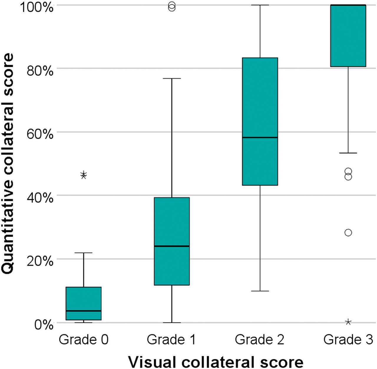
  • Fig 3.
    • Download figure
    • Open in new tab
    • Download powerpoint
    Fig 3.

    Distribution of quantitative collateral scores per visual collateral score, ranging from absent collaterals (0% filling of the occluded territory) to good collaterals (100% filling of the occluded territory). The quantitative collateral score was significantly different among all visual collateral score groups, except for absent collaterals (grade 0) versus poor collaterals (grade 1).

  • Fig 4.
    • Download figure
    • Open in new tab
    • Download powerpoint
    Fig 4.

    Receiver operating characteristic curve analysis of visual and quantitative collateral scores for discriminating favorable outcome (mRS 0–2) with areas under the curve of, respectively, 0.65 and 0.68 (A) and large infarct (FIV of >90 mL) with areas under the curve of, respectively, 0.69 and 0.71 (B).

  • Fig 5.
    • Download figure
    • Open in new tab
    • Download powerpoint
    Fig 5.

    Bar graphs depict the proportion of functional independence (mRS 0–2) by quantitative collateral score strata for CTA image acquisition in the early arterial phase (A) and arteriovenous phase (B), and by visual collateral scores in the early arterial phase (C) and arteriovenous phase (D).

Tables

  • Figures
    • View popup
    Table 1:

    Baseline characteristics

    CharacteristicAll (N = 442)
    Age in years, median [IQR]66 [54–76]
    Female sex, % (n)41.4 (183)
    Left hemisphere infarct, % (n)52.9 (234)
    NIHSS score at baseline, median [IQR]18 [14–22]
    Alteplase (tPA) delivered, % (n)88.9 (392)
    Allocated to endovascular therapy, % (n)46.8 (207)
    Atrial fibrillation, % (n)26.7 (118)
    Myocardial infarction, % (n)14.9 (66)
    Peripheral arterial disease, % (n)5.2 (23)
    Diabetes mellitus, % (n)13.6 (60)
    Hypertension, % (n)45.4 (200)
    History of ischemic stroke, % (n)10.6 (47)
    Tobacco use, % (n)26.3 (116)
    Use of statins, % (n)79.4 (350)
    Onset to randomization in minutes, median [IQR]201 [150–256]
    Prestroke modified Rankin Scale score, % (n)
        079.9 (353)
        110.4 (46)
        ≥25.7 (25)
    ASPECTS at baseline, % (n)
        0–42.2 (10)
        5–711.2 (49)
        8–1086.6 (381)
    Occlusion location, % (n)
        ICA0.7 (3)
        ICA-T27.4 (121)
        M163.3 (280)
        M28.6 (38)
    • View popup
    Table 2:

    Spearman rank ρ (95% CI) of collateral measures with outcomes for all studies and per CTA acquisition phase

    All Studies (N = 442)Early Arterial (n = 91)Peak Arterial (n = 56)Equilibrium (n = 123)Peak Venous (n = 114)Late Venous (n = 58)
    Follow-up infarct volume
        Visual collateral score−.44 (−.53 to −.36)a−.35 (−.55 to −.13)a−.46 (−.65 to −.21a−.51 (−.64 to −.35)a−.49 (−.64 to −.29a−.47 (−.66 to −.24)a
        Quantitative collateral score−.46 (−.54 to −.37)a−.45 (−.62 to −.24)a−.46 (−.66 to −.19)a−.51 (−.65 to −.34)a−.41 (−.56 to −.20)a−.52 (−.70 to −.29)a
    mRS at 90 days
        Visual collateral score−.31 (−.39 to −.22)a−.19 (−.38 to .03)−.22 (−.49 to −.04)−.28 (−.49 to −.15)a−.29 (−.45 to −.09)a−.41 (−.63 to −.19)a
        Quantitative collateral score−.40 (−.48 to −.32)a−.35 (−.53 to −.13)a−.34 (−.55 to −.10)a−.38 (−.58 to −.25)a−.30 (−.42 to −.08)a−.37 (−.59 to −.12)a
    • ↵a Significant correlation at P < .01.

    • View popup
    Table 3:

    Results of adjusted and unadjusted regression analyses for the effect of collateral capacity on follow-up infarct volume

    AdjustedUnadjusted
    β Log-Transformed (95% CI)aExp. (β)bP Valueβ Log-Transformed (95% CI)aExp. (β)bP Value
    Visual collateral score per 1 point−0.49 (−0.61 to −0.37)0.60<.001−0.59 (−0.71 to −0.47)0.54<.001
    Quantitative collateral score per 10%−0.13 (−0.16 to −0.099)0.88<.001−0.14 (−0.18 to −0.11)0.87<.001
    • Note:—Exp. (β) indicates exponent of β.

    • ↵a Due to the non-normal distribution of follow-up infarct volume, a log +1 transformation was performed to best fit the assumptions associated with the linear regression model.

    • ↵b Exponent of β was calculated to determine the relative difference of follow-up infarct volume with an increase in collateral scores.

    • View popup
    Table 4:

    Results of adjusted and unadjusted regression analyses for the effect of collateral capacity on modified Rankin Scale

    AdjustedUnadjusted
    Odds Ratio (95% CI)P ValueOdds Ratio (95% CI)P Value
    Visual collateral score per 1 point0.61 (0.50–0.75)<.0010.51 (0.42–0.62)<.001
    Quantitative collateral score per 10%0.81 (0.77–0.86)<.0010.85 (0.81–0.90)<.001
    • View popup
    Table 5:

    P values of interaction analysis for primary and secondary outcome measures

    FIV (Continuous)FIV ≤90 mL vs FIV >90 mLmRS (Ordinal)mRS 0–2 vs 3–6
    vCS-unadjusted analysis.40.43.23.11
    vCS-adjusted analysis.60.54.10.075
    qCS-unadjusted analysis.15.75.048a.049a
    qCS-adjusted analysis.33.48.081.18
    • ↵a P < .05, 2-sided.

Next
Back to top
Advertisement
Print
Download PDF
Email Article

Thank you for your interest in spreading the word on American Journal of Neuroradiology.

NOTE: We only request your email address so that the person you are recommending the page to knows that you wanted them to see it, and that it is not junk mail. We do not capture any email address.

Enter multiple addresses on separate lines or separate them with commas.
Value of Quantitative Collateral Scoring on CT Angiography in Patients with Acute Ischemic Stroke
(Your Name) has sent you a message from American Journal of Neuroradiology
(Your Name) thought you would like to see the American Journal of Neuroradiology web site.
CAPTCHA
This question is for testing whether or not you are a human visitor and to prevent automated spam submissions.
Cite this article
A.M.M. Boers, R. Sales Barros, I.G.H. Jansen, O.A. Berkhemer, L.F.M. Beenen, B.K. Menon, D.W.J. Dippel, A. van der Lugt, W.H. van Zwam, Y.B.W.E.M. Roos, R.J. van Oostenbrugge, C.H. Slump, C.B.L.M. Majoie, H.A. Marquering
Value of Quantitative Collateral Scoring on CT Angiography in Patients with Acute Ischemic Stroke
American Journal of Neuroradiology Apr 2018, DOI: 10.3174/ajnr.A5623

Citation Manager Formats

  • BibTeX
  • Bookends
  • EasyBib
  • EndNote (tagged)
  • EndNote 8 (xml)
  • Medlars
  • Mendeley
  • Papers
  • RefWorks Tagged
  • Ref Manager
  • RIS
  • Zotero
0 Responses
Respond to this article
Share
Bookmark this article
Value of Quantitative Collateral Scoring on CT Angiography in Patients with Acute Ischemic Stroke
A.M.M. Boers, R. Sales Barros, I.G.H. Jansen, O.A. Berkhemer, L.F.M. Beenen, B.K. Menon, D.W.J. Dippel, A. van der Lugt, W.H. van Zwam, Y.B.W.E.M. Roos, R.J. van Oostenbrugge, C.H. Slump, C.B.L.M. Majoie, H.A. Marquering
American Journal of Neuroradiology Apr 2018, DOI: 10.3174/ajnr.A5623
del.icio.us logo Twitter logo Facebook logo Mendeley logo
  • Tweet Widget
  • Facebook Like
  • Google Plus One
Purchase

Jump to section

  • Article
    • Abstract
    • ABBREVIATIONS:
    • Materials and Methods
    • Results
    • Discussion
    • Conclusions
    • Footnotes
    • References
  • Figures & Data
  • Info & Metrics
  • Responses
  • References
  • PDF

Related Articles

  • No related articles found.
  • PubMed
  • Google Scholar

Cited By...

  • Arterial collateral status and treatment effect of intravenous alteplase thrombolysis prior to endovascular treatment in patients with anterior circulation large vessel occlusion: prespecified analysis of the MR CLEAN-NO IV trial
  • Time Since Stroke Onset, Quantitative Collateral Score, and Functional Outcome After Endovascular Treatment for Acute Ischemic Stroke
  • MRI-based computational model generation for cerebral perfusion simulations in health and ischaemic stroke
  • On the sensitivity analysis of porous finite element models for cerebral perfusion estimation
  • Crossref
  • Google Scholar

This article has not yet been cited by articles in journals that are participating in Crossref Cited-by Linking.

More in this TOC Section

Neurointervention

  • A Retrospective Study in Tentorial DAVFs
  • Proximal Protection Devices for Carotid Stenting
  • Rescue Reentry in Carotid Near-Occlusion
Show more Neurointervention

Adult Brain

  • Diagnostic Neuroradiology of Monoclonal Antibodies
  • ML for Glioma Molecular Subtype Prediction
  • NCCT vs. MRI for Brain Atrophy in Acute Stroke
Show more Adult Brain

Similar Articles

Advertisement

Indexed Content

  • Current Issue
  • Accepted Manuscripts
  • Article Preview
  • Past Issues
  • Editorials
  • Editor's Choice
  • Fellows' Journal Club
  • Letters to the Editor
  • Video Articles

Cases

  • Case Collection
  • Archive - Case of the Week
  • Archive - Case of the Month
  • Archive - Classic Case

More from AJNR

  • Trainee Corner
  • Imaging Protocols
  • MRI Safety Corner
  • Book Reviews

Multimedia

  • AJNR Podcasts
  • AJNR Scantastics

Resources

  • Turnaround Time
  • Submit a Manuscript
  • Submit a Video Article
  • Submit an eLetter to the Editor/Response
  • Manuscript Submission Guidelines
  • Statistical Tips
  • Fast Publishing of Accepted Manuscripts
  • Graphical Abstract Preparation
  • Imaging Protocol Submission
  • Evidence-Based Medicine Level Guide
  • Publishing Checklists
  • Author Policies
  • Become a Reviewer/Academy of Reviewers
  • News and Updates

About Us

  • About AJNR
  • Editorial Board
  • Editorial Board Alumni
  • Alerts
  • Permissions
  • Not an AJNR Subscriber? Join Now
  • Advertise with Us
  • Librarian Resources
  • Feedback
  • Terms and Conditions
  • AJNR Editorial Board Alumni

American Society of Neuroradiology

  • Not an ASNR Member? Join Now

© 2025 by the American Society of Neuroradiology All rights, including for text and data mining, AI training, and similar technologies, are reserved.
Print ISSN: 0195-6108 Online ISSN: 1936-959X

Powered by HighWire