Advertisement

Main menu

  • Home
  • Content
    • Current Issue
    • Accepted Manuscripts
    • Article Preview
    • Past Issue Archive
    • Video Articles
    • AJNR Case Collection
    • Case of the Week Archive
    • Case of the Month Archive
    • Classic Case Archive
  • Special Collections
    • AJNR Awards
    • Low-Field MRI
    • Alzheimer Disease
    • ASNR Foundation Special Collection
    • Photon-Counting CT
    • View All
  • Multimedia
    • AJNR Podcasts
    • AJNR SCANtastic
    • Trainee Corner
    • MRI Safety Corner
    • Imaging Protocols
  • For Authors
    • Submit a Manuscript
    • Submit a Video Article
    • Submit an eLetter to the Editor/Response
    • Manuscript Submission Guidelines
    • Statistical Tips
    • Fast Publishing of Accepted Manuscripts
    • Graphical Abstract Preparation
    • Imaging Protocol Submission
    • Author Policies
  • About Us
    • About AJNR
    • Editorial Board
    • Editorial Board Alumni
  • More
    • Become a Reviewer/Academy of Reviewers
    • Subscribers
    • Permissions
    • Alerts
    • Feedback
    • Advertisers
    • ASNR Home

User menu

  • Alerts
  • Log in

Search

  • Advanced search
American Journal of Neuroradiology

American Journal of Neuroradiology

ASHNR American Society of Functional Neuroradiology ASHNR American Society of Pediatric Neuroradiology ASSR
  • Alerts
  • Log in

Advanced Search

  • Home
  • Content
    • Current Issue
    • Accepted Manuscripts
    • Article Preview
    • Past Issue Archive
    • Video Articles
    • AJNR Case Collection
    • Case of the Week Archive
    • Case of the Month Archive
    • Classic Case Archive
  • Special Collections
    • AJNR Awards
    • Low-Field MRI
    • Alzheimer Disease
    • ASNR Foundation Special Collection
    • Photon-Counting CT
    • View All
  • Multimedia
    • AJNR Podcasts
    • AJNR SCANtastic
    • Trainee Corner
    • MRI Safety Corner
    • Imaging Protocols
  • For Authors
    • Submit a Manuscript
    • Submit a Video Article
    • Submit an eLetter to the Editor/Response
    • Manuscript Submission Guidelines
    • Statistical Tips
    • Fast Publishing of Accepted Manuscripts
    • Graphical Abstract Preparation
    • Imaging Protocol Submission
    • Author Policies
  • About Us
    • About AJNR
    • Editorial Board
    • Editorial Board Alumni
  • More
    • Become a Reviewer/Academy of Reviewers
    • Subscribers
    • Permissions
    • Alerts
    • Feedback
    • Advertisers
    • ASNR Home
  • Follow AJNR on Twitter
  • Visit AJNR on Facebook
  • Follow AJNR on Instagram
  • Join AJNR on LinkedIn
  • RSS Feeds

AJNR Awards, New Junior Editors, and more. Read the latest AJNR updates

Research ArticleSPINE IMAGING AND SPINE IMAGE-GUIDED INTERVENTIONS

Automated Detection of Cervical Spinal Stenosis and Cord Compression via Vision Transformer and Rules-Based Classification

David L. Payne, Xuan Xu, Farshid Faraji, Kevin John, Katherine Ferra Pradas, Vahni Vishala Bernard, Lev Bangiyev and Prateek Prasanna
American Journal of Neuroradiology February 2024, DOI: https://doi.org/10.3174/ajnr.A8141
David L. Payne
aFrom the Department of Radiology (D.L.P., F.F., K.J., K.F.P., V.V.B., L.B.), Stony Brook University Hospital, Stony Brook, New York
bDepartment of Biomedical Informatics (D.L.P., X.X., F.F., K.J., P.P.), Stony Brook University, Stony Brook, New York
  • Find this author on Google Scholar
  • Find this author on PubMed
  • Search for this author on this site
  • ORCID record for David L. Payne
Xuan Xu
bDepartment of Biomedical Informatics (D.L.P., X.X., F.F., K.J., P.P.), Stony Brook University, Stony Brook, New York
  • Find this author on Google Scholar
  • Find this author on PubMed
  • Search for this author on this site
Farshid Faraji
aFrom the Department of Radiology (D.L.P., F.F., K.J., K.F.P., V.V.B., L.B.), Stony Brook University Hospital, Stony Brook, New York
bDepartment of Biomedical Informatics (D.L.P., X.X., F.F., K.J., P.P.), Stony Brook University, Stony Brook, New York
  • Find this author on Google Scholar
  • Find this author on PubMed
  • Search for this author on this site
  • ORCID record for Farshid Faraji
Kevin John
aFrom the Department of Radiology (D.L.P., F.F., K.J., K.F.P., V.V.B., L.B.), Stony Brook University Hospital, Stony Brook, New York
bDepartment of Biomedical Informatics (D.L.P., X.X., F.F., K.J., P.P.), Stony Brook University, Stony Brook, New York
  • Find this author on Google Scholar
  • Find this author on PubMed
  • Search for this author on this site
  • ORCID record for Kevin John
Katherine Ferra Pradas
aFrom the Department of Radiology (D.L.P., F.F., K.J., K.F.P., V.V.B., L.B.), Stony Brook University Hospital, Stony Brook, New York
  • Find this author on Google Scholar
  • Find this author on PubMed
  • Search for this author on this site
Vahni Vishala Bernard
aFrom the Department of Radiology (D.L.P., F.F., K.J., K.F.P., V.V.B., L.B.), Stony Brook University Hospital, Stony Brook, New York
  • Find this author on Google Scholar
  • Find this author on PubMed
  • Search for this author on this site
Lev Bangiyev
aFrom the Department of Radiology (D.L.P., F.F., K.J., K.F.P., V.V.B., L.B.), Stony Brook University Hospital, Stony Brook, New York
  • Find this author on Google Scholar
  • Find this author on PubMed
  • Search for this author on this site
  • ORCID record for Lev Bangiyev
Prateek Prasanna
bDepartment of Biomedical Informatics (D.L.P., X.X., F.F., K.J., P.P.), Stony Brook University, Stony Brook, New York
  • Find this author on Google Scholar
  • Find this author on PubMed
  • Search for this author on this site
  • ORCID record for Prateek Prasanna
  • Article
  • Figures & Data
  • Info & Metrics
  • Responses
  • References
  • PDF
Loading

Article Figures & Data

Figures

  • Tables
  • FIG 1.
    • Download figure
    • Open in new tab
    • Download powerpoint
    FIG 1.

    Flow chart of patient inclusion.

  • FIG 2.
    • Download figure
    • Open in new tab
    • Download powerpoint
    FIG 2.

    Representative GRE images of the cervical spine (top) with insets (middle) and examples of spinal canal segmentations (bottom) performed by trained radiology physicians demonstrating examples of no/mild stenosis (A), moderate stenosis (B), and severe stenosis/cord compression (C).
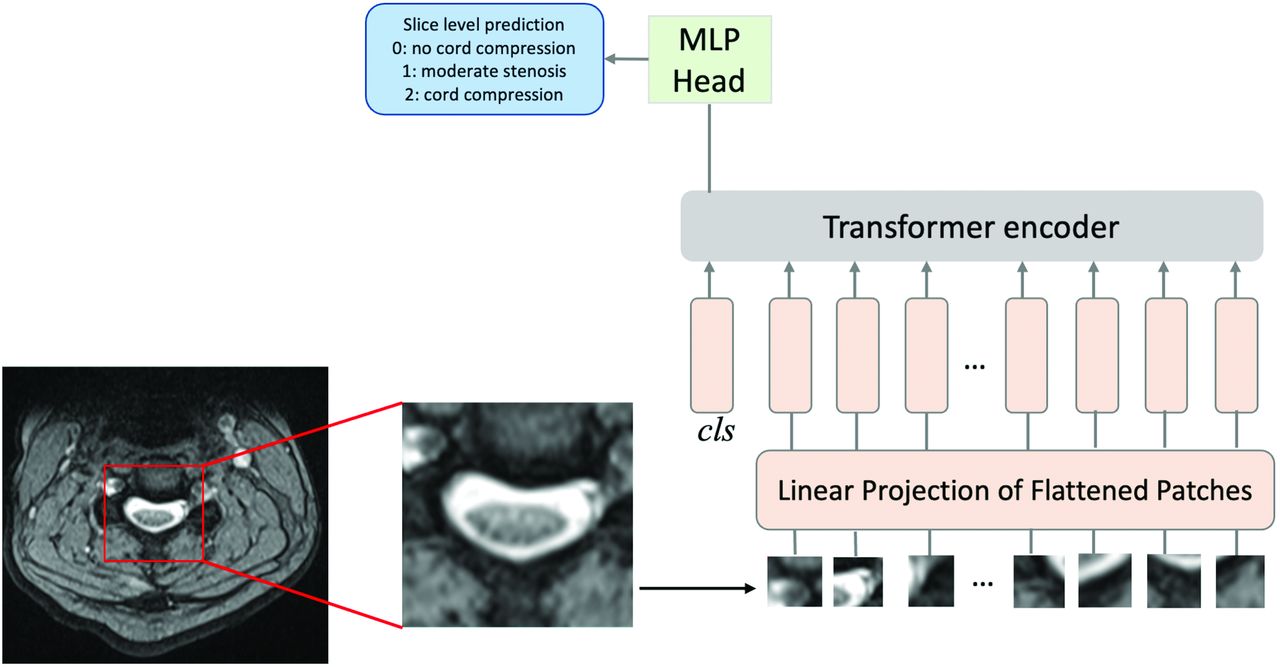
  • FIG 3.
    • Download figure
    • Open in new tab
    • Download powerpoint
    FIG 3.

    Vision transformer analysis pipeline. Images are divided into flattened patches, which are then fed into the transformer encoder along with positional encoding. For each section, a prediction of severity is generated.

  • FIG 4.
    • Download figure
    • Open in new tab
    • Download powerpoint
    FIG 4.

    Receiver operating characteristic curves comparing patient-level performance of ViT, DenseNet121, and ResNet50 in detecting cord compression.

  • FIG 5.
    • Download figure
    • Open in new tab
    • Download powerpoint
    FIG 5.

    False-positive and -negative cases. A, False-positive: Diffuse congenital moderate stenosis with small superimposed annular bulge, incorrectly categorized as positive for cord compression. B, False-negative: Large annular bulge with severe spinal stenosis and cord compression and abnormal T2 hyperintense signal within the cord, incorrectly categorized as negative for cord compression.

Tables

  • Figures
    • View popup
    Table 1:

    Patient demographics and clinical setting in the training/validation and testing cohorts

    CharacteristicTraining/Validation (n = 113)Testing (n = 29)
    Demographics
     Male56 (49.5%)11 (37.9%)
     Age57.553.1
    Clinical setting
     ER64 (56.6%)18 (62.1%)
     Inpatient16 (14.2%)3 (10.3%)
     Outpatient33 (29.2%)8 (27.6%)
     History of trauma49 (43.4%)14 (48.3%)
    • View popup
    Table 2:

    Section-level and patient-level test results for ViT, ResNet50, and DenseNet121 models

    ViTResNet50DenseNet121
    Section-level classification accuracy82%72%78%
    Patient-level accuracy93%62%62%
    Patient-level sensitivity0.901.00.8
    Patient-level specificity0.950.420.52
    Patient-level PPV0.900.470.47
    Patient-level NPV0.951.00.83
PreviousNext
Back to top
Advertisement
Print
Download PDF
Email Article

Thank you for your interest in spreading the word on American Journal of Neuroradiology.

NOTE: We only request your email address so that the person you are recommending the page to knows that you wanted them to see it, and that it is not junk mail. We do not capture any email address.

Enter multiple addresses on separate lines or separate them with commas.
Automated Detection of Cervical Spinal Stenosis and Cord Compression via Vision Transformer and Rules-Based Classification
(Your Name) has sent you a message from American Journal of Neuroradiology
(Your Name) thought you would like to see the American Journal of Neuroradiology web site.
CAPTCHA
This question is for testing whether or not you are a human visitor and to prevent automated spam submissions.
Cite this article
David L. Payne, Xuan Xu, Farshid Faraji, Kevin John, Katherine Ferra Pradas, Vahni Vishala Bernard, Lev Bangiyev, Prateek Prasanna
Automated Detection of Cervical Spinal Stenosis and Cord Compression via Vision Transformer and Rules-Based Classification
American Journal of Neuroradiology Feb 2024, DOI: 10.3174/ajnr.A8141

Citation Manager Formats

  • BibTeX
  • Bookends
  • EasyBib
  • EndNote (tagged)
  • EndNote 8 (xml)
  • Medlars
  • Mendeley
  • Papers
  • RefWorks Tagged
  • Ref Manager
  • RIS
  • Zotero
0 Responses
Respond to this article
Share
Bookmark this article
Automated Detection of Cervical Spinal Stenosis and Cord Compression via Vision Transformer and Rules-Based Classification
David L. Payne, Xuan Xu, Farshid Faraji, Kevin John, Katherine Ferra Pradas, Vahni Vishala Bernard, Lev Bangiyev, Prateek Prasanna
American Journal of Neuroradiology Feb 2024, DOI: 10.3174/ajnr.A8141
del.icio.us logo Twitter logo Facebook logo Mendeley logo
  • Tweet Widget
  • Facebook Like
  • Google Plus One
Purchase

Jump to section

  • Article
    • Abstract
    • ABBREVIATIONS:
    • MATERIALS AND METHODS
    • RESULTS
    • DISCUSSION
    • CONCLUSIONS
    • Footnotes
    • REFERENCES
  • Figures & Data
  • Info & Metrics
  • Responses
  • References
  • PDF

Related Articles

  • No related articles found.
  • PubMed
  • Google Scholar

Cited By...

  • Performance of GPT-4 on the American College of Radiology In-Service Examination
  • Crossref
  • Google Scholar

This article has not yet been cited by articles in journals that are participating in Crossref Cited-by Linking.

More in this TOC Section

  • Management Outcomes For VO Spine Biopsy
  • Characteristics of SIH Type I Culprit Lesions
  • Advanced Imaging of Type 2 Spinal CSF Leaks
Show more Spine Imaging and Spine Image-Guided Interventions

Similar Articles

Advertisement

Indexed Content

  • Current Issue
  • Accepted Manuscripts
  • Article Preview
  • Past Issues
  • Editorials
  • Editor's Choice
  • Fellows' Journal Club
  • Letters to the Editor
  • Video Articles

Cases

  • Case Collection
  • Archive - Case of the Week
  • Archive - Case of the Month
  • Archive - Classic Case

More from AJNR

  • Trainee Corner
  • Imaging Protocols
  • MRI Safety Corner
  • Book Reviews

Multimedia

  • AJNR Podcasts
  • AJNR Scantastics

Resources

  • Turnaround Time
  • Submit a Manuscript
  • Submit a Video Article
  • Submit an eLetter to the Editor/Response
  • Manuscript Submission Guidelines
  • Statistical Tips
  • Fast Publishing of Accepted Manuscripts
  • Graphical Abstract Preparation
  • Imaging Protocol Submission
  • Evidence-Based Medicine Level Guide
  • Publishing Checklists
  • Author Policies
  • Become a Reviewer/Academy of Reviewers
  • News and Updates

About Us

  • About AJNR
  • Editorial Board
  • Editorial Board Alumni
  • Alerts
  • Permissions
  • Not an AJNR Subscriber? Join Now
  • Advertise with Us
  • Librarian Resources
  • Feedback
  • Terms and Conditions
  • AJNR Editorial Board Alumni

American Society of Neuroradiology

  • Not an ASNR Member? Join Now

© 2025 by the American Society of Neuroradiology All rights, including for text and data mining, AI training, and similar technologies, are reserved.
Print ISSN: 0195-6108 Online ISSN: 1936-959X

Powered by HighWire