Advertisement

Main menu

  • Home
  • Content
    • Current Issue
    • Accepted Manuscripts
    • Article Preview
    • Past Issue Archive
    • Video Articles
    • AJNR Case Collection
    • Case of the Week Archive
    • Case of the Month Archive
    • Classic Case Archive
  • Special Collections
    • AJNR Awards
    • Low-Field MRI
    • Alzheimer Disease
    • ASNR Foundation Special Collection
    • Photon-Counting CT
    • View All
  • Multimedia
    • AJNR Podcasts
    • AJNR SCANtastic
    • Trainee Corner
    • MRI Safety Corner
    • Imaging Protocols
  • For Authors
    • Submit a Manuscript
    • Submit a Video Article
    • Submit an eLetter to the Editor/Response
    • Manuscript Submission Guidelines
    • Statistical Tips
    • Fast Publishing of Accepted Manuscripts
    • Graphical Abstract Preparation
    • Imaging Protocol Submission
    • Author Policies
  • About Us
    • About AJNR
    • Editorial Board
    • Editorial Board Alumni
  • More
    • Become a Reviewer/Academy of Reviewers
    • Subscribers
    • Permissions
    • Alerts
    • Feedback
    • Advertisers
    • ASNR Home

User menu

  • Alerts
  • Log in

Search

  • Advanced search
American Journal of Neuroradiology

American Journal of Neuroradiology

ASHNR American Society of Functional Neuroradiology ASHNR American Society of Pediatric Neuroradiology ASSR
  • Alerts
  • Log in

Advanced Search

  • Home
  • Content
    • Current Issue
    • Accepted Manuscripts
    • Article Preview
    • Past Issue Archive
    • Video Articles
    • AJNR Case Collection
    • Case of the Week Archive
    • Case of the Month Archive
    • Classic Case Archive
  • Special Collections
    • AJNR Awards
    • Low-Field MRI
    • Alzheimer Disease
    • ASNR Foundation Special Collection
    • Photon-Counting CT
    • View All
  • Multimedia
    • AJNR Podcasts
    • AJNR SCANtastic
    • Trainee Corner
    • MRI Safety Corner
    • Imaging Protocols
  • For Authors
    • Submit a Manuscript
    • Submit a Video Article
    • Submit an eLetter to the Editor/Response
    • Manuscript Submission Guidelines
    • Statistical Tips
    • Fast Publishing of Accepted Manuscripts
    • Graphical Abstract Preparation
    • Imaging Protocol Submission
    • Author Policies
  • About Us
    • About AJNR
    • Editorial Board
    • Editorial Board Alumni
  • More
    • Become a Reviewer/Academy of Reviewers
    • Subscribers
    • Permissions
    • Alerts
    • Feedback
    • Advertisers
    • ASNR Home
  • Follow AJNR on Twitter
  • Visit AJNR on Facebook
  • Follow AJNR on Instagram
  • Join AJNR on LinkedIn
  • RSS Feeds

AJNR Awards, New Junior Editors, and more. Read the latest AJNR updates

Research ArticleULTRA-HIGH-FIELD MRI/IMAGING OF EPILEPSY/DEMYELINATING DISEASES/INFLAMMATION/INFECTION

Epstein-Barr Virus–Associated Smooth-Muscle Tumor of the Brain

Afra Alfalahi, Abdelsimar Il Omar, Krystal Fox, Julian Spears, Malika Sharma, Aditya Bharatha, David G. Munoz and Suradech Suthiphosuwan
American Journal of Neuroradiology May 2024, DOI: https://doi.org/10.3174/ajnr.A8258
Afra Alfalahi
aFrom the Division of Diagnostic Neuroradiology (A.A., A.B., S.S.), St. Michael’s Hospital-Unity Health Toronto, University of Toronto, Toronto, Ontario, Canada
dDivision of Diagnostic Neuroradiology (A.A.), Department of Medical Imaging, University Health Network, University of Toronto, Toronto, Ontario, Canada
  • Find this author on Google Scholar
  • Find this author on PubMed
  • Search for this author on this site
  • ORCID record for Afra Alfalahi
Abdelsimar Il Omar
bDepartment of Medical Imaging, Division of Neurosurgery (A.I.O., K.F., J.S.), Department of Surgery, St. Michael’s Hospital-Unity Health Toronto, University of Toronto, Toronto, Ontario, Canada
eDivision of Neurosurgery (A.I.O.), Department of Surgery, McMaster University, Hamilton, Ontario, Canada
  • Find this author on Google Scholar
  • Find this author on PubMed
  • Search for this author on this site
Krystal Fox
bDepartment of Medical Imaging, Division of Neurosurgery (A.I.O., K.F., J.S.), Department of Surgery, St. Michael’s Hospital-Unity Health Toronto, University of Toronto, Toronto, Ontario, Canada
  • Find this author on Google Scholar
  • Find this author on PubMed
  • Search for this author on this site
  • ORCID record for Krystal Fox
Julian Spears
bDepartment of Medical Imaging, Division of Neurosurgery (A.I.O., K.F., J.S.), Department of Surgery, St. Michael’s Hospital-Unity Health Toronto, University of Toronto, Toronto, Ontario, Canada
  • Find this author on Google Scholar
  • Find this author on PubMed
  • Search for this author on this site
  • ORCID record for Julian Spears
Malika Sharma
cDepartment of Infectious Diseases (M.S.), St. Michael’s Hospital-Unity Health Toronto, University of Toronto, Toronto, Ontario, Canada
  • Find this author on Google Scholar
  • Find this author on PubMed
  • Search for this author on this site
Aditya Bharatha
aFrom the Division of Diagnostic Neuroradiology (A.A., A.B., S.S.), St. Michael’s Hospital-Unity Health Toronto, University of Toronto, Toronto, Ontario, Canada
  • Find this author on Google Scholar
  • Find this author on PubMed
  • Search for this author on this site
  • ORCID record for Aditya Bharatha
David G. Munoz
fDepartment of Laboratory Medicine (D.G.M.), St. Michael’s Hospital-Unity Health Toronto, University of Toronto, Toronto, Ontario, Canada
  • Find this author on Google Scholar
  • Find this author on PubMed
  • Search for this author on this site
  • ORCID record for David G. Munoz
Suradech Suthiphosuwan
aFrom the Division of Diagnostic Neuroradiology (A.A., A.B., S.S.), St. Michael’s Hospital-Unity Health Toronto, University of Toronto, Toronto, Ontario, Canada
  • Find this author on Google Scholar
  • Find this author on PubMed
  • Search for this author on this site
  • ORCID record for Suradech Suthiphosuwan
  • Article
  • Figures & Data
  • Info & Metrics
  • Responses
  • References
  • PDF
Loading

Article Figures & Data

Figures

  • FIG 1.
    • Download figure
    • Open in new tab
    • Download powerpoint
    FIG 1.

    Tumor appearance on MR imaging. MR images show a right posterior fossa mass, which had isointense signal on pregadolinium T1WI (A) and hypointense signal on T2WI (B) and FLAIR (C) with perilesional edema (B and C). The mass does not show diffusion restriction on DWI (D). There are a few blooming foci within the tumor seen on SWI (E). The mass shows intense enhancement with multifocal, small, nonenhancing areas of necrosis on axial postgadolinium T1WI (F). Sagittal postgadolinium T1WI (G) demonstrates the tumor attached to the inferior surface of the right tentorium cerebelli (arrow). On DSC MR perfusion (H), the tumor had low rCBV.
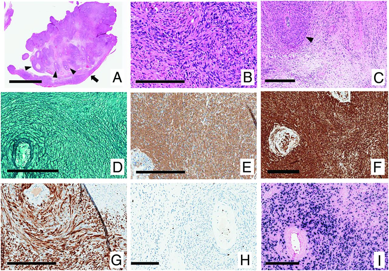
  • FIG 2.
    • Download figure
    • Open in new tab
    • Download powerpoint
    FIG 2.

    Histopathology. H&E-stained sections A, B, and C (magnification A, bar 4 mm; B, bar 200 um; and C, bar 300 um). A, The attachment to the dura mater is demonstrated at the lower right (arrow). B, Cells have spindle-shaped nuclei with blunted ends and wavy eosinophilic cytoplasm of uncertain limits and are arranged in intersecting streams and whorls. The tendency to surround blood vessels is seen in A and C (arrowheads). Reticulin stains demonstrate wrapping of single cells, with no reticulin-free areas (D, bar 200 um). Immunohistochemistry shows expression of smooth-muscle actin (E, bar 200 um) and myosin (F, bar 200 um) with only a subset of cells expressing desmin (G, bar 200 um). The Ki-67 proliferation index was approximately 2%; high only in the lining of blood vessels (H, bar 200 um). EBV-encoded RNA in situ hybridization was intensely positive (I, bar 200 um).

PreviousNext
Back to top
Advertisement
Print
Download PDF
Email Article

Thank you for your interest in spreading the word on American Journal of Neuroradiology.

NOTE: We only request your email address so that the person you are recommending the page to knows that you wanted them to see it, and that it is not junk mail. We do not capture any email address.

Enter multiple addresses on separate lines or separate them with commas.
Epstein-Barr Virus–Associated Smooth-Muscle Tumor of the Brain
(Your Name) has sent you a message from American Journal of Neuroradiology
(Your Name) thought you would like to see the American Journal of Neuroradiology web site.
CAPTCHA
This question is for testing whether or not you are a human visitor and to prevent automated spam submissions.
Cite this article
Afra Alfalahi, Abdelsimar Il Omar, Krystal Fox, Julian Spears, Malika Sharma, Aditya Bharatha, David G. Munoz, Suradech Suthiphosuwan
Epstein-Barr Virus–Associated Smooth-Muscle Tumor of the Brain
American Journal of Neuroradiology May 2024, DOI: 10.3174/ajnr.A8258

Citation Manager Formats

  • BibTeX
  • Bookends
  • EasyBib
  • EndNote (tagged)
  • EndNote 8 (xml)
  • Medlars
  • Mendeley
  • Papers
  • RefWorks Tagged
  • Ref Manager
  • RIS
  • Zotero
0 Responses
Respond to this article
Share
Bookmark this article
Epstein-Barr Virus–Associated Smooth-Muscle Tumor of the Brain
Afra Alfalahi, Abdelsimar Il Omar, Krystal Fox, Julian Spears, Malika Sharma, Aditya Bharatha, David G. Munoz, Suradech Suthiphosuwan
American Journal of Neuroradiology May 2024, DOI: 10.3174/ajnr.A8258
del.icio.us logo Twitter logo Facebook logo Mendeley logo
  • Tweet Widget
  • Facebook Like
  • Google Plus One
Purchase

Jump to section

  • Article
    • SUMMARY:
    • ABBREVIATIONS:
    • DISCUSSION
    • Footnotes
    • References
  • Figures & Data
  • Info & Metrics
  • Responses
  • References
  • PDF

Related Articles

  • No related articles found.
  • PubMed
  • Google Scholar

Cited By...

  • No citing articles found.
  • Crossref
  • Google Scholar

This article has not yet been cited by articles in journals that are participating in Crossref Cited-by Linking.

More in this TOC Section

  • DL Image Reconstruction in T2-Weighted TSE at 7T
  • Automated Lesion Segmentation Software in MS
  • Interpretation Errors in Adults with MRE
Show more Ultra-High-Field MRI/Imaging of Epilepsy/Demyelinating Diseases/Inflammation/Infection

Similar Articles

Advertisement

Indexed Content

  • Current Issue
  • Accepted Manuscripts
  • Article Preview
  • Past Issues
  • Editorials
  • Editor's Choice
  • Fellows' Journal Club
  • Letters to the Editor
  • Video Articles

Cases

  • Case Collection
  • Archive - Case of the Week
  • Archive - Case of the Month
  • Archive - Classic Case

More from AJNR

  • Trainee Corner
  • Imaging Protocols
  • MRI Safety Corner
  • Book Reviews

Multimedia

  • AJNR Podcasts
  • AJNR Scantastics

Resources

  • Turnaround Time
  • Submit a Manuscript
  • Submit a Video Article
  • Submit an eLetter to the Editor/Response
  • Manuscript Submission Guidelines
  • Statistical Tips
  • Fast Publishing of Accepted Manuscripts
  • Graphical Abstract Preparation
  • Imaging Protocol Submission
  • Evidence-Based Medicine Level Guide
  • Publishing Checklists
  • Author Policies
  • Become a Reviewer/Academy of Reviewers
  • News and Updates

About Us

  • About AJNR
  • Editorial Board
  • Editorial Board Alumni
  • Alerts
  • Permissions
  • Not an AJNR Subscriber? Join Now
  • Advertise with Us
  • Librarian Resources
  • Feedback
  • Terms and Conditions
  • AJNR Editorial Board Alumni

American Society of Neuroradiology

  • Not an ASNR Member? Join Now

© 2025 by the American Society of Neuroradiology All rights, including for text and data mining, AI training, and similar technologies, are reserved.
Print ISSN: 0195-6108 Online ISSN: 1936-959X

Powered by HighWire