Advertisement

Main menu

  • Home
  • Content
    • Current Issue
    • Accepted Manuscripts
    • Article Preview
    • Past Issue Archive
    • Video Articles
    • AJNR Case Collection
    • Case of the Week Archive
    • Case of the Month Archive
    • Classic Case Archive
  • Special Collections
    • AJNR Awards
    • Low-Field MRI
    • Alzheimer Disease
    • ASNR Foundation Special Collection
    • Photon-Counting CT
    • View All
  • Multimedia
    • AJNR Podcasts
    • AJNR SCANtastic
    • Trainee Corner
    • MRI Safety Corner
    • Imaging Protocols
  • For Authors
    • Submit a Manuscript
    • Submit a Video Article
    • Submit an eLetter to the Editor/Response
    • Manuscript Submission Guidelines
    • Statistical Tips
    • Fast Publishing of Accepted Manuscripts
    • Graphical Abstract Preparation
    • Imaging Protocol Submission
    • Author Policies
  • About Us
    • About AJNR
    • Editorial Board
    • Editorial Board Alumni
  • More
    • Become a Reviewer/Academy of Reviewers
    • Subscribers
    • Permissions
    • Alerts
    • Feedback
    • Advertisers
    • ASNR Home

User menu

  • Alerts
  • Log in

Search

  • Advanced search
American Journal of Neuroradiology

American Journal of Neuroradiology

ASHNR American Society of Functional Neuroradiology ASHNR American Society of Pediatric Neuroradiology ASSR
  • Alerts
  • Log in

Advanced Search

  • Home
  • Content
    • Current Issue
    • Accepted Manuscripts
    • Article Preview
    • Past Issue Archive
    • Video Articles
    • AJNR Case Collection
    • Case of the Week Archive
    • Case of the Month Archive
    • Classic Case Archive
  • Special Collections
    • AJNR Awards
    • Low-Field MRI
    • Alzheimer Disease
    • ASNR Foundation Special Collection
    • Photon-Counting CT
    • View All
  • Multimedia
    • AJNR Podcasts
    • AJNR SCANtastic
    • Trainee Corner
    • MRI Safety Corner
    • Imaging Protocols
  • For Authors
    • Submit a Manuscript
    • Submit a Video Article
    • Submit an eLetter to the Editor/Response
    • Manuscript Submission Guidelines
    • Statistical Tips
    • Fast Publishing of Accepted Manuscripts
    • Graphical Abstract Preparation
    • Imaging Protocol Submission
    • Author Policies
  • About Us
    • About AJNR
    • Editorial Board
    • Editorial Board Alumni
  • More
    • Become a Reviewer/Academy of Reviewers
    • Subscribers
    • Permissions
    • Alerts
    • Feedback
    • Advertisers
    • ASNR Home
  • Follow AJNR on Twitter
  • Visit AJNR on Facebook
  • Follow AJNR on Instagram
  • Join AJNR on LinkedIn
  • RSS Feeds

AJNR Awards, New Junior Editors, and more. Read the latest AJNR updates

Research ArticleNEUROIMAGING PHYSICS/FUNCTIONAL NEUROIMAGING/CT AND MRI TECHNOLOGY

Fractional Anisotropy is a More Sensitive Diagnostic Biomarker Than Mean Kurtosis for Patients with Parkinson Disease with Cognitive Dysfunction: A Diffusional Kurtosis Map Tract-Based Spatial Statistics Study

Bingbing Gao, Mingrui Qu, Yuhan Jiang, Wanyao Li, Man Wang, Chenhui Pei, Dandan Zheng, Chao Yang and Yanwei Miao
American Journal of Neuroradiology July 2024, DOI: https://doi.org/10.3174/ajnr.A8297
Bingbing Gao
aFrom the Department of Radiology (B.G., M.Q., Y.J., W.l., M.W., C.Y., Y.M), The First Affiliated Hospital of Dalian Medical University, Dalian, China
  • Find this author on Google Scholar
  • Find this author on PubMed
  • Search for this author on this site
  • ORCID record for Bingbing Gao
Mingrui Qu
aFrom the Department of Radiology (B.G., M.Q., Y.J., W.l., M.W., C.Y., Y.M), The First Affiliated Hospital of Dalian Medical University, Dalian, China
  • Find this author on Google Scholar
  • Find this author on PubMed
  • Search for this author on this site
  • ORCID record for Mingrui Qu
Yuhan Jiang
aFrom the Department of Radiology (B.G., M.Q., Y.J., W.l., M.W., C.Y., Y.M), The First Affiliated Hospital of Dalian Medical University, Dalian, China
  • Find this author on Google Scholar
  • Find this author on PubMed
  • Search for this author on this site
  • ORCID record for Yuhan Jiang
Wanyao Li
aFrom the Department of Radiology (B.G., M.Q., Y.J., W.l., M.W., C.Y., Y.M), The First Affiliated Hospital of Dalian Medical University, Dalian, China
  • Find this author on Google Scholar
  • Find this author on PubMed
  • Search for this author on this site
Man Wang
aFrom the Department of Radiology (B.G., M.Q., Y.J., W.l., M.W., C.Y., Y.M), The First Affiliated Hospital of Dalian Medical University, Dalian, China
  • Find this author on Google Scholar
  • Find this author on PubMed
  • Search for this author on this site
Chenhui Pei
bDepartment of Neurology (C.P.), The First Affiliated Hospital of Dalian Medical University, Dalian, China
  • Find this author on Google Scholar
  • Find this author on PubMed
  • Search for this author on this site
  • ORCID record for Chenhui Pei
Dandan Zheng
cPhilips Healthcare, Clinical &Technique Support (D.Z.), Beijing, China
  • Find this author on Google Scholar
  • Find this author on PubMed
  • Search for this author on this site
Chao Yang
aFrom the Department of Radiology (B.G., M.Q., Y.J., W.l., M.W., C.Y., Y.M), The First Affiliated Hospital of Dalian Medical University, Dalian, China
  • Find this author on Google Scholar
  • Find this author on PubMed
  • Search for this author on this site
  • ORCID record for Chao Yang
Yanwei Miao
aFrom the Department of Radiology (B.G., M.Q., Y.J., W.l., M.W., C.Y., Y.M), The First Affiliated Hospital of Dalian Medical University, Dalian, China
  • Find this author on Google Scholar
  • Find this author on PubMed
  • Search for this author on this site
  • Article
  • Figures & Data
  • Supplemental
  • Info & Metrics
  • Responses
  • References
  • PDF
Loading

Article Figures & Data

Figures

  • Tables
  • FIG 1.
    • Download figure
    • Open in new tab
    • Download powerpoint
    FIG 1.

    Post hoc analyses results between patients with PD-MCI and PDD. Green represents the mean FA skeleton of all subjects. Yellow and red represent regions with significant statistical values (P < . 05, TFCE-corrected).
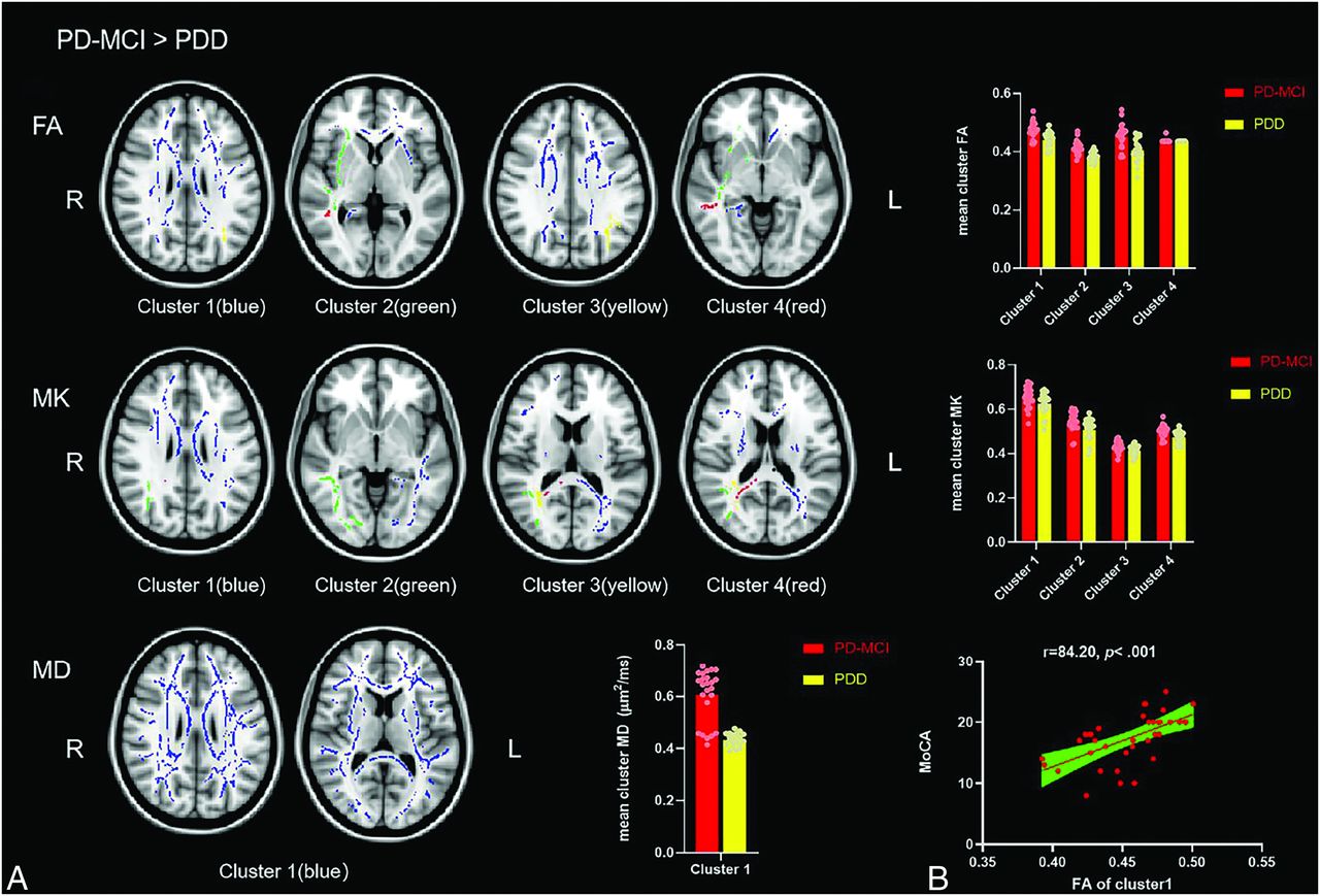
  • FIG 2.
    • Download figure
    • Open in new tab
    • Download powerpoint
    FIG 2.

    A, Voxel clusters of decreased FA, MK, and MD of the PDD group more than PD-MCI group. The colorful regions (blue, green, yellow, red) represent regions with significant statistical values (P < . 05, TFCE-corrected). B, Positive correlation between the FA of cluster 1 and the MoCA. FA has 4 independent clusters, MK has 4 independent clusters, and MD has 1 independent cluster. The bar plots represent the corresponding mean diffusion metrics for each group.

  • FIG 3.
    • Download figure
    • Open in new tab
    • Download powerpoint
    FIG 3.

    The ROC curves of significantly different clusters of DKI metrics and the combined ROC model. The combined model was fitted with cluster 1, 2, and 3 of the FA map and cluster 2 and 3 of the MK map.

Tables

  • Figures
    • View popup
    Table 1:

    Clinical data of PD-MCI and PDD groups

    PD-MCI (n = 19)PDD (n = 18)t/χ2/zP
    Age of PD diagnosed (mean) (yr)63.34 (SD, 5.53)65.73 (SD, 5.98)−1.262.215a
    Age of PD-MCI or PDD diagnosed (mean) (yr)64.33 (SD, 7.96)71.10 (SD, 6.26)−3.060.004a**
    Sex, M/F11/86/122.245.134b
    Disease duration (yr)2 (range, 2.00–3.50)3.943 (SD, 2.59)−2.004.052c
    Education (mean) (yr)11.42 (SD, 3.61)10.22 (SD, 3.20)1.065.294a
    H-Y stage (No.) (%)––−0.836.403c
     16 (31.58)5 (27.78)––
     1.5NA1 (5.56)––
     28 (42.11)4 (22.22)––
     2.52 (10.53)2 (11.11)––
     33 (15.79)5 (27.78)––
     5NA1 (5.56)––
    MMSE (mean)26.36 (SD, 1.70)19.61 (SD, 3.98)6.765<.001a**
    MoCA (mean)19.89 (SD, 2.57)14.61 (SD, 3.51)5.230<.001a**
    • Note:—** indicates significant difference; NA, not applicable; en dash, no raw data or analyzed data.

    • ↵a Independent t test.

    • b χ2 tests.

    • c Mann-Whitney test.

    • View popup
    Table 2:

    Cluster sizes and locations for voxels with significantly reduced MD values in the PDD group compared with PD-MCI group (PD-MCI > PDD)a

    Cluster No.JHU White Matter Tractography AtlasJHU ICBM-DTI-81 White-Matter LabelsVoxels and MNI CoordinatesP
    1Forceps minor: 3.07203Body of corpus callosum: 4.2185751534 (64/152/81).002
    • Note:—MNI indicates the Montreal Neurological Institute.

    • a The value after each region indicates the percentage probability of the cluster belonging to the given atlas label. Only regions with probability >3% were included.

    • View popup
    Table 3:

    Comparison between significantly different clusters and the combined ROC curve model

    ClustersAUCStandard Error95% CICompared with the Combined Model
    χ2P
    FA cluster 10.810.070.660.953.32.068
    FA cluster 20.850.060.730.981.80.180
    FA cluster 30.760.080.600.923.77.052
    FA cluster 40.530.030.470.5847.79<.001
    MK cluster 10.630.090.450.818.45.003
    MK cluster 20.690.090.520.876.99.008
    MK cluster 30.700.090.520.877.12.007
    MK cluster 40.680.090.500.869.01.002
    MD cluster 10.650.090.460.8310.55.001
    Combined0.910.050.821.00––
    • Note:—The en dash indicates no analyzed data.

    • View popup
    Table 4:

    Predictors of MoCA scores in patients with PD with cognitive dysfunction: univariate and multiple linear regression analysis

    CharacteristicUnivariate AnalysisaMultiple Analysisb
    Coefficient (95% CI)PCoefficient (95% CI)P
    Age of PD diagnosed−0.31 (−0.52∼−0.09).006**
    Sex−3.64 (−6.08∼−1.20).005**
    Education0.39 (0.01∼0.77).042*0.38 (0.08∼0.68).014*
    Duration−0.20 (−0.82∼0.41).503
    Age of PD-MCI or PDD diagnosed−0.35 (−0.56∼−0.14).002**
    FA cluster 185.16 (46.48∼123.84)<.001***84.20 (48.30∼120.11)<.001***
    FA cluster 2112.82 (59.30∼166.35)<.001***
    FA cluster 335.08 (4.73∼65.43).025*
    FA cluster 4200.32 (−81.12∼481.76).157
    MK cluster 135.08 (10.44∼59.73).007**
    MK cluster 242.35 (18.44∼66.26).001**
    MK cluster 385.27 (32.95∼137.59).002**
    MK cluster 470.63 (24.81∼116.44).004**
    MD cluster 183.00 (29.99∼136.01).003**
    • Note:—Single asterisk indicates P < .05; double asterisks, P < .005; triple asterisks, P < .001.

    • ↵a Univariate analysis based on the complete cases without missing value.

    • b Multiple analyses based on imputed values in predictors.

PreviousNext
Back to top
Advertisement
Print
Download PDF
Email Article

Thank you for your interest in spreading the word on American Journal of Neuroradiology.

NOTE: We only request your email address so that the person you are recommending the page to knows that you wanted them to see it, and that it is not junk mail. We do not capture any email address.

Enter multiple addresses on separate lines or separate them with commas.
Fractional Anisotropy is a More Sensitive Diagnostic Biomarker Than Mean Kurtosis for Patients with Parkinson Disease with Cognitive Dysfunction: A Diffusional Kurtosis Map Tract-Based Spatial Statistics Study
(Your Name) has sent you a message from American Journal of Neuroradiology
(Your Name) thought you would like to see the American Journal of Neuroradiology web site.
CAPTCHA
This question is for testing whether or not you are a human visitor and to prevent automated spam submissions.
Cite this article
Bingbing Gao, Mingrui Qu, Yuhan Jiang, Wanyao Li, Man Wang, Chenhui Pei, Dandan Zheng, Chao Yang, Yanwei Miao
Fractional Anisotropy is a More Sensitive Diagnostic Biomarker Than Mean Kurtosis for Patients with Parkinson Disease with Cognitive Dysfunction: A Diffusional Kurtosis Map Tract-Based Spatial Statistics Study
American Journal of Neuroradiology Jul 2024, DOI: 10.3174/ajnr.A8297

Citation Manager Formats

  • BibTeX
  • Bookends
  • EasyBib
  • EndNote (tagged)
  • EndNote 8 (xml)
  • Medlars
  • Mendeley
  • Papers
  • RefWorks Tagged
  • Ref Manager
  • RIS
  • Zotero
0 Responses
Respond to this article
Share
Bookmark this article
Fractional Anisotropy is a More Sensitive Diagnostic Biomarker Than Mean Kurtosis for Patients with Parkinson Disease with Cognitive Dysfunction: A Diffusional Kurtosis Map Tract-Based Spatial Statistics Study
Bingbing Gao, Mingrui Qu, Yuhan Jiang, Wanyao Li, Man Wang, Chenhui Pei, Dandan Zheng, Chao Yang, Yanwei Miao
American Journal of Neuroradiology Jul 2024, DOI: 10.3174/ajnr.A8297
del.icio.us logo Twitter logo Facebook logo Mendeley logo
  • Tweet Widget
  • Facebook Like
  • Google Plus One
Purchase

Jump to section

  • Article
    • Abstract
    • ABBREVIATIONS:
    • MATERIALS AND METHODS
    • RESULTS
    • DISCUSSION
    • CONCLUSIONS
    • Acknowledgments
    • Footnotes
    • References
  • Figures & Data
  • Supplemental
  • Info & Metrics
  • Responses
  • References
  • PDF

Related Articles

  • PubMed
  • Google Scholar

Cited By...

  • No citing articles found.
  • Crossref
  • Google Scholar

This article has not yet been cited by articles in journals that are participating in Crossref Cited-by Linking.

More in this TOC Section

  • A Comprehensive Review of IA Imaging Modalities
  • Synthetic MRI based on 3D-QALAS MR Quantification
  • Delta wave MRI
Show more Neuroimaging Physics/Functional Neuroimaging/CT and MRI Technology

Similar Articles

Advertisement

Indexed Content

  • Current Issue
  • Accepted Manuscripts
  • Article Preview
  • Past Issues
  • Editorials
  • Editor's Choice
  • Fellows' Journal Club
  • Letters to the Editor
  • Video Articles

Cases

  • Case Collection
  • Archive - Case of the Week
  • Archive - Case of the Month
  • Archive - Classic Case

More from AJNR

  • Trainee Corner
  • Imaging Protocols
  • MRI Safety Corner
  • Book Reviews

Multimedia

  • AJNR Podcasts
  • AJNR Scantastics

Resources

  • Turnaround Time
  • Submit a Manuscript
  • Submit a Video Article
  • Submit an eLetter to the Editor/Response
  • Manuscript Submission Guidelines
  • Statistical Tips
  • Fast Publishing of Accepted Manuscripts
  • Graphical Abstract Preparation
  • Imaging Protocol Submission
  • Evidence-Based Medicine Level Guide
  • Publishing Checklists
  • Author Policies
  • Become a Reviewer/Academy of Reviewers
  • News and Updates

About Us

  • About AJNR
  • Editorial Board
  • Editorial Board Alumni
  • Alerts
  • Permissions
  • Not an AJNR Subscriber? Join Now
  • Advertise with Us
  • Librarian Resources
  • Feedback
  • Terms and Conditions
  • AJNR Editorial Board Alumni

American Society of Neuroradiology

  • Not an ASNR Member? Join Now

© 2025 by the American Society of Neuroradiology All rights, including for text and data mining, AI training, and similar technologies, are reserved.
Print ISSN: 0195-6108 Online ISSN: 1936-959X

Powered by HighWire