Advertisement

Main menu

  • Home
  • Content
    • Current Issue
    • Accepted Manuscripts
    • Article Preview
    • Past Issue Archive
    • AJNR Case Collection
    • Case of the Week Archive
    • Classic Case Archive
    • Case of the Month Archive
  • Special Collections
    • Spinal CSF Leak Articles (Jan 2020-June 2024)
    • 2024 AJNR Journal Awards
    • Most Impactful AJNR Articles
  • Multimedia
    • AJNR Podcast
    • AJNR Scantastics
    • Video Articles
  • For Authors
    • Submit a Manuscript
    • Author Policies
    • Fast publishing of Accepted Manuscripts
    • Graphical Abstract Preparation
    • Manuscript Submission Guidelines
    • Imaging Protocol Submission
    • Submit a Case for the Case Collection
  • About Us
    • About AJNR
    • Editorial Board
  • More
    • Become a Reviewer/Academy of Reviewers
    • Subscribers
    • Permissions
    • Alerts
    • Feedback
    • Advertisers
    • ASNR Home
  • Other Publications
    • ajnr

User menu

  • Alerts
  • Log in

Search

  • Advanced search
American Journal of Neuroradiology

American Journal of Neuroradiology

ASHNR American Society of Functional Neuroradiology ASHNR American Society of Pediatric Neuroradiology ASSR
  • Alerts
  • Log in

Advanced Search

  • Home
  • Content
    • Current Issue
    • Accepted Manuscripts
    • Article Preview
    • Past Issue Archive
    • AJNR Case Collection
    • Case of the Week Archive
    • Classic Case Archive
    • Case of the Month Archive
  • Special Collections
    • Spinal CSF Leak Articles (Jan 2020-June 2024)
    • 2024 AJNR Journal Awards
    • Most Impactful AJNR Articles
  • Multimedia
    • AJNR Podcast
    • AJNR Scantastics
    • Video Articles
  • For Authors
    • Submit a Manuscript
    • Author Policies
    • Fast publishing of Accepted Manuscripts
    • Graphical Abstract Preparation
    • Manuscript Submission Guidelines
    • Imaging Protocol Submission
    • Submit a Case for the Case Collection
  • About Us
    • About AJNR
    • Editorial Board
  • More
    • Become a Reviewer/Academy of Reviewers
    • Subscribers
    • Permissions
    • Alerts
    • Feedback
    • Advertisers
    • ASNR Home
  • Follow AJNR on Twitter
  • Visit AJNR on Facebook
  • Follow AJNR on Instagram
  • Join AJNR on LinkedIn
  • RSS Feeds

Welcome to the new AJNR, Updated Hall of Fame, and more. Read the full announcements.


AJNR is seeking candidates for the position of Associate Section Editor, AJNR Case Collection. Read the full announcement.


OtherSPINE

Depiction of a Postoperative Pseudomeningocele with Digital Subtraction Myelography

C. Douglas Phillips, George J. Kaptain and Nasser Razack
American Journal of Neuroradiology February 2002, 23 (2) 337-338;
C. Douglas Phillips
  • Find this author on Google Scholar
  • Find this author on PubMed
  • Search for this author on this site
George J. Kaptain
  • Find this author on Google Scholar
  • Find this author on PubMed
  • Search for this author on this site
Nasser Razack
  • Find this author on Google Scholar
  • Find this author on PubMed
  • Search for this author on this site
  • Article
  • Figures & Data
  • Info & Metrics
  • Responses
  • References
  • PDF
Loading

Abstract

Summary: Digital subtraction myelography is described for its utility in the detection of dural leaks associated with pseudomeningoceles. Although myelography, CT, and MR imaging have been described as effective means for diagnosing pseudomeningocele, this complicated entity can be difficult to diagnose.

Postsurgical pseudomeningoceles are relatively rare complications of spinal surgery that have received little attention in both the neurosurgical and radiologic literature (1). They result from an inadvertent meningeal tear or inadequate closure during spinal surgery (1–3). CSF extravasates from a dural-arachnoid tear and is contained within the wound. Sequelae such as wound swelling, headache, and focal neurologic may result (4). Often, radicular pain can be precipitated or aggravated by maneuvers that increase intracranial and intraspinal pressure, such as coughing, sneezing, or jugular compression (5).

We describe a case of recurrent pseudomeningocele that was not clearly defined at myelography, postmyelographic CT, or MR imaging. An experienced neurosurgical team performed an interval surgical procedure before digital subtraction angiography; the procedure did not demonstrate the leak. Diagnosis was confirmed at digital subtraction myelography, a novel study that we offer as an additional technique for use in the diagnosis of pseudomeningocele.

Description of Technique

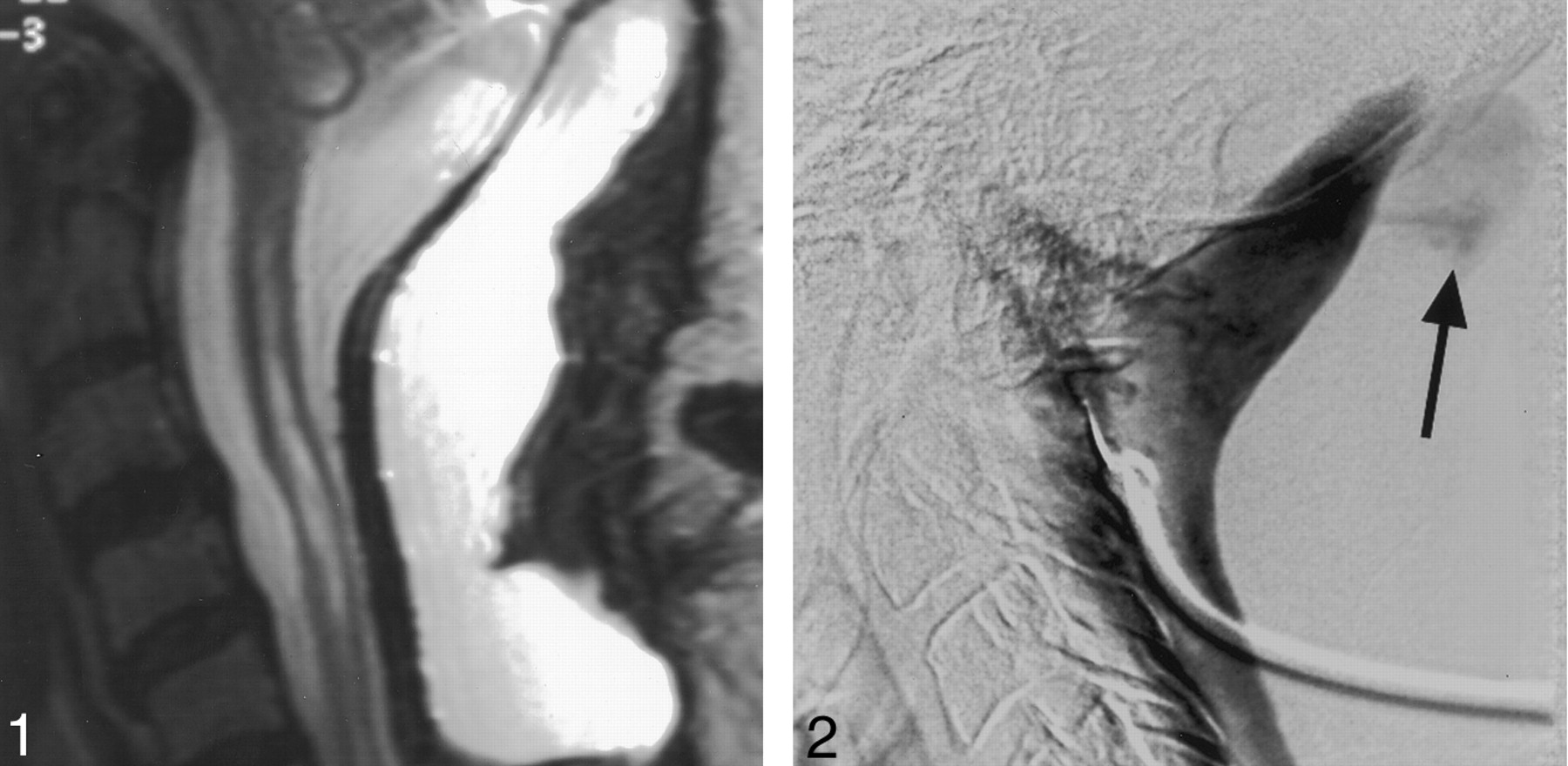
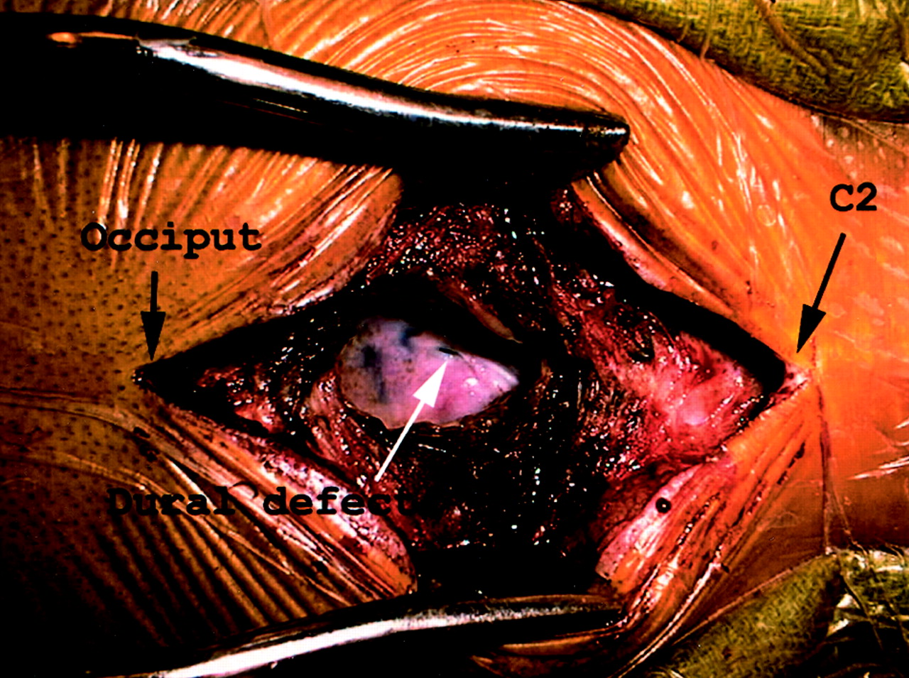
After MR imaging (Fig 1), myelography, and CT failed to depict a dural defect associated with a pseudomeningocele, digital subtraction myelography was performed. The patient was taken to the angiography suite (our current myelography suite does not have a cine or rapid-sequence capability). The patient was positioned laterally and underwent a C1-C2 puncture. A rapid subtraction acquisition was performed. After a mask image was obtained, a total of 5 mL of contrast material (Omnipaque 180; Nycomed Amersham, Princeton, NJ) was injected with a hand syringe at a rate of approximately 1 mL/s. Subtraction images were acquired at a rate of one frame per second. Extravasation of contrast material through a high cervical dural defect was immediately identified (Fig 2). Postmyelographic CT images were less convincing but revealed the large fluid collection. The patient then was taken to the operating room, and the level of contrast material extravasation was surgically explored. The dural defect was intraoperatively identified at this level (Fig 3).

Fig 1.
  • Download figure
  • Open in new tab
  • Download powerpoint
Fig 1.

Sagittal T2-weighted (5400/112 [TR/TE]) cervical MR image depicts fluid dorsal to the posterior fossa and the cervical thecal sac, but no dural leak is identified.

Fig 2.
  • Download figure
  • Open in new tab
  • Download powerpoint
Fig 2.

Axial postmyelographic CT demonstrates the pseudomeningocele (arrow) but fails to localize the dural defect.

Fig 3.
  • Download figure
  • Open in new tab
  • Download powerpoint
Fig 3.

Intraoperative photograph shows the dural defect. Surgical retractors extend from the occiput (patient’s left) to the level of C2 (patient’s right).

Discussion

Pseudomeningocele should be suspected if the surgeon is aware of a dural tear during the time of the original surgery (5). The diagnosis of this entity is debated in the literature. Initially, myelography was recommended as the technique of choice to establish the diagnosis (5). Other studies have shown myelography to be ineffective (6) and have suggested that CT and MR imaging are the techniques of choice for depicting this entity (2, 7). The diagnosis is further complicated in that a communication between the pseudomeningocele and the subarachnoid space may not exist (2). Often, dural tears leading to pseudomeningoceles are not identified intraoperatively (5). Although myelography, CT, and MR imaging have been described as effective for diagnosing a pseudomeningocele, it remains difficult to localize.

References

  1. ↵
    D’Andrea F, Maiuri F, Corriero G, Gambardella A, La Tessa G, Gangemi M. Postoperative lumbar arachnoidal diverticula. Surg Neurol 1985;23:287–290
    PubMed
  2. ↵
    Teplick JG, Peyster RG, Teplick SK, Goodman LR, Haskin ME. CT identification of postlaminectomy pseudomeningocele. AJR Am J Roentgenol 1983;140:1203–1206
    PubMed
  3. ↵
    Kim YW, Unger JD, Grinsell PJ. Post-operative pseudodiverticular (spurious meningoceles) of the cervical subarachnoid space. Acta Radiol Diagn 1974;15:16–20
  4. ↵
    Kaar GF, Briggs M, Bashir SH. Thecal repair in post-surgical pseudomeningocele. Br J Neurosurg 1994;8:703–708
    PubMed
  5. ↵
    Rinaldi I, Hodges TO. Iatrogenic lumbar meningocele: report of three cases. J Neurol Neurosurg Psychiatry 1970;33:484–492
    Abstract/FREE Full Text
  6. ↵
    Rinaldi I, Peach WF Jr. Postoperative lumbar meningocele. Report of two cases. J Neurosurg 1969;30:504–507
    CrossRefPubMed
  7. ↵
    Murayama S, Numaguchi Y, Whitecloud TS, Brent CR. Magnetic resonance imaging of post-surgical pseudomeningocele. Comput Med Imaging Graph 1989;13:335–339
    PubMed
  • Received January 29, 2001.
  • Accepted after revision May 22, 2001.
  • Copyright © American Society of Neuroradiology
View Abstract
PreviousNext
Back to top

In this issue

American Journal of Neuroradiology: 23 (2)
American Journal of Neuroradiology
Vol. 23, Issue 2
1 Feb 2002
  • Table of Contents
  • Index by author
Advertisement
Print
Download PDF
Email Article

Thank you for your interest in spreading the word on American Journal of Neuroradiology.

NOTE: We only request your email address so that the person you are recommending the page to knows that you wanted them to see it, and that it is not junk mail. We do not capture any email address.

Enter multiple addresses on separate lines or separate them with commas.
Depiction of a Postoperative Pseudomeningocele with Digital Subtraction Myelography
(Your Name) has sent you a message from American Journal of Neuroradiology
(Your Name) thought you would like to see the American Journal of Neuroradiology web site.
CAPTCHA
This question is for testing whether or not you are a human visitor and to prevent automated spam submissions.
Cite this article
C. Douglas Phillips, George J. Kaptain, Nasser Razack
Depiction of a Postoperative Pseudomeningocele with Digital Subtraction Myelography
American Journal of Neuroradiology Feb 2002, 23 (2) 337-338;

Citation Manager Formats

  • BibTeX
  • Bookends
  • EasyBib
  • EndNote (tagged)
  • EndNote 8 (xml)
  • Medlars
  • Mendeley
  • Papers
  • RefWorks Tagged
  • Ref Manager
  • RIS
  • Zotero
0 Responses
Respond to this article
Share
Bookmark this article
Depiction of a Postoperative Pseudomeningocele with Digital Subtraction Myelography
C. Douglas Phillips, George J. Kaptain, Nasser Razack
American Journal of Neuroradiology Feb 2002, 23 (2) 337-338;
del.icio.us logo Twitter logo Facebook logo Mendeley logo
  • Tweet Widget
  • Facebook Like
  • Google Plus One

Jump to section

  • Article
    • Abstract
    • Description of Technique
    • Discussion
    • References
  • Figures & Data
  • Info & Metrics
  • Responses
  • References
  • PDF

Related Articles

  • No related articles found.
  • PubMed
  • Google Scholar

Cited By...

  • CSF-venous fistula in spontaneous intracranial hypotension
  • Neuroimaging in Superficial Siderosis: An In-Depth Look
  • Localization of a Rapid CSF Leak with Digital Subtraction Myelography
  • Crossref
  • Google Scholar

This article has not yet been cited by articles in journals that are participating in Crossref Cited-by Linking.

More in this TOC Section

  • Bern Score Validity for SIH
  • MP2RAGE 7T in MS Lesions of the Cervical Spine
  • Deep Learning for STIR Spine MRI Quality
Show more Spine

Similar Articles

Advertisement

Indexed Content

  • Current Issue
  • Accepted Manuscripts
  • Article Preview
  • Past Issues
  • Editorials
  • Editors Choice
  • Fellow Journal Club
  • Letters to the Editor

Cases

  • Case Collection
  • Archive - Case of the Week
  • Archive - Case of the Month
  • Archive - Classic Case

Special Collections

  • Special Collections

Resources

  • News and Updates
  • Turn around Times
  • Submit a Manuscript
  • Author Policies
  • Manuscript Submission Guidelines
  • Evidence-Based Medicine Level Guide
  • Publishing Checklists
  • Graphical Abstract Preparation
  • Imaging Protocol Submission
  • Submit a Case
  • Become a Reviewer/Academy of Reviewers
  • Get Peer Review Credit from Publons

Multimedia

  • AJNR Podcast
  • AJNR SCANtastic
  • Video Articles

About Us

  • About AJNR
  • Editorial Board
  • Not an AJNR Subscriber? Join Now
  • Alerts
  • Feedback
  • Advertise with us
  • Librarian Resources
  • Permissions
  • Terms and Conditions

American Society of Neuroradiology

  • Not an ASNR Member? Join Now

© 2025 by the American Society of Neuroradiology All rights, including for text and data mining, AI training, and similar technologies, are reserved.
Print ISSN: 0195-6108 Online ISSN: 1936-959X

Powered by HighWire