Advertisement

Main menu

  • Home
  • Content
    • Current Issue
    • Accepted Manuscripts
    • Article Preview
    • Past Issue Archive
    • AJNR Case Collection
    • Case of the Week Archive
    • Classic Case Archive
    • Case of the Month Archive
  • Special Collections
    • Spinal CSF Leak Articles (Jan 2020-June 2024)
    • 2024 AJNR Journal Awards
    • Most Impactful AJNR Articles
  • Multimedia
    • AJNR Podcast
    • AJNR Scantastics
    • Video Articles
  • For Authors
    • Submit a Manuscript
    • Author Policies
    • Fast publishing of Accepted Manuscripts
    • Graphical Abstract Preparation
    • Manuscript Submission Guidelines
    • Imaging Protocol Submission
    • Submit a Case for the Case Collection
  • About Us
    • About AJNR
    • Editorial Board
  • More
    • Become a Reviewer/Academy of Reviewers
    • Subscribers
    • Permissions
    • Alerts
    • Feedback
    • Advertisers
    • ASNR Home
  • Other Publications
    • ajnr

User menu

  • Alerts
  • Log in

Search

  • Advanced search
American Journal of Neuroradiology

American Journal of Neuroradiology

ASHNR American Society of Functional Neuroradiology ASHNR American Society of Pediatric Neuroradiology ASSR
  • Alerts
  • Log in

Advanced Search

  • Home
  • Content
    • Current Issue
    • Accepted Manuscripts
    • Article Preview
    • Past Issue Archive
    • AJNR Case Collection
    • Case of the Week Archive
    • Classic Case Archive
    • Case of the Month Archive
  • Special Collections
    • Spinal CSF Leak Articles (Jan 2020-June 2024)
    • 2024 AJNR Journal Awards
    • Most Impactful AJNR Articles
  • Multimedia
    • AJNR Podcast
    • AJNR Scantastics
    • Video Articles
  • For Authors
    • Submit a Manuscript
    • Author Policies
    • Fast publishing of Accepted Manuscripts
    • Graphical Abstract Preparation
    • Manuscript Submission Guidelines
    • Imaging Protocol Submission
    • Submit a Case for the Case Collection
  • About Us
    • About AJNR
    • Editorial Board
  • More
    • Become a Reviewer/Academy of Reviewers
    • Subscribers
    • Permissions
    • Alerts
    • Feedback
    • Advertisers
    • ASNR Home
  • Follow AJNR on Twitter
  • Visit AJNR on Facebook
  • Follow AJNR on Instagram
  • Join AJNR on LinkedIn
  • RSS Feeds

Welcome to the new AJNR, Updated Hall of Fame, and more. Read the full announcements.


AJNR is seeking candidates for the position of Associate Section Editor, AJNR Case Collection. Read the full announcement.


  • Getting new auth cookie, if you see this message a lot, tell someone!
  • Getting new auth cookie, if you see this message a lot, tell someone!
  • Getting new auth cookie, if you see this message a lot, tell someone!
OtherBRAIN

3T High-Spatial-Resolution Contrast-Enhanced MR Angiography of the Intracranial Venous System with Parallel Imaging

M. Lettau, K. Sartor, S. Heiland and S. Hähnel
American Journal of Neuroradiology January 2009, 30 (1) 185-187; DOI: https://doi.org/10.3174/ajnr.A1147
M. Lettau
  • Find this author on Google Scholar
  • Find this author on PubMed
  • Search for this author on this site
K. Sartor
  • Find this author on Google Scholar
  • Find this author on PubMed
  • Search for this author on this site
S. Heiland
  • Find this author on Google Scholar
  • Find this author on PubMed
  • Search for this author on this site
S. Hähnel
  • Find this author on Google Scholar
  • Find this author on PubMed
  • Search for this author on this site
  • Article
  • Figures & Data
  • Info & Metrics
  • Responses
  • References
  • PDF
Loading

Abstract

SUMMARY: The diagnostic image quality of contrast-enhanced (CE) 3D MR venography (MRV) was prospectively compared with that of 2D time-of-flight (TOF) MRV and contrast-enhanced 3D magnetization-prepared rapid acquisition of gradient echo (MPRAGE) sequences for the visualization of the intracranial venous system at 3T in 22 patients. CE MRV provides high-quality images and was shown to be superior to TOF MRV and MPRAGE sequences in visualizing the normal intracranial venous system.

Contrast-enhanced (CE) MR venography (MRV) with 1.5T MR imaging has proved to be superior to time-of-flight (TOF)1–3 as well as phase-contrast1 MRV to visualize the intracranial venous system. Because small intracranial vein thrombosis is often associated with venous infarction or hemorrhage, MRV requires spatial resolution in submillimeter dimension. CE MRV with parallel imaging has been performed for imaging of the intracranial venous system with 1.5T MR imaging4 and 3T MR imaging.5 The magnetization-prepared rapid acquisition of gradient echo (MPRAGE) sequences have already been implemented to visualize the intracranial venous system with 1.5T MR imaging.6,7 The goal of our study was to compare CE MRV, TOF MRV, and MPRAGE sequences using 3T MR imaging with parallel imaging for the visualization of the intracranial venous system.

Technique

Patients

From October 2006 through February 2007, a total of 22 consecutive patients (8 men, 14 women; mean age, 42.4 years; age range, 17–70 years) were examined prospectively. There was a broad spectrum of clinical indications. In 12 patients, cerebral venous thrombosis was suspected. Our study was approved by the responsible ethics commission.

MR Examinations

We performed all MR imaging studies using a 3T system (Trio; Siemens Medical Systems, Erlangen, Germany) with an 8-element head coil. After routine MR imaging, we performed the following protocol: TOF MRV, nonenhanced MPRAGE sequence, bolus-detection sequence, and nonenhanced CE MRV acquisition. Also, after a single injection of contrast medium, a contrast-enhanced CE MRV acquisition and a contrast-enhanced MPRAGE sequence were performed. The imaging parameters are listed in Table 1. As contrast agent, gadopentetate dimeglumine (Magnevist; Bayer Schering Pharma, Berlin, Germany) at a dose of 0.1 mmoL/kg was injected intravenously at a rate of 3 mL/s with use of an injector.

View this table:
  • View inline
  • View popup
Table 1.

Imaging parameters in CE MRV, TOF MRV, and MPRAGE sequences

Image Analysis

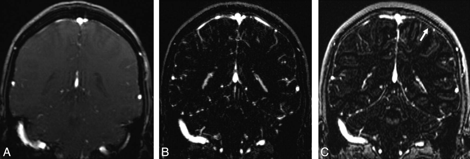
The source images, subtraction images, 2D multiplanar reconstructed (MPR) images as well as 3D maximum intensity projection (MIP) angiograms were interpreted prospectively by 2 experienced neuroradiologists. The image quality of 34 predefined venous structures was graded as follows: intense and continuous = 3, faint and continuous = 2, noncontinuous = 1, and invisible = 0. The grades assigned were the result of a consensus among the observers. In addition, the number of bridging veins seen on coronal images (Fig 1) was compared.

Fig 1.
  • Download figure
  • Open in new tab
  • Download powerpoint
Fig 1.

Coronal 2D MPR images obtained from 2D TOF MRV (A), CE MRV (B), and MPRAGE sequences (C). The section thickness is 2.5 mm (A), 0.7 mm (B), and 1.2 mm (C). The number of bridging veins in both hemispheres was compared. On the MPRAGE sequences (C), the bridging veins could not easily be distinguished from dural enhancement (arrow).

Statistical Analysis

We analyzed the differences in the number of bridging veins using the Wilcoxon matched-pairs signed-rank test. The nonparametric sign test was used to analyze differences in the image quality of the cerebral veins and dural sinuses. A commercially available statistical software package (SPSS 14.0; SPSS, Chicago, Ill) was used.

Results

We found significantly higher image quality scores on the CE MRV compared with the TOF MRV or MPRAGE sequences and on the MPRAGE sequences compared with the TOF MRV (Table 2 and 3). The number of detected bridging veins was significantly higher with the CE MRV compared with the TOF MRV or MPRAGE sequences (Table 3). In all of the 22 patients, MR imaging results did not show signs of cerebral venous thrombosis.

View this table:
  • View inline
  • View popup
Table 2.

Comparison (P < .05) of the image quality of unpaired cerebral sinuses and veins in TOF MRV, CE MRV, and MPRAGE sequences

View this table:
  • View inline
  • View popup
Table 3.

Comparison (P < .05) of the image quality of paired sinuses and veins and bridging veins in TOF MRV, CE MRV, and MPRAGE sequences

Discussion

Visualization of the normal intracranial venous system at 3T was superior on the CE MRV compared with the TOF MRV and MPRAGE sequences. A crucial aspect of MRV is spatial resolution. In contrast to the results of Kirchhof et al,1 who used imaging at 1.5T, in our study, in which imaging at 3T with a higher spatial resolution of CE MRV was used, most of the defined different venous structures were visualized more effectively by CE MRV compared with TOF MRV. The section thickness of CE MRV in our study is lower than previously reported in other studies with use of 1.5T1–4 or 3T5 imaging. Another crucial aspect of CE MRV is the synchronization of the acquisition with the arrival of the contrast agent. Parallel imaging techniques increase the time efficiency of spatial signal intensity encoding but are generally associated with a reduction in the signal-to-noise ratio. Hu et al4 demonstrated that, at 1.5T, CE MRV accelerated by 4 with a shorter acquisition time was superior to an unaccelerated counterpart. Nael et al5 reported a CE MRV accelerated by 6 on a 32-channel 3T MR imaging system. We used a CE MRV accelerated by 2 on an 8-channel system for comparison with twofold accelerated TOF MRV and MPRAGE sequences.

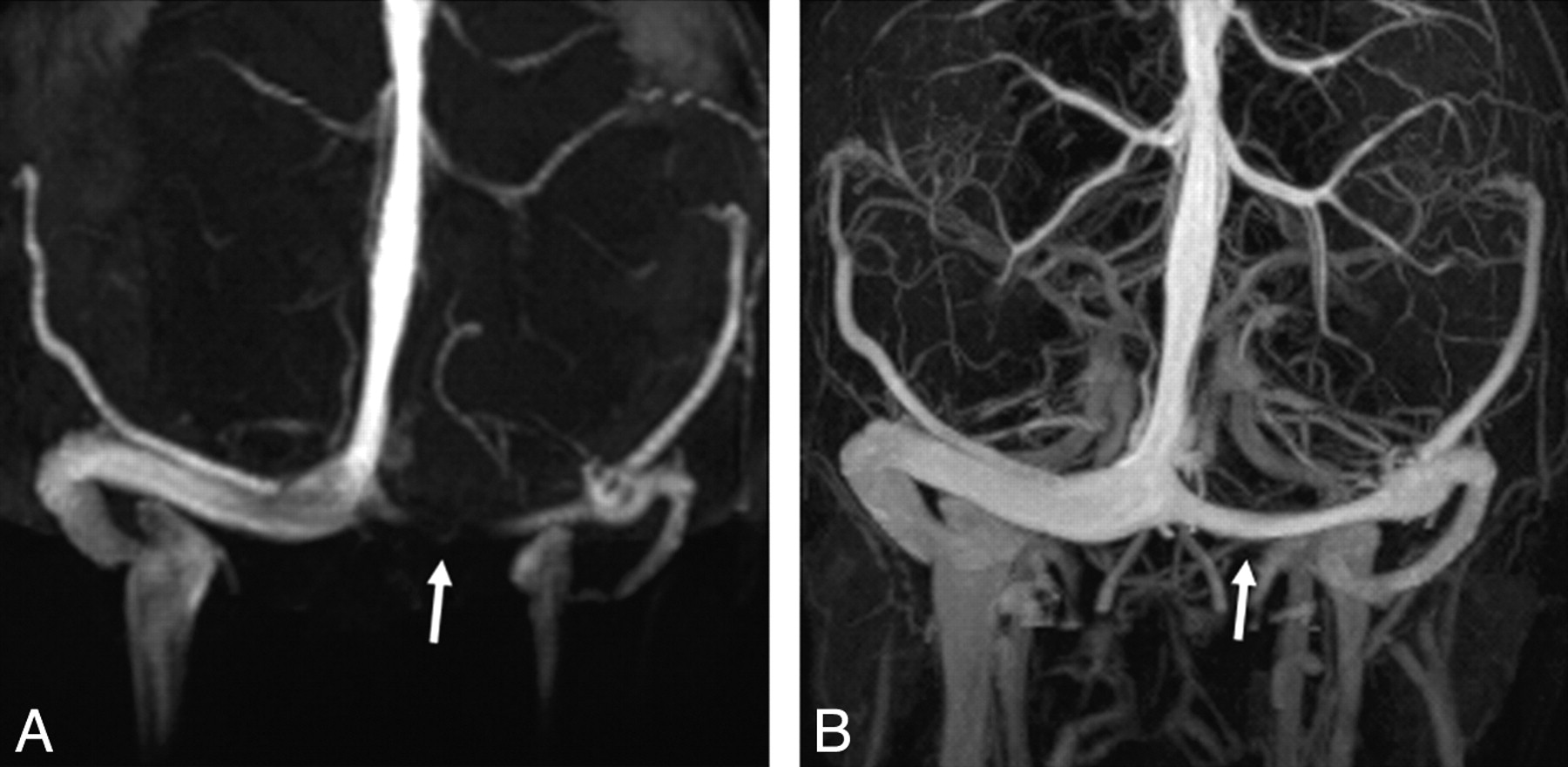
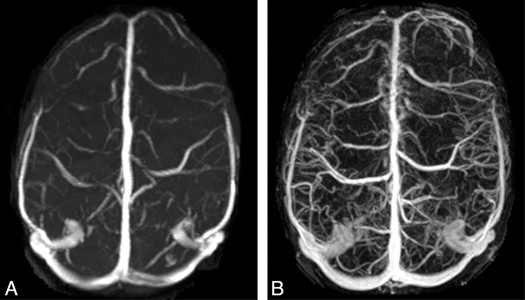
Well-known disadvantages of TOF MRV are signal intensity loss because of in-plane saturation effect as well as the slow and turbulent flow. In our study, the transverse and sigmoid sinuses were visualized best by CE MRV. As with Ayanzen et al,8 we observed flow gaps in the transverse sinuses with TOF MRV in several patients (Fig 2). In isolated cortical vein thrombosis, the diagnosis can be extremely difficult. In our study, the superficial veins were visualized best by CE MRV (Fig 3).

Fig 2.
  • Download figure
  • Open in new tab
  • Download powerpoint
Fig 2.

3D MIP images obtained from 2D TOF MRV (A), and CE MRV (B). Note the flow gap in the left transverse sinus on 2D TOF MRV (A) and the intense and continuous signal intensity on CE MRV (B) images (arrows).

Fig 3.
  • Download figure
  • Open in new tab
  • Download powerpoint
Fig 3.

3D MIP images obtained from 2D TOF MRV (A), and CE MRV (B). The superficial veins were detected best by CE MRV (B).

At 1.5T, Liang et al7 showed MPRAGE sequences to be superior to TOF MRV. In our study, disadvantages of the MPRAGE sequences were a signal intensity loss at the upper and lower end of the head coil in every patient and dural contrast enhancement, as some venous structures could not easily be distinguished from dural enhancement (Fig 1).

Our study had the following limitations: There was low statistical impact because of a small number of patients. Furthermore, cerebral venous thrombosis could not be detected in any of the participants; thus, the drawing of conclusions from these data about the performance of CE MRV and MPRAGE sequences in the detection of venous thrombosis remains difficult. Technical disadvantages, such as a relatively long TE on TOF MRV sequences, anisotropic voxels, and low acceleration factors at parallel imaging were limitations to this study. Furthermore, the performance of contrast-enhanced CE MR acquisition before contrast-enhanced MPRAGE sequence in all patients was considered a possible disadvantage.

References

  1. ↵
    Kirchhof K, Welzel T, Jansen O, et al. More reliable noninvasive visualization of the cerebral veins and dural sinuses: comparison of three MR angiographic techniques. Radiology 2002;224:804–10
    CrossRefPubMed
  2. Farb IF, Scott JN, Willinsky RA, et al. Intracranial venous system: gadolinium-enhanced three-dimensional MR venography with auto-triggered elliptic centric-ordered sequence-initial experience. Radiology 2003;226:203–09
    PubMed
  3. ↵
    Rollins N, Ison C, Reyes T, et al. Cerebral MR venography in children: comparison of 2D time-of-flight and gadolinium-enhanced 3D gradient-echo techniques. Radiology 2005;235:1011–17
    CrossRefPubMed
  4. ↵
    Hu HH, Campeau NG, Huston J, et al. High-spatial-resolution contrast-enhanced MR angiography of the intracranial venous system with fourfold accelerated two-dimensional sensitivity encoding. Radiology 2007;243:853–61
    CrossRefPubMed
  5. ↵
    Nael K, Fenchel M, Salamon N, et al. Three-dimensional cerebral contrast-enhanced magnetic resonance venography at 3.0 Tesla: initial results using highly accelerated parallel acquisition. Invest Radiol 2006;41:763–68
    CrossRefPubMed
  6. ↵
    Stevenson J, Knopp EA, Litt AW. MP-RAGE subtraction venography: a new technique. J Magn Reson Imaging 1995;5:239–41
    PubMed
  7. ↵
    Liang L, Korogi Y, Sugahara T, et al. Evaluation of the intracranial dural sinuses with a 3D contrast-enhanced MP-RAGE sequence: prospective comparison with 2D-TOF MR venography and digital subtraction angiography. AJNR Am J Neuroradiol 2001;22:481–92
    Abstract/FREE Full Text
  8. ↵
    Ayanzen RH, Bird CR, Keller PJ, et al. Cerebral MR venography: normal anatomy and potential diagnostic pitfalls. AJNR Am J Neuroradiol 2000;21:74–78
    Abstract/FREE Full Text
  • Received January 4, 2008.
  • Accepted after revision February 26, 2008.
  • Copyright © American Society of Neuroradiology
View Abstract
PreviousNext
Back to top

In this issue

American Journal of Neuroradiology: 30 (1)
American Journal of Neuroradiology
Vol. 30, Issue 1
January 2009
  • Table of Contents
  • Index by author
Advertisement
Print
Download PDF
Email Article

Thank you for your interest in spreading the word on American Journal of Neuroradiology.

NOTE: We only request your email address so that the person you are recommending the page to knows that you wanted them to see it, and that it is not junk mail. We do not capture any email address.

Enter multiple addresses on separate lines or separate them with commas.
3T High-Spatial-Resolution Contrast-Enhanced MR Angiography of the Intracranial Venous System with Parallel Imaging
(Your Name) has sent you a message from American Journal of Neuroradiology
(Your Name) thought you would like to see the American Journal of Neuroradiology web site.
CAPTCHA
This question is for testing whether or not you are a human visitor and to prevent automated spam submissions.
Cite this article
M. Lettau, K. Sartor, S. Heiland, S. Hähnel
3T High-Spatial-Resolution Contrast-Enhanced MR Angiography of the Intracranial Venous System with Parallel Imaging
American Journal of Neuroradiology Jan 2009, 30 (1) 185-187; DOI: 10.3174/ajnr.A1147

Citation Manager Formats

  • BibTeX
  • Bookends
  • EasyBib
  • EndNote (tagged)
  • EndNote 8 (xml)
  • Medlars
  • Mendeley
  • Papers
  • RefWorks Tagged
  • Ref Manager
  • RIS
  • Zotero
0 Responses
Respond to this article
Share
Bookmark this article
3T High-Spatial-Resolution Contrast-Enhanced MR Angiography of the Intracranial Venous System with Parallel Imaging
M. Lettau, K. Sartor, S. Heiland, S. Hähnel
American Journal of Neuroradiology Jan 2009, 30 (1) 185-187; DOI: 10.3174/ajnr.A1147
del.icio.us logo Twitter logo Facebook logo Mendeley logo
  • Tweet Widget
  • Facebook Like
  • Google Plus One
Purchase

Jump to section

  • Article
    • Abstract
    • Technique
    • Results
    • Discussion
    • References
  • Figures & Data
  • Info & Metrics
  • Responses
  • References
  • PDF

Related Articles

  • No related articles found.
  • PubMed
  • Google Scholar

Cited By...

  • Depiction of the Superior Petrosal Vein Complex by 3D Contrast-Enhanced MR Angiography
  • Prevalence of dural venous sinus stenosis and hypoplasia in a generalized population
  • Visualization of the Internal Cerebral Veins on MR Phase-Sensitive Imaging: Comparison with 3D Gadolinium-Enhanced MR Venography and Fast-Spoiled Gradient Recalled Imaging
  • Diagnosis and Management of Cerebral Venous Thrombosis: A Statement for Healthcare Professionals From the American Heart Association/American Stroke Association
  • Crossref (20)
  • Google Scholar

This article has been cited by the following articles in journals that are participating in Crossref Cited-by Linking.

  • Diagnosis and Management of Cerebral Venous Thrombosis
    Gustavo Saposnik, Fernando Barinagarrementeria, Robert D. Brown, Cheryl D. Bushnell, Brett Cucchiara, Mary Cushman, Gabrielle deVeber, Jose M. Ferro, Fong Y. Tsai
    Stroke 2011 42 4
  • Prevalence of dural venous sinus stenosis and hypoplasia in a generalized population
    Christopher R Durst, David A Ornan, Michael A Reardon, Prachi Mehndiratta, Sugoto Mukherjee, Robert M Starke, Max Wintermark, Avery Evans, Mary E Jensen, R Webster Crowley, John Gaughen, Kenneth C Liu
    Journal of NeuroInterventional Surgery 2016 8 11
  • Cerebral Venous and Dural Sinus Thrombosis*
    Jennifer Linn, Hartmut Brückmann
    Clinical Neuroradiology 2010 20 1
  • Accuracy of magnetic resonance venography in diagnosing cerebral venous sinus thrombosis
    Liansheng Gao, Weilin Xu, Tao Li, Xiaobo Yu, Shenglong Cao, Hangzhe Xu, Feng Yan, Gao Chen
    Thrombosis Research 2018 167
  • Time-resolved MR angiography of the intracranial venous system: an alternative MR venography technique
    Hasan Yiğit, Aynur Turan, Elif Ergün, Pınar Koşar, Uğur Koşar
    European Radiology 2012 22 5
  • 3-T contrast-enhanced MR angiography with parallel imaging in cerebral venous and sinus thrombosis
    M. Lettau, M. Laible, R.J. Barrows, S. Heiland, M. Bendszus, S. Hähnel
    Journal of Neuroradiology 2011 38 5
  • 3D Fast Spin-Echo T1 Black-Blood Imaging for the Preoperative Detection of Venous Sinus Invasion by Meningioma
    Dongdong Wang, Yiping Lu, Bo Yin, Mingyu Chen, Daoying Geng, Li Liu, Jianbo Wen, Ping Zhong, Yuan Li
    Clinical Neuroradiology 2019 29 1
  • Depiction of the Superior Petrosal Vein Complex by 3D Contrast-Enhanced MR Angiography
    B. Bender, T.-K. Hauser, A. Korn, U. Klose, M. Tatagiba, U. Ernemann, F.H. Ebner
    American Journal of Neuroradiology 2018 39 12
  • Non–Contrast-Enhanced Magnetic Resonance Angiography Using T2-Weighted 3-Dimensional Fat-Suppressed Turbo Spin Echo (SPACE)
    Qiong Song, Mengsu Zeng, Caizhong Chen, Jing Ma, Hong Yun, Shengxiang Rao, Xiuzhong Yao
    Journal of Computer Assisted Tomography 2012 36 6
  • Visualization of the Internal Cerebral Veins on MR Phase-Sensitive Imaging: Comparison with 3D Gadolinium-Enhanced MR Venography and Fast-Spoiled Gradient Recalled Imaging
    J. Sun, J. Wang, L. Jie, H. Wang, X. Gong
    American Journal of Neuroradiology 2011 32 10

More in this TOC Section

  • Progression of Microstructural Damage in Spinocerebellar Ataxia Type 2: A Longitudinal DTI Study
  • Usefulness of Quantitative Susceptibility Mapping for the Diagnosis of Parkinson Disease
  • Evaluating the Effects of White Matter Multiple Sclerosis Lesions on the Volume Estimation of 6 Brain Tissue Segmentation Methods
Show more Brain

Similar Articles

Advertisement

Indexed Content

  • Current Issue
  • Accepted Manuscripts
  • Article Preview
  • Past Issues
  • Editorials
  • Editors Choice
  • Fellow Journal Club
  • Letters to the Editor

Cases

  • Case Collection
  • Archive - Case of the Week
  • Archive - Case of the Month
  • Archive - Classic Case

Special Collections

  • Special Collections

Resources

  • News and Updates
  • Turn around Times
  • Submit a Manuscript
  • Author Policies
  • Manuscript Submission Guidelines
  • Evidence-Based Medicine Level Guide
  • Publishing Checklists
  • Graphical Abstract Preparation
  • Imaging Protocol Submission
  • Submit a Case
  • Become a Reviewer/Academy of Reviewers
  • Get Peer Review Credit from Publons

Multimedia

  • AJNR Podcast
  • AJNR SCANtastic
  • Video Articles

About Us

  • About AJNR
  • Editorial Board
  • Not an AJNR Subscriber? Join Now
  • Alerts
  • Feedback
  • Advertise with us
  • Librarian Resources
  • Permissions
  • Terms and Conditions

American Society of Neuroradiology

  • Not an ASNR Member? Join Now

© 2025 by the American Society of Neuroradiology All rights, including for text and data mining, AI training, and similar technologies, are reserved.
Print ISSN: 0195-6108 Online ISSN: 1936-959X

Powered by HighWire