Advertisement

Main menu

  • Home
  • Content
    • Current Issue
    • Accepted Manuscripts
    • Article Preview
    • Past Issue Archive
    • AJNR Case Collection
    • Case of the Week Archive
    • Classic Case Archive
    • Case of the Month Archive
  • Special Collections
    • Spinal CSF Leak Articles (Jan 2020-June 2024)
    • 2024 AJNR Journal Awards
    • Most Impactful AJNR Articles
  • Multimedia
    • AJNR Podcast
    • AJNR Scantastics
    • Video Articles
  • For Authors
    • Submit a Manuscript
    • Author Policies
    • Fast publishing of Accepted Manuscripts
    • Graphical Abstract Preparation
    • Manuscript Submission Guidelines
    • Imaging Protocol Submission
    • Submit a Case for the Case Collection
  • About Us
    • About AJNR
    • Editorial Board
  • More
    • Become a Reviewer/Academy of Reviewers
    • Subscribers
    • Permissions
    • Alerts
    • Feedback
    • Advertisers
    • ASNR Home
  • Other Publications
    • ajnr

User menu

  • Alerts
  • Log in

Search

  • Advanced search
American Journal of Neuroradiology

American Journal of Neuroradiology

ASHNR American Society of Functional Neuroradiology ASHNR American Society of Pediatric Neuroradiology ASSR
  • Alerts
  • Log in

Advanced Search

  • Home
  • Content
    • Current Issue
    • Accepted Manuscripts
    • Article Preview
    • Past Issue Archive
    • AJNR Case Collection
    • Case of the Week Archive
    • Classic Case Archive
    • Case of the Month Archive
  • Special Collections
    • Spinal CSF Leak Articles (Jan 2020-June 2024)
    • 2024 AJNR Journal Awards
    • Most Impactful AJNR Articles
  • Multimedia
    • AJNR Podcast
    • AJNR Scantastics
    • Video Articles
  • For Authors
    • Submit a Manuscript
    • Author Policies
    • Fast publishing of Accepted Manuscripts
    • Graphical Abstract Preparation
    • Manuscript Submission Guidelines
    • Imaging Protocol Submission
    • Submit a Case for the Case Collection
  • About Us
    • About AJNR
    • Editorial Board
  • More
    • Become a Reviewer/Academy of Reviewers
    • Subscribers
    • Permissions
    • Alerts
    • Feedback
    • Advertisers
    • ASNR Home
  • Follow AJNR on Twitter
  • Visit AJNR on Facebook
  • Follow AJNR on Instagram
  • Join AJNR on LinkedIn
  • RSS Feeds

Welcome to the new AJNR, Updated Hall of Fame, and more. Read the full announcements.


AJNR is seeking candidates for the position of Associate Section Editor, AJNR Case Collection. Read the full announcement.


Getting new auth cookie, if you see this message a lot, tell someone!
Research ArticleInterventional
Open Access

In Vitro and In Vivo Imaging Characteristics Assessment of Polymeric Coils Compared with Standard Platinum Coils for the Treatment of Intracranial Aneurysms

P. Mordasini, A.K. Kraehenbuehl, J.V. Byrne, S. Vandenberghe, M. Reinert, H. Hoppe and J. Gralla
American Journal of Neuroradiology November 2013, 34 (11) 2177-2183; DOI: https://doi.org/10.3174/ajnr.A3589
P. Mordasini
aFrom the Institute of Diagnostic and Interventional Neuroradiology (P.M., J.G.)
  • Find this author on Google Scholar
  • Find this author on PubMed
  • Search for this author on this site
A.K. Kraehenbuehl
cDepartment of Neurosurgery (A.K.K., M.R.)
  • Find this author on Google Scholar
  • Find this author on PubMed
  • Search for this author on this site
J.V. Byrne
eNeurovascular and Neuroradiology Research Unit (J.V.B.), Nuffield Department of Surgery, The John Radcliffe Hospital, University of Oxford, Oxford, United Kingdom.
  • Find this author on Google Scholar
  • Find this author on PubMed
  • Search for this author on this site
S. Vandenberghe
dARTORG Center for Biomedical Engineering Research (S.V.), University of Bern, Bern, Switzerland
  • Find this author on Google Scholar
  • Find this author on PubMed
  • Search for this author on this site
M. Reinert
cDepartment of Neurosurgery (A.K.K., M.R.)
  • Find this author on Google Scholar
  • Find this author on PubMed
  • Search for this author on this site
H. Hoppe
bInstitute of Diagnostic, Pediatric, and Interventional Radiology (H.H.), Inselspital
  • Find this author on Google Scholar
  • Find this author on PubMed
  • Search for this author on this site
J. Gralla
aFrom the Institute of Diagnostic and Interventional Neuroradiology (P.M., J.G.)
  • Find this author on Google Scholar
  • Find this author on PubMed
  • Search for this author on this site
  • Article
  • Figures & Data
  • Supplemental
  • Info & Metrics
  • Responses
  • References
  • PDF
Loading

Article Figures & Data

Figures

  • Fig 1.
    • Download figure
    • Open in new tab
    • Download powerpoint
    Fig 1.

    A, Schematic illustration of the polymeric coil system (above) and the delivery/detacher device (below). The coil system consists of a proximal shaft (open arrow, ink marker) attached to a hemostatic valve and a distal 65-cm-long radiopaque polymeric strand with an outer diameter of 0.018 inch (asterisk). The delivery/detacher device (total length, 270 cm) is introduced through the hemostatic valve into the inner lumen of the coil system. The delivery/detacher device consists of the microwire (arrowhead) and the heater coil attached to its distal tip (arrow). B, Fluoroscopy of an in vitro silicon aneurysm model showing the radiopaque polymeric strand (black asterisk) within the microcatheter placed in the aneurysm (microcatheter tip, open arrow) and its unfolded distal part within the aneurysm sac (white asterisk). Note the radiopaque section of the heater coil (arrow) at the distal tip of the delivery/detacher device (arrowhead) running within the coil strand. The polymeric strand can be detached at any point by advancing the radiopaque section of the heater coil at the level of the microcatheter tip and activating the thermal detachment. C, Photographic depiction of the silicon aneurysm model after introduction of the polymeric coil.
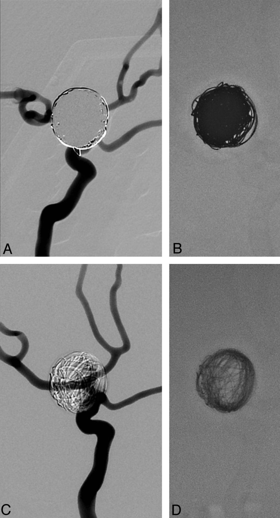
  • Fig 2.
    • Download figure
    • Open in new tab
    • Download powerpoint
    Fig 2.

    DSA of the in vitro silicon aneurysm model coiled with a calculated packing attenuation of 20%. Note the transparency of the polymeric coils (C) with persistent visualization of the superimposed vessel anatomy, which is completely obscured by standard platinum coils (A). Fluoroscopic images show the difference in radio-opacity of polymeric (D) and standard platinum coils (B).

  • Fig 3.
    • Download figure
    • Open in new tab
    • Download powerpoint
    Fig 3.

    Flat panel CT images of standard platinum coils (A and B) and polymeric coils (C and D). Note significant artifact reduction of polymeric coils compared with standard platinum coils; however, visualization of the peri-aneurysmal area is still significantly impaired.

  • Fig 4.
    • Download figure
    • Open in new tab
    • Download powerpoint
    Fig 4.

    CT scan of standard platinum coils (A and B) and polymeric coils (C and D). Note significant reduction of beam-hardening artifacts compared with standard platinum coils. In the aneurysm embolized with polymeric coils, individual coils as well as residual aneurysm lumen and neck (arrow) can be distinguished.
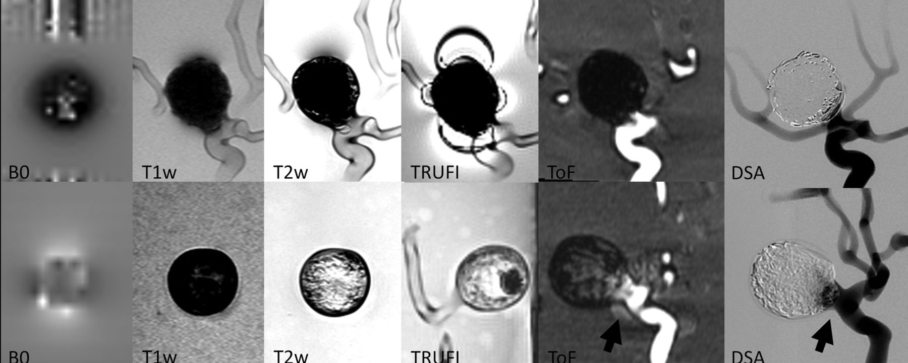
  • Fig 5.
    • Download figure
    • Open in new tab
    • Download powerpoint
    Fig 5.

    MR images and corresponding DSA of standard platinum coils (upper row) and polymeric coils (lower row) in vitro. Less magnetic field distortion and artifact production are seen with polymeric coils with the use of MRI. Individual coils can be distinguished in T2WI, TRUFI (true fast imaging with steady state precession), and TOF images as compared with standard coils. Note the visualization of residual flow within the aneurysm sac and the neck area in the aneurysm treated with polymeric coils in the TOF and DSA (arrows) compared with standard platinum coils at the same packing attenuation.
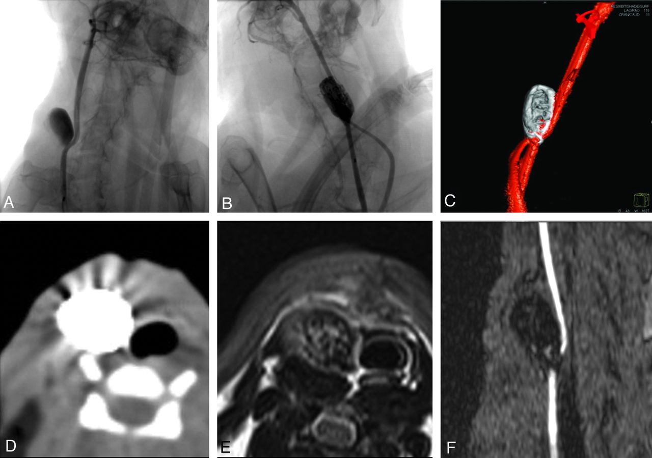
  • Fig 6.
    • Download figure
    • Open in new tab
    • Download powerpoint
    Fig 6.

    DSA demonstrates the anatomy of the venous patch carotid artery bifurcation model with the anastomosis of the left carotid artery proximal to the aneurysm (A and B). Posttreatment DSA (B) and 3D rotational angiography (C) show complete obliteration of the aneurysm (B). Note the persistent visibility of the right carotid artery despite superimposition by the coil mass. Note the minor effect of beam-hardening artifacts on the surrounding soft tissue on CT imaging (D). MR images demonstrate visibility of individual coil loops in the T2WI (E) and the lack of intra-aneurysmal flow in the TOF image (F).

  • Fig 7.
    • Download figure
    • Open in new tab
    • Download powerpoint
    Fig 7.

    CT images of a coiled carotid bifurcation aneurysm. Note the reduced artifact production and visibility of individual coils on the unenhanced CT scan (A and B). CTA shows complete occlusion of the aneurysm and minimal artifact production (C and D).

PreviousNext
Back to top

In this issue

American Journal of Neuroradiology: 34 (11)
American Journal of Neuroradiology
Vol. 34, Issue 11
1 Nov 2013
  • Table of Contents
  • Index by author
  • Complete Issue (PDF)
Advertisement
Print
Download PDF
Email Article

Thank you for your interest in spreading the word on American Journal of Neuroradiology.

NOTE: We only request your email address so that the person you are recommending the page to knows that you wanted them to see it, and that it is not junk mail. We do not capture any email address.

Enter multiple addresses on separate lines or separate them with commas.
In Vitro and In Vivo Imaging Characteristics Assessment of Polymeric Coils Compared with Standard Platinum Coils for the Treatment of Intracranial Aneurysms
(Your Name) has sent you a message from American Journal of Neuroradiology
(Your Name) thought you would like to see the American Journal of Neuroradiology web site.
CAPTCHA
This question is for testing whether or not you are a human visitor and to prevent automated spam submissions.
Cite this article
P. Mordasini, A.K. Kraehenbuehl, J.V. Byrne, S. Vandenberghe, M. Reinert, H. Hoppe, J. Gralla
In Vitro and In Vivo Imaging Characteristics Assessment of Polymeric Coils Compared with Standard Platinum Coils for the Treatment of Intracranial Aneurysms
American Journal of Neuroradiology Nov 2013, 34 (11) 2177-2183; DOI: 10.3174/ajnr.A3589

Citation Manager Formats

  • BibTeX
  • Bookends
  • EasyBib
  • EndNote (tagged)
  • EndNote 8 (xml)
  • Medlars
  • Mendeley
  • Papers
  • RefWorks Tagged
  • Ref Manager
  • RIS
  • Zotero
0 Responses
Respond to this article
Share
Bookmark this article
In Vitro and In Vivo Imaging Characteristics Assessment of Polymeric Coils Compared with Standard Platinum Coils for the Treatment of Intracranial Aneurysms
P. Mordasini, A.K. Kraehenbuehl, J.V. Byrne, S. Vandenberghe, M. Reinert, H. Hoppe, J. Gralla
American Journal of Neuroradiology Nov 2013, 34 (11) 2177-2183; DOI: 10.3174/ajnr.A3589
del.icio.us logo Twitter logo Facebook logo Mendeley logo
  • Tweet Widget
  • Facebook Like
  • Google Plus One
Purchase

Jump to section

  • Article
    • Abstract
    • ABBREVIATION:
    • Materials and Methods
    • Results
    • Discussion
    • Conclusions
    • Footnotes
    • REFERENCES
  • Figures & Data
  • Supplemental
  • Info & Metrics
  • Responses
  • References
  • PDF

Related Articles

  • PubMed
  • Google Scholar

Cited By...

  • Three-dimensional printing of anatomically accurate, patient specific intracranial aneurysm models
  • Crossref (6)
  • Google Scholar

This article has been cited by the following articles in journals that are participating in Crossref Cited-by Linking.

  • Three-dimensional printing of anatomically accurate, patient specific intracranial aneurysm models
    Jeff R Anderson, Walker L Thompson, Abdulaziz K Alkattan, Orlando Diaz, Richard Klucznik, Yi J Zhang, Gavin W Britz, Robert G Grossman, Christof Karmonik
    Journal of NeuroInterventional Surgery 2016 8 5
  • Stereolithographic models in the interdisciplinary planning of treatment for complex intracranial aneurysms
    Lukas Andereggen, Jan Gralla, Robert H. Andres, Stefan Weber, Gerhard Schroth, Jürgen Beck, Hans Rudolf Widmer, Michael Reinert, Andreas Raabe, Matthias Peterhans
    Acta Neurochirurgica 2016 158 9
  • Improving the accuracy of computational fluid dynamics simulations of coiled cerebral aneurysms using finite element modeling
    Patrick Fillingham, Julia Romero Bhathal, Laurel M.M. Marsh, Michael C. Barbour, Mehmet Kurt, Ciprian N. Ionita, Jason M. Davies, Alberto Aliseda, Michael R. Levitt
    Journal of Biomechanics 2023 157
  • High-flow venous pouch aneurysm in the rabbit carotid artery: A model for large aneurysms
    Anna K Krähenbühl, Jan Gralla, Janine Abu-Isa, Pasquale Mordasini, Hans R Widmer, Andreas Raabe, Michael Reinert
    Interventional Neuroradiology 2015 21 3
  • Implant-specific follow-up imaging of treated intracranial aneurysms: TOF-MRA vs. metal artifact reduced intravenous flat panel computed tomography angiography (FPCTA)
    N.H. Hänsel, G.A. Schubert, B. Scholz, O. Nikoubashman, A.E. Othman, M. Wiesmann, R. Pjontek, M.A. Brockmann
    Clinical Radiology 2018 73 2
  • Single Component Polymers, Polymer Blends, and Polymer Composites for Interventional Endovascular Embolization of Intracranial Aneurysms
    Hasan Hüseyin Karadeli, Emel Kuram
    Macromolecular Bioscience 2024 24 5

More in this TOC Section

  • SAVE vs. Solumbra Techniques for Thrombectomy
  • CT Perfusion&Reperfusion in Acute Ischemic Stroke
  • Delayed Reperfusion Post-Thrombectomy&Thrombolysis
Show more Interventional

Similar Articles

Advertisement

Indexed Content

  • Current Issue
  • Accepted Manuscripts
  • Article Preview
  • Past Issues
  • Editorials
  • Editors Choice
  • Fellow Journal Club
  • Letters to the Editor

Cases

  • Case Collection
  • Archive - Case of the Week
  • Archive - Case of the Month
  • Archive - Classic Case

Special Collections

  • Special Collections

Resources

  • News and Updates
  • Turn around Times
  • Submit a Manuscript
  • Author Policies
  • Manuscript Submission Guidelines
  • Evidence-Based Medicine Level Guide
  • Publishing Checklists
  • Graphical Abstract Preparation
  • Imaging Protocol Submission
  • Submit a Case
  • Become a Reviewer/Academy of Reviewers
  • Get Peer Review Credit from Publons

Multimedia

  • AJNR Podcast
  • AJNR SCANtastic
  • Video Articles

About Us

  • About AJNR
  • Editorial Board
  • Not an AJNR Subscriber? Join Now
  • Alerts
  • Feedback
  • Advertise with us
  • Librarian Resources
  • Permissions
  • Terms and Conditions

American Society of Neuroradiology

  • Not an ASNR Member? Join Now

© 2025 by the American Society of Neuroradiology All rights, including for text and data mining, AI training, and similar technologies, are reserved.
Print ISSN: 0195-6108 Online ISSN: 1936-959X

Powered by HighWire