Advertisement

Main menu

  • Home
  • Content
    • Current Issue
    • Accepted Manuscripts
    • Article Preview
    • Past Issue Archive
    • AJNR Case Collection
    • Case of the Week Archive
    • Classic Case Archive
    • Case of the Month Archive
  • Special Collections
    • Spinal CSF Leak Articles (Jan 2020-June 2024)
    • 2024 AJNR Journal Awards
    • Most Impactful AJNR Articles
  • Multimedia
    • AJNR Podcast
    • AJNR Scantastics
    • Video Articles
  • For Authors
    • Submit a Manuscript
    • Author Policies
    • Fast publishing of Accepted Manuscripts
    • Graphical Abstract Preparation
    • Manuscript Submission Guidelines
    • Imaging Protocol Submission
    • Submit a Case for the Case Collection
  • About Us
    • About AJNR
    • Editorial Board
  • More
    • Become a Reviewer/Academy of Reviewers
    • Subscribers
    • Permissions
    • Alerts
    • Feedback
    • Advertisers
    • ASNR Home
  • Other Publications
    • ajnr

User menu

  • Alerts
  • Log in

Search

  • Advanced search
American Journal of Neuroradiology

American Journal of Neuroradiology

ASHNR American Society of Functional Neuroradiology ASHNR American Society of Pediatric Neuroradiology ASSR
  • Alerts
  • Log in

Advanced Search

  • Home
  • Content
    • Current Issue
    • Accepted Manuscripts
    • Article Preview
    • Past Issue Archive
    • AJNR Case Collection
    • Case of the Week Archive
    • Classic Case Archive
    • Case of the Month Archive
  • Special Collections
    • Spinal CSF Leak Articles (Jan 2020-June 2024)
    • 2024 AJNR Journal Awards
    • Most Impactful AJNR Articles
  • Multimedia
    • AJNR Podcast
    • AJNR Scantastics
    • Video Articles
  • For Authors
    • Submit a Manuscript
    • Author Policies
    • Fast publishing of Accepted Manuscripts
    • Graphical Abstract Preparation
    • Manuscript Submission Guidelines
    • Imaging Protocol Submission
    • Submit a Case for the Case Collection
  • About Us
    • About AJNR
    • Editorial Board
  • More
    • Become a Reviewer/Academy of Reviewers
    • Subscribers
    • Permissions
    • Alerts
    • Feedback
    • Advertisers
    • ASNR Home
  • Follow AJNR on Twitter
  • Visit AJNR on Facebook
  • Follow AJNR on Instagram
  • Join AJNR on LinkedIn
  • RSS Feeds

Welcome to the new AJNR, Updated Hall of Fame, and more. Read the full announcements.


AJNR is seeking candidates for the position of Associate Section Editor, AJNR Case Collection. Read the full announcement.


Research ArticleAdult Brain
Open Access

Pressing Issues in COVID-19: Probable Cause to Seize SARS-CoV-2 for Its Preferential Involvement of Posterior Circulation Manifesting as Severe Posterior Reversible Encephalopathy Syndrome and Posterior Strokes

F. D’Amore, G. Vinacci, E. Agosti, L.P. Cariddi, A.V. Terrana, F.A. Vizzari, M. Mauri and A. Giorgianni
American Journal of Neuroradiology October 2020, 41 (10) 1800-1803; DOI: https://doi.org/10.3174/ajnr.A6679
F. D’Amore
aFrom the Departments of Neuroradiology (F.D., A.V.T., F.A.V., A.G.)
  • Find this author on Google Scholar
  • Find this author on PubMed
  • Search for this author on this site
  • ORCID record for F. D’Amore
G. Vinacci
cDepartments of Radiology (G.V.)
  • Find this author on Google Scholar
  • Find this author on PubMed
  • Search for this author on this site
  • ORCID record for G. Vinacci
E. Agosti
dNeurosurgery (E.A.), University of Insubria, Hospital of Circolo and Macchi Foundation, Varese, Lombardia, Italy
  • Find this author on Google Scholar
  • Find this author on PubMed
  • Search for this author on this site
  • ORCID record for E. Agosti
L.P. Cariddi
bNeurology and Stroke Unit (L.P.C., M.M.), Hospital of Circolo and Macchi Foundation, Varese, Lombardia, Italy
eClinical and Experimental Medical Humanities (L.P.C.), Center of Research in Medical Pharmacology, Univeristy of Insubria, Varese, Italy.
  • Find this author on Google Scholar
  • Find this author on PubMed
  • Search for this author on this site
  • ORCID record for L.P. Cariddi
A.V. Terrana
aFrom the Departments of Neuroradiology (F.D., A.V.T., F.A.V., A.G.)
  • Find this author on Google Scholar
  • Find this author on PubMed
  • Search for this author on this site
  • ORCID record for A.V. Terrana
F.A. Vizzari
aFrom the Departments of Neuroradiology (F.D., A.V.T., F.A.V., A.G.)
  • Find this author on Google Scholar
  • Find this author on PubMed
  • Search for this author on this site
  • ORCID record for F.A. Vizzari
M. Mauri
bNeurology and Stroke Unit (L.P.C., M.M.), Hospital of Circolo and Macchi Foundation, Varese, Lombardia, Italy
  • Find this author on Google Scholar
  • Find this author on PubMed
  • Search for this author on this site
  • ORCID record for M. Mauri
A. Giorgianni
aFrom the Departments of Neuroradiology (F.D., A.V.T., F.A.V., A.G.)
  • Find this author on Google Scholar
  • Find this author on PubMed
  • Search for this author on this site
  • ORCID record for A. Giorgianni
  • Article
  • Figures & Data
  • Supplemental
  • Info & Metrics
  • Responses
  • References
  • PDF
Loading

Abstract

SUMMARY: Since December 2019, a novel Severe Acute Respiratory Syndrome coronavirus 2 from China has rapidly spread worldwide. Although respiratory involvement is the mainstay of coronavirus disease 2019 (COVID-19), systemic involvement has recently drawn more attention. In particular, a number of recent articles have shed light on the nervous system as one of the possible targets. At our institution, we observed 15 patients with acute brain vascular manifestations; most interesting, we had a higher prevalence of the posterior circulation acute impairment. In our series, 7 patients had acute posterior cerebral injury: 1, hemorrhagic posterior reversible encephalopathy syndrome; 5, posterior circulation ischemic stroke; and 1, parieto-occipital hemorrhagic stroke. On the basis of our evidence and previous basic science reports, we believe a common etiopathogenetic thread may connect ischemic/hemorrhagic events of the posterior circulation and posterior reversible encephalopathy syndrome in the setting of COVID-19.

ABBREVIATIONS:

COVID-19
coronavirus disease 2019
PRES
posterior reversible encephalopathy syndrome
SARS-CoV-2
Severe Acute Respiratory Syndrome coronavirus 2

Last December, the first cases of a novel viral coronavirus disease 2019 (COVID-19) caused by the Severe Acute Respiratory Syndrome coronavirus 2 (SARS-CoV-2) were identified in China. Since then, the virus has spread so quickly worldwide that the World Health Organization declared COVID-19 a pandemic on March 11, 2020.1 Italy first faced the largest outbreak besides Asia, with Lombardy outnumbering all other regions in Italy in terms of COVID-19 infections and deaths. As of May 21, >5 million SARS-CoV-2 infections have been confirmed worldwide, with the highest number of cases (>1.5 million) and deaths (almost 100,000) registered in the United States.2 From the beginning of this medical emergency, our hospital has admitted >1100 patients who tested positive for SARS-CoV-2.3 Although it is well-known that SARS-CoV-2 has a preferential pulmonary and gastrointestinal tropism, current evidence points to multiorgan involvement. A growing number of studies have reported SARS-CoV-2 involvement of the CNS, showing a wide variety of neurologic symptoms.4 According to a Chinese study, the incidence of neurologic symptoms was about 36% in a cohort of patients with COVID-19.5

Case Series

From February 21 to the submission of this article, we collected patients positive for SARS-CoV-2 presenting with neurologic impairment who underwent CT and CTA and/or MR imaging (On-line Table). Demographic, clinical, and anamnestic data (age, sex, and clinical data) were obtained from clinical records. All imaging examinations were separately reviewed by 3 board-certified Neuroradiologists (F.D., A.V.T., A.G., with, respectively, 7, 6, and 10 years of experience in neuroimaging).

A total of 27 patients satisfied the inclusion criteria. Among these, 15 (7 men, 8 women; mean age, 68 years; range, 21–88 years) had acute imaging findings (On-line Table), of whom, 7 patients had acute posterior circulation ischemic-hemorrhagic events. The remainder showed miscellaneous findings: 1 case with supratentorial posttraumatic SAH, 2 cases with spontaneous acute or chronic convexities subdural hemorrhage, 1 posttraumatic, 2 with spontaneous intraparenchymal supratentorial hemorrhage (with negative findings on CTA), 1 with multicompartmental bleeding of unknown etiology, and 1 patient who developed clinical and imaging findings in keeping with encephalitis. CT did not show any acute findings in 12 cases. In all patients with a baseline brain CT scan positive for acute changes, 6 underwent CTA, and MR imaging was available in 4 cases; all had CT follow-up, except those who died.

The subgroup of posterior circulation acute events comprised 7 patients (3 men, 4 women; mean age, 72 years, range, 57–88 years) representing 47% (7/15) of the total positive cases (On-line Table). In 5 patients, acute ischemic changes were found (Fig 1), of which 2 cases evolved into hemorrhagic transformation. One patient showed right parieto-occipital hemorrhagic stroke at baseline; subsequent CTA ruled out vascular malformations or acute thrombosis. In the other patient, imaging at baseline revealed extensive bilateral subcortical edema with multifocal hemorrhages, predominantly in the occipital lobes. Follow-up imaging showed progressive resorption of edema and hemorrhages, in keeping with posterior reversible encephalopathy syndrome (PRES) (Fig 2).

FIG 1.
  • Download figure
  • Open in new tab
  • Download powerpoint
FIG 1.

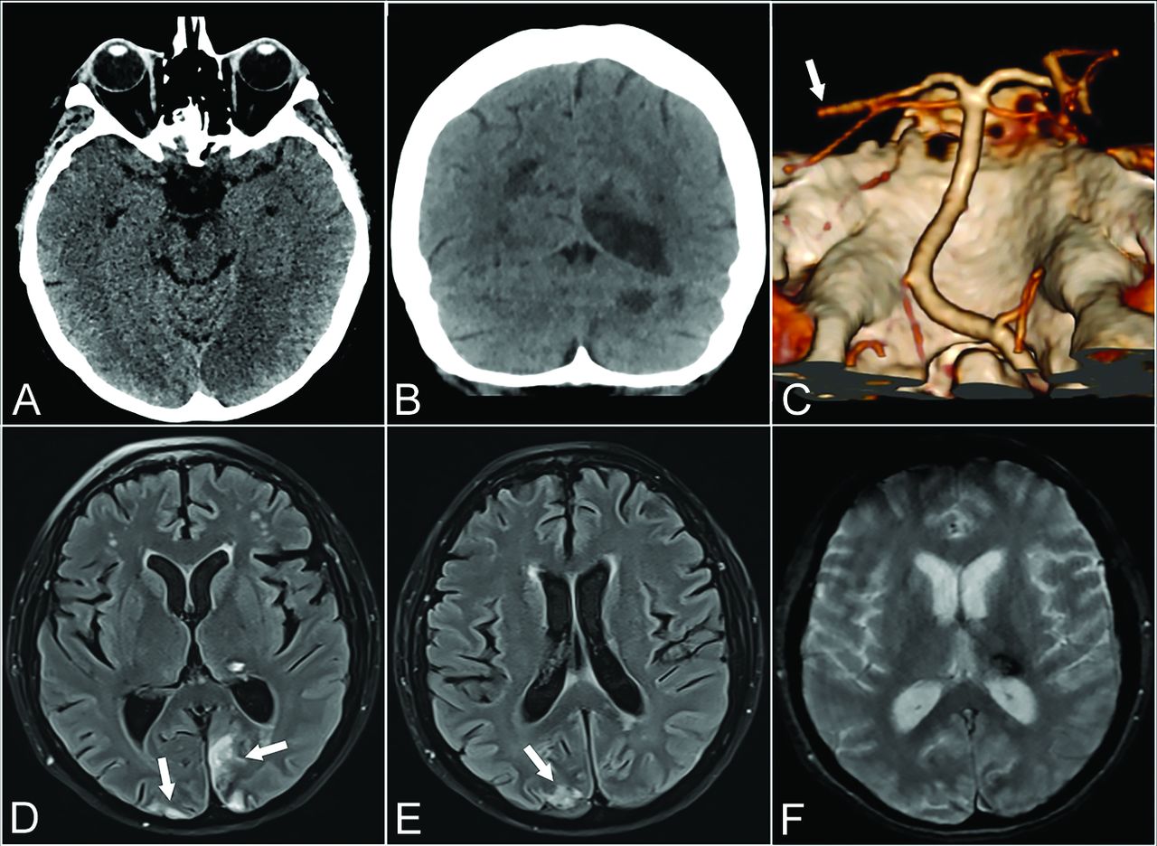
Patient 1. A 57-year-old woman with psychomotor retardation, left gaze deviation, and right superior and inferior limb paresis. Axial CT at baseline shows medial occipital loss of gray-white differentiation consistent with acute ischemia (A). Ten days later, coronal CT shows additional well-defined left cerebellar and right parieto-occipital hypoattenuating areas in keeping with subacute ischemia (B). Left distal posterior cerebral artery occlusion on CTA VR (Volume Rendering) reformats from the same day (white arrow, C). Axial T2 FLAIR (D and E) and axial T2* gradient recalled-echo (F) 6 weeks later show focal left thalamic signal drop-out, consistent with hemorrhage. Early chronic ischemic changes are also noted in the parieto-occipital lobes (white arrows, D and E).

FIG 2.
  • Download figure
  • Open in new tab
  • Download powerpoint
FIG 2.

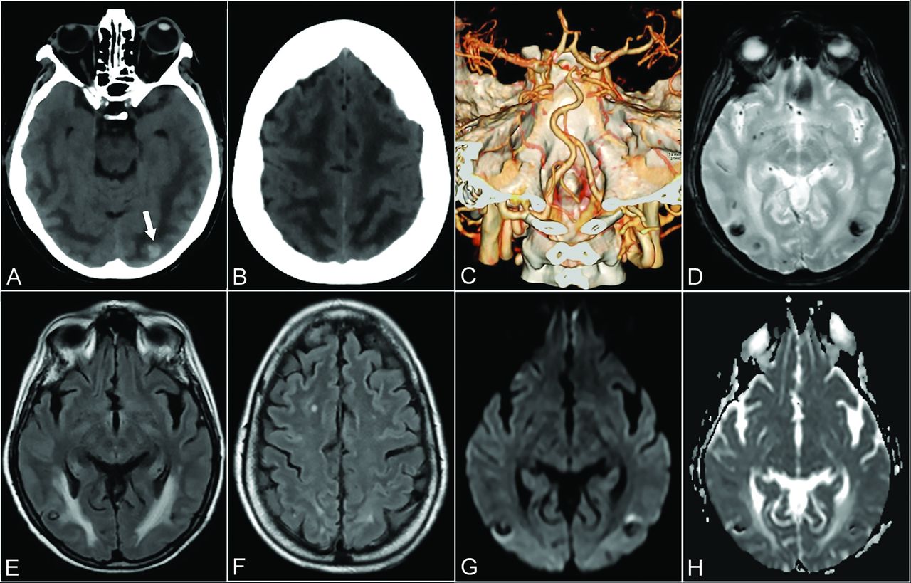
Patient 6. A 64-year-old woman presenting with drowsiness and reduction in visual acuity. Baseline axial CT shows ill-defined, bilateral hemispheric, white matter hypoattenuation, sparing the adjacent cortex, tapering off in the polar aspect of the frontal lobes, consistent with vasogenic edema (A and B). A small hyperattenuating lesion is noted in the left occipital lobe, suggestive of focal bleeding (arrow, A). CTA VR reformatted imaging was unremarkable in appearance (C). Three weeks later axial T2* gradient recalled-echo (D), axial T2 FLAIR (E and F), DWI (G), and an ADC map (H) demonstrate a partial resorption of edema and the presence of bilateral focal predominantly hypointense lesions (E) characterized by blooming (D) and signal blackout (G and H) in keeping with focal hemorrhage.

DISCUSSION

Our series shows a high prevalence of posterior circulation vascular events, not directly referable to a secondary cause as opposed to the heterogeneous etiology in the remaining cases. We speculate that SARS-CoV-2 may have played a role leading to acute brain injury, with a preferential involvement of the posterior circulation, which may be more susceptible to the SARS-CoV-2 infection effects.

Although SARS-CoV-2 targets primarily the lung parenchyma, earlier evidence in the literature highlighted its neurotrophic and neuroinvasive tendencies.6 SARS-CoV-2 invades the CNS through different pathways, from peripheral nerves, especially via the olfactory nerves, through the bloodstream by infecting endothelial cells and altering the BBB, by lymphatic pathways, and, last, by the disruption of the gastrointestinal environment, which may indirectly alter BBB integrity.7 SARS-CoV-2 can interact with host cells through the angiotensin-converting enzyme 2 cell membrane receptor expressed in a wide variety of cells such as airway epithelia, lung parenchyma, kidney cells, small intestine cells, as well as neurons and vascular endothelia.6 Once it binds to neuronal angiotensin-converting enzyme 2 receptors, it is able to activate a self-reinforcing inflammatory response through a cytokine storm, which ultimately results in irreversible neuronal damage.8 Angiotensin-converting en-zyme 2 receptors are expressed in cerebral endothelial cells that take part in hormone formation, the sympathoadrenal system, and vascular autoregulation.9 Endothelial disruption as a consequence of endotheliitis induced by SARS-CoV-2 can contribute to the pathophysiology of brain damage.6 Infective lymphocytic endotheliitis has been proved by postmortem analysis.10 The neuroinvasive potential is also supported by clinical experience as reported by several recent studies. For example, a Chinese study5 reported various manifestations of CNS involvement in 53 patients with COVID-19 in a sample of 214 patients. In another study, SARS-CoV-2 was found in the CSF in a patient diagnosed with encephalitis, therefore confirming its direct CNS invasion.11 Other studies have also highlighted imaging features of COVID-19 brain involvement.12-15

As recently described, ischemic stroke is a known complication in patients with COVID-19. In our series, we found 5 ischemic strokes of the posterior circulation in line with other studies. Mao et al5 described 4 cases of acute stroke, while others described the onset of acute ischemic stroke in 5 younger patients.15 Beyrouti et al12 described 6 cases of acute stroke, 4 of which were posterior strokes. Goldberg and Goldberg16 recently showed a case of ischemic stroke with occlusion of the MCA and bilateral anterior cerebral arteries.

Ischemic stroke could be related both to the endotheliitis process10 and hypercoagulability status, which can evolve to embolism.17 Endothelium inflammation may shift vascular equilibrium toward vasoconstriction, leading to ischemia.18 We also reported 1 parieto-occipital hemorrhage, 1 case of complicated hemorrhagic PRES, and the hemorrhagic transformation of 2 ischemic strokes. Intracranial hemorrhage in patients with COVID-19 has already been reported.5,19 In our series, we also noted 4 hemorrhagic strokes involving the anterior circulation.

One patient in our cohort presented with clinical and imaging features of a severe form of PRES. A recent case report has shown 2 cases of hemorrhagic PRES.20 Our patient presented with extensive edema on a baseline CT scan with the presence of hemorrhagic lesions which is a severe and rare manifastation of PRES (Fig 2).21 PRES is a multifactorial clinical-radiologic entity characterized by reversible subcortical vasogenic edema. Although not completely elucidated, several pathogenetic theories entailing rapid increase of arterial blood pressure beyond the autoregulatory potential, the release of vasoconstrictors, and endogenous or exogenous stimulants in addition to T-cell-derived cytokine release converge to an abnormal activation and consequent disruption of endothelial cell homeostasis.22,23 Moreover, infective agents promote polymorphonuclear leukocyte activation and lead to additional release of mediators, resulting in increased vessel permeability, which leads to the development of interstitial edema.23 Most interesting, the well-known frequent association of PRES with posterior circulation involvement has been ascribed to its lack of sympathetic innervation in contrast to the anterior circulation.22,24 We ruled out other potential PRES mimickers, such as hypoxic-ischemic encephalopathy, meningoencephalitis, and acute toxic leukoencephalopathy in light of clinical and laboratory data and imaging follow-up. The improvement of pneumonia and laboratory parameters as well as the negative results on subsequent nasopharyngeal swabs paralleled by neurologic symptom resolution may strengthen the hypothesis of SARS-CoV-2 as a primary causative agent. Therefore, endothelial dysfunction, possibly at the capillary level, set off by SARS-CoV-2 infection, may be the etiopathogenesis of PRES.

In our cohort, we found that the posterior circulation was the most common primary site of acute vascular injury. The endothelial disruption related to SARS-CoV-2 could be a common feature acting as a promoting and synergistic agent underlying the preferential involvement of the posterior circulation, presumably due to its intrinsic hemodynamic weakness compared with the anterior circulation. Further studies with larger sample sizes are needed to support our hypothesis.

Indicates open access to non-subscribers at www.ajnr.org

References

  1. 1.↵
    WHO Director-General’s opening remarks at the media briefing on COVID-19, March 18, 2020. https://www.who.int/dg/speeches/detail/who-director-general-s-opening-remarks-at-the-media-briefing-on-covid-19—11-march-2020. Accessed May 21, 2020
  2. 2.↵
    COVID-19 Map: Johns Hopkins Coronavirus Resource Center. https://coronavirus.jhu.edu/map.html. Accessed May 21, 2020
  3. 3.↵
    Oltre millecento pazienti accolti nelle strutture dedicate all’emergenza Covid-19 dell’ASST Sette Laghi - asstsettelaghi. https://www.asst-settelaghi.it/contenuto-web/-/asset_publisher/shmOK1pliUE0/content/oltre-millecento-pazienti-accolti-nelle-strutture-dedicate-all-emergenza-covid-19-dell-asst-sette-laghi. Accessed May 21, 2020
  4. 4.↵
    1. Ahmad I,
    2. Rathore FA
    . Neurological manifestations and complications of COVID-19: a literature review. J Clin Neurosci 2020;77:8–12 doi:10.1016/j.jocn.2020.05.017 pmid:32409215
    CrossRefPubMed
  5. 5.↵
    1. Mao L,
    2. Jin H,
    3. Wang M, et al
    . Neurologic manifestations of hospitalized patients with coronavirus disease 2019 in Wuhan, China. JAMA Neurol 2020 Apr 10. [Epub ahead of Print] doi:10.1001/jamaneurol.2020.1127 pmid:32275288
    CrossRefPubMed
  6. 6.↵
    1. Baig AM,
    2. Khaleeq A,
    3. Ali U, et al
    . Evidence of the COVID-19 virus targeting the CNS: tissue distribution, host–virus interaction, and proposed neurotropic mechanisms. ACS Chem Neurosci 2020;11:995–98 doi:10.1021/acschemneuro.0c00122 pmid:32167747
    CrossRefPubMed
  7. 7.↵
    1. Li Z,
    2. Liu T,
    3. Yang N, et al
    . Neurological manifestations of patients with COVID-19: potential routes of SARS-CoV-2 neuroinvasion from the periphery to the brain. Front Med 2020 May 4. [Epub ahead of Print] doi:10.1007/s11684-020-0786-5 pmid:32367431
    CrossRefPubMed
  8. 8.↵
    1. Mehta P,
    2. McAuley DF,
    3. Brown M, et al
    ; HLH Across Speciality Collaboration, UK. COVID-19: consider cytokine storm syndromes and immunosuppression. Lancet 2020;395:1033–34 doi:10.1016/S0140-6736(20)30628-0 pmid:32192578
    CrossRefPubMed
  9. 9.↵
    1. Saavedra JM
    . Brain angiotensin II: new developments, unanswered questions and therapeutic opportunities. Cell Mol Neurobiol 2005;25:485–512 doi:10.1007/s10571-005-4011-5 pmid:16075377
  10. 10.↵
    1. Varga Z,
    2. Flammer AJ,
    3. Steiger P, et al
    . Endothelial cell infection and endotheliitis in COVID-19. Lancet 2020;395:1417–18 doi:10.1016/S0140-6736(20)30937-5 pmid:32325026
    CrossRefPubMed
  11. 11.↵
    1. Moriguchi T,
    2. Harii N,
    3. Goto J, et al
    . A first case of meningitis/encephalitis associated with SARS-Coronavirus-2. Int J Infect Dis 2020; 94:55–58 doi:10.1016/j.ijid.2020.03.062
    CrossRefPubMed
  12. 12.↵
    1. Beyrouti R,
    2. Adams ME,
    3. Benjamin L, et al
    . Characteristics of ischaemic stroke associated with COVID-19. J Neurol Neurosurg Psychiatry 2020 Apr 30. [Epub ahead of Print] doi:10.1136/jnnp-2020-323586 pmid:32354768
    FREE Full Text
  13. 13.
    1. Helms J,
    2. Kremer S,
    3. Merdji H, et al
    . Neurologic features in severe SARS-CoV-2 infection. N Engl J Med 2020;382:2268–62 doi:10.1056/NEJMc2008597 pmid:32294339
    CrossRefPubMed
  14. 14.
    1. Giorgianni A,
    2. Vinacci G,
    3. Agosti E, et al
    . Neuroradiological features in COVID-19 patients: first evidence in a complex scenario. J Neuroradiol 2020 May 15. [Epub ahead of Print] doi:10.1016/j.neurad.2020.05.005 pmid:32417159
    CrossRefPubMed
  15. 15.↵
    1. Oxley TJ,
    2. Mocco J,
    3. Majidi S, et al
    . Large-vessel stroke as a presenting feature of Covid-19 in the young. N Engl J Med 2020;382:e60 doi:10.1056/NEJMc2009787
    CrossRefPubMed
  16. 16.↵
    1. Goldberg MF,
    2. Goldberg MF
    . Cerebrovascular disease in COVID-19. AJNR Am J Neuroradiol 2020 May 14. [Epub ahead of Print] doi:10.3174/ajnr.A6588 pmid:32409316
    CrossRefPubMed
  17. 17.↵
    1. Marietta M,
    2. Ageno W,
    3. Artoni A, et al
    . COVID-19 and haemostasis: a position paper from Italian Society on Thrombosis and Haemostasis (SISET). Blood Transfus 2020;18:167–69 doi:10.2450/2020.0083-20 pmid:32281926
    CrossRefPubMed
  18. 18.↵
    1. Bonetti PO,
    2. Lerman LO,
    3. Lerman A
    . Endothelial dysfunction: a marker of atherosclerotic risk. Arterioscler Thromb Vasc Biol 2003;23:168–75 doi:10.1161/01.atv.0000051384.43104.fc pmid:12588755
    Abstract/FREE Full Text
  19. 19.↵
    1. Sharifi-Razavi A,
    2. Karimi N,
    3. Rouhani N
    . COVID-19 and intracerebral haemorrhage: causative or coincidental? New Microbes New Infect 2020;35:100669 doi:10.1016/j.nmni.2020.100669 pmid:32322398
    CrossRefPubMed
  20. 20.↵
    1. Franceschi AM,
    2. Ahmed O,
    3. Giliberto L
    . Hemorrhagic posterior reversible encephalopathy syndrome as a manifestation of COVID-19 infection AJNR Am J Neuroradiol 2020 May 21. [Epub ahead of Print] doi:10.3174/ajnr.A6595] pmid:32439646
    CrossRefPubMed
  21. 21.↵
    1. Hugonnet E,
    2. Da Ines D,
    3. Boby H, et al
    . Posterior reversible encephalopathy syndrome (PRES): features on CT and MR imaging. Diagn Interv Imaging 2013;94:45–52 doi:10.1016/j.diii.2012.02.005 pmid:22835573
    CrossRefPubMed
  22. 22.↵
    1. Hinduja A
    . Posterior reversible encephalopathy syndrome: clinical features and outcome. Front Neurol 2020;11:71 doi:10.3389/fneur.2020.00071 pmid:32117030
    CrossRefPubMed
  23. 23.↵
    1. Fugate JE,
    2. Rabinstein AA
    . Posterior reversible encephalopathy syndrome: clinical and radiological manifestations, pathophysiology, and outstanding questions. Lancet Neurol 2015;14:914–25 doi:10.1016/S1474-4422(15)00111-8 pmid:26184985
    CrossRefPubMed
  24. 24.↵
    1. Gao B,
    2. Lyu C,
    3. Lerner A, et al
    . Controversy of posterior reversible encephalopathy syndrome: what have we learnt in the last 20 years? J Neurol Neurosurg Psychiatry 2018;89:14–20 doi:10.1136/jnnp-2017-316225 pmid:28794149
    Abstract/FREE Full Text
  • Received May 21, 2020.
  • Accepted after revision June 3, 2020.
  • © 2020 by American Journal of Neuroradiology
View Abstract
PreviousNext
Back to top

In this issue

American Journal of Neuroradiology: 41 (10)
American Journal of Neuroradiology
Vol. 41, Issue 10
1 Oct 2020
  • Table of Contents
  • Index by author
  • Complete Issue (PDF)
Advertisement
Print
Download PDF
Email Article

Thank you for your interest in spreading the word on American Journal of Neuroradiology.

NOTE: We only request your email address so that the person you are recommending the page to knows that you wanted them to see it, and that it is not junk mail. We do not capture any email address.

Enter multiple addresses on separate lines or separate them with commas.
Pressing Issues in COVID-19: Probable Cause to Seize SARS-CoV-2 for Its Preferential Involvement of Posterior Circulation Manifesting as Severe Posterior Reversible Encephalopathy Syndrome and Posterior Strokes
(Your Name) has sent you a message from American Journal of Neuroradiology
(Your Name) thought you would like to see the American Journal of Neuroradiology web site.
CAPTCHA
This question is for testing whether or not you are a human visitor and to prevent automated spam submissions.
Cite this article
F. D’Amore, G. Vinacci, E. Agosti, L.P. Cariddi, A.V. Terrana, F.A. Vizzari, M. Mauri, A. Giorgianni
Pressing Issues in COVID-19: Probable Cause to Seize SARS-CoV-2 for Its Preferential Involvement of Posterior Circulation Manifesting as Severe Posterior Reversible Encephalopathy Syndrome and Posterior Strokes
American Journal of Neuroradiology Oct 2020, 41 (10) 1800-1803; DOI: 10.3174/ajnr.A6679

Citation Manager Formats

  • BibTeX
  • Bookends
  • EasyBib
  • EndNote (tagged)
  • EndNote 8 (xml)
  • Medlars
  • Mendeley
  • Papers
  • RefWorks Tagged
  • Ref Manager
  • RIS
  • Zotero
0 Responses
Respond to this article
Share
Bookmark this article
Pressing Issues in COVID-19: Probable Cause to Seize SARS-CoV-2 for Its Preferential Involvement of Posterior Circulation Manifesting as Severe Posterior Reversible Encephalopathy Syndrome and Posterior Strokes
F. D’Amore, G. Vinacci, E. Agosti, L.P. Cariddi, A.V. Terrana, F.A. Vizzari, M. Mauri, A. Giorgianni
American Journal of Neuroradiology Oct 2020, 41 (10) 1800-1803; DOI: 10.3174/ajnr.A6679
del.icio.us logo Twitter logo Facebook logo Mendeley logo
  • Tweet Widget
  • Facebook Like
  • Google Plus One
Purchase

Jump to section

  • Article
    • Abstract
    • ABBREVIATIONS:
    • DISCUSSION
    • References
  • Figures & Data
  • Supplemental
  • Info & Metrics
  • Responses
  • References
  • PDF

Related Articles

  • PubMed
  • Google Scholar

Cited By...

  • No citing articles found.
  • Crossref (20)
  • Google Scholar

This article has been cited by the following articles in journals that are participating in Crossref Cited-by Linking.

  • COVID-19 and cerebrovascular diseases: a comprehensive overview
    Georgios Tsivgoulis, Lina Palaiodimou, Ramin Zand, Vasileios Arsenios Lioutas, Christos Krogias, Aristeidis H. Katsanos, Ashkan Shoamanesh, Vijay K. Sharma, Shima Shahjouei, Claudio Baracchini, Charalambos Vlachopoulos, Rossetos Gournellis, Petros P. Sfikakis, Else Charlotte Sandset, Andrei V. Alexandrov, Sotirios Tsiodras
    Therapeutic Advances in Neurological Disorders 2020 13
  • Brain dysfunction in COVID‐19 and CAR‐T therapy: cytokine storm‐associated encephalopathy
    Umberto Pensato, Lorenzo Muccioli, Ilaria Cani, Damir Janigro, Pier Luigi Zinzani, Maria Guarino, Pietro Cortelli, Francesca Bisulli
    Annals of Clinical and Translational Neurology 2021 8 4
  • Neuroimaging findings of brain MRI and CT in patients with COVID-19: A systematic review and meta-analysis
    Yangsean Choi, Min Kyoung Lee
    European Journal of Radiology 2020 133
  • Presentation, Treatment Response and Short-Term Outcomes in Paediatric Multisystem Inflammatory Syndrome Temporally Associated with SARS-CoV-2 (PIMS-TS)
    Susanna Felsenstein, Emily Willis, Hannah Lythgoe, Liza McCann, Andrew Cleary, Kamran Mahmood, David Porter, Jessica Jones, Janet McDonagh, Alice Chieng, Giulia Varnier, Stephen Hughes, Mary Boullier, Fiona Ryan, Olumoyin Awogbemi, Giridhar Soda, Phuoc Duong, Clare Pain, Phil Riley, Christian M. Hedrich
    Journal of Clinical Medicine 2020 9 10
  • Brain MRI findings in COVID-19 patients with PRES: A systematic review
    Rubaya Yeahia, Javin Schefflein, Patrick Chiarolanzio, Anna Rozenstein, William Gomes, Sana Ali, Hasit Mehta, Fawaz Al-Mufti, Andrew McClelland, Edwin Gulko
    Clinical Imaging 2022 81
  • A review of neuroradiological abnormalities in patients with coronavirus disease 2019 (COVID-19)
    Bahar Bahranifard, Somayeh Mehdizadeh, Ali Hamidi, Alireza Khosravi, Ramin Emami, Kamran Mirzaei, Reza Nemati, Fatemeh Nemati, Majid Assadi, Ali Gholamrezanezhad
    The Neuroradiology Journal 2022 35 1
  • The association of posterior reversible encephalopathy syndrome with COVID-19: A systematic review
    Sadaf Iftikhar, Aqeeb Ur Rehman, Muhammad Zain Ameer, Ahmad Nawaz, Muhammad Aemaz Ur Rehman, Hareem Farooq, Abyaz Asmar, Muhammad Ebaad Ur Rehman
    Annals of Medicine and Surgery 2021 72
  • Acute Seizures Occurring in Association With SARS-CoV-2
    Sean T. Hwang, Ahmad A. Ballout, Usman Mirza, Anup N. Sonti, Arif Husain, Claudia Kirsch, Ruben Kuzniecky, Souhel Najjar
    Frontiers in Neurology 2020 11
  • Posterior reversible encephalopathy syndrome and reversible cerebral vasoconstriction syndrome in patients with COVID-19 infection: is there a link? A systematic review and case report analysis
    Adriano Bonura, Gianmarco Iaccarino, Sergio Soeren Rossi, Fioravante Capone, Francesco Motolese, Rosalinda Calandrelli, Vincenzo Di Lazzaro, Fabio Pilato
    Journal of Neurology 2023 270 6
  • Acute Neurological Complications of Coronavirus Disease
    Sanders Chang, Michael Schecht, Rajan Jain, Puneet Belani
    Neuroimaging Clinics of North America 2023 33 1

More in this TOC Section

  • Diagnostic Neuroradiology of Monoclonal Antibodies
  • ML for Glioma Molecular Subtype Prediction
  • Segmentation of Brain Metastases with BLAST
Show more Adult Brain

Similar Articles

Advertisement

Indexed Content

  • Current Issue
  • Accepted Manuscripts
  • Article Preview
  • Past Issues
  • Editorials
  • Editors Choice
  • Fellow Journal Club
  • Letters to the Editor

Cases

  • Case Collection
  • Archive - Case of the Week
  • Archive - Case of the Month
  • Archive - Classic Case

Special Collections

  • Special Collections

Resources

  • News and Updates
  • Turn around Times
  • Submit a Manuscript
  • Author Policies
  • Manuscript Submission Guidelines
  • Evidence-Based Medicine Level Guide
  • Publishing Checklists
  • Graphical Abstract Preparation
  • Imaging Protocol Submission
  • Submit a Case
  • Become a Reviewer/Academy of Reviewers
  • Get Peer Review Credit from Publons

Multimedia

  • AJNR Podcast
  • AJNR SCANtastic
  • Video Articles

About Us

  • About AJNR
  • Editorial Board
  • Not an AJNR Subscriber? Join Now
  • Alerts
  • Feedback
  • Advertise with us
  • Librarian Resources
  • Permissions
  • Terms and Conditions

American Society of Neuroradiology

  • Not an ASNR Member? Join Now

© 2025 by the American Society of Neuroradiology All rights, including for text and data mining, AI training, and similar technologies, are reserved.
Print ISSN: 0195-6108 Online ISSN: 1936-959X

Powered by HighWire