
Article Figures & Data
Figures
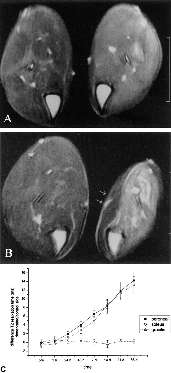
- Fig 1.
Axial T2-weighted double fast spin-echo images (2000/17/96), obtained 4 mm below the knee joint, and time course graph.
A, Obtained 48 hours after transection of the left sciatic nerve. A slight signal intensity increase in the denervated muscles of the lower leg is already present.
B, Obtained 2 months after transection of the left sciatic nerve. The signal intensity increase becomes more prominent. The gracilis muscle (innervated by the obturator nerve, arrows) is not affected.
C, Time course of the difference in T2 TR between the peroneal, soleus, and gracilis muscles of the left and right lower legs.
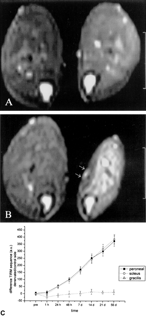
- Fig 2.
Axial TIRM images (3250/60; inversion time, 150), obtained 4 mm below the knee joint, and time course graph.
A, Obtained 48 hours after transection of the left sciatic nerve. A marked signal intensity increase in the denervated muscles is already present.
B, Obtained 2 months after transection of the left sciatic nerve. Marked signal intensity further increases.
C, Time course of the difference in signal intensity (in arbitrary units) between the left and right lower legs.
- Fig 3.
Axial T1-weighted spin-echo image (460/14), obtained 4 mm below the knee joint, and time course graph.
A, Obtained 48 hours after transection of the left sciatic nerve. No evidence of atrophy is seen.
B, Obtained 2 months after transection of the left sciatic nerve. Marked atrophy of the denervated muscles and hypertrophy of the contralateral leg can be seen.
C, Time course of the difference in circumference of the left and right lower legs.


