
Article Figures & Data
Figures
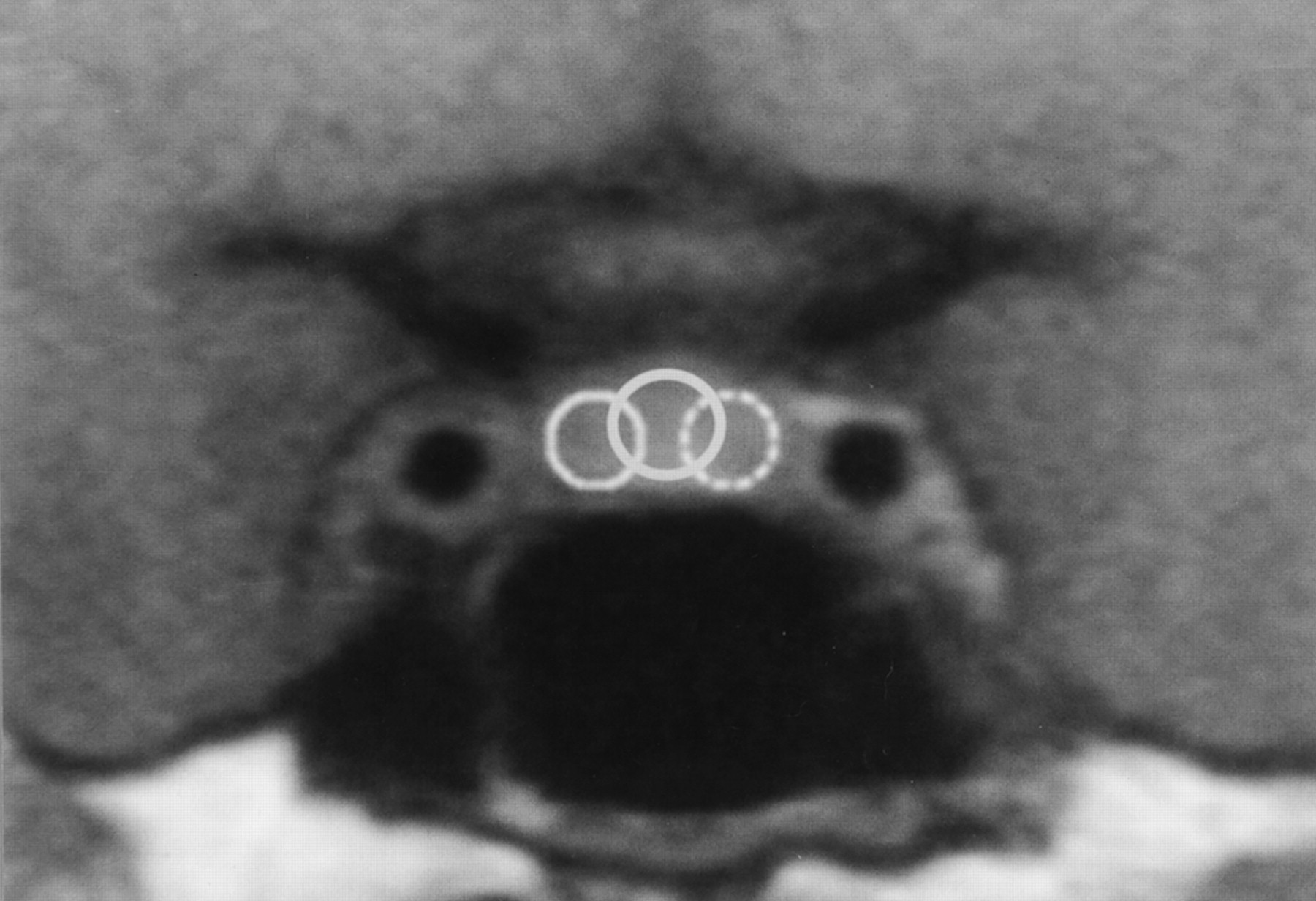
- Fig 1.
ROIs on a coronal dynamic image. The ROIs were located at the center and bilateral peripheral areas of the anterior pituitary gland for dynamic curve analyses.
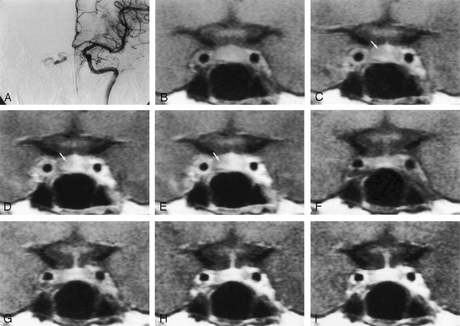
- Fig 2.
Images in a 50-year-old woman with a dural AVF draining into the right cavernous sinus (case 7).
A, Digital subtraction angiogram reveals the AVF draining into the right cavernous sinus.
B–E, Pretreatment dynamic MR images. In phase 0 (B) and in the first (C), second (D), and third (E) phases, the enhancement of the right side of the pituitary gland is delayed (arrow in C–E).
F–I, Posttreatment dynamic MR images in phase 0 (F) and in the first (G), second (H), and third (I) phases. After treatment of the disease, the pituitary gland is symmetrically enhancing on both the right and left sides, and the height of the pituitary gland decreased from 7 to 5 mm. Part of the posterior pituitary gland is seen as a hyperintense area in phase 0.
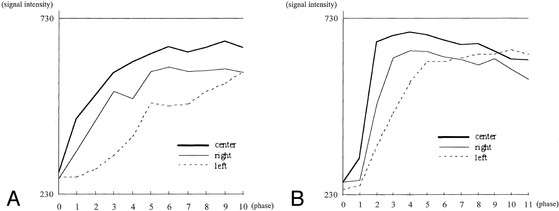
- Fig 3.
Dynamic curves in a 50-year-old woman with a dural AVF draining into the right cavernous sinus (case 7).
A, Pretreatment dynamic curves. The three ROIs were located at the center and bilateral peripheral areas of the pituitary gland (Fig 1). On the right side of the pituitary gland, the peak of the enhancement was not found within the imaging time before treatment.
B, Posttreatment dynamic curves. On the right side, the peak-enhanced phase was shortened and found within the 10 phases.
- Fig 4.
Images in a 62-year-old woman with a dural AVF draining into the left cavernous sinus (case 6).
A, Digital subtraction angiogram reveals the AVF draining into the left cavernous sinus.
B–E, Pretreatment dynamic MR images of the anterior pituitary gland. In phase 0 (B) and in the first (C), second (D), and third (E) phases, the enhancement of the left side of the pituitary gland is delayed (arrow in C–E). In phase 0, the left cavernous sinus is already enhancing because of the AVFs, but no enhancement can be seen in the pituitary gland; this is why this image was defined as phase 0.
F–I, Posttreatment dynamic MR images in phase 0 (F) and in the first (G), second (H), and third (I) phases. After treatment of the disease, the laterality of the enhancement on early dynamic images became less prominent, and the height of the pituitary gland decreased from 8 to 7 mm. In this case only, the laterality of the enhancement somewhat remained after treatment (arrow in G).
- Fig 5.
Dynamic curves in a 62-year-old woman with a dural AVF draining into the left cavernous sinus (case 6).
A, Pretreatment dynamic curves. The peaks of the dynamic curves were in phase 9 for the central area, phase 6 for the right side, and out of the imaging time for the left side.
B, Posttreatment dynamic curves. The peaks of the dynamic curves are shortened at both the central and bilateral peripheral areas of the pituitary gland.


Tables
Patient No./Age, y/Sex Fistula* Dilated Draining Vein* Embolization Radiation Therapy Cavernous Sinus Coil Packing External Carotid Artery 1/68/F D/D SOV, CV/SOV, CV No Yes Yes 2/58/F D/– IPS/IPS Yes Yes Yes 3/68/F D/D IPS, SOV/IPS, SOV No Yes Yes 4/62/F –/D None/SOV No No Yes 5/77/F D/D SOV, IPS/SOV, CV No Yes Yes 6/62/F –/D CV/SOV, SPS, CV Yes Yes No 7/50/F D/– SOV/none Yes Yes No 8/69/F D/– SOV, IPS, SPS/none Yes Yes No 9/62/F D/– SOV, SPS/none Yes Yes No * Data are for the right side/left side. CV indicates cortical vein; IPS, inferior petrosal vein; SOV, superior ophthalmic vein; SPS, superior petrosal vein; D, type D fistula; –, no fistula.
Patient Side of Fistula on Angiography Laterality of Enhancement, Delayed Side Phase with Homogenous Enhancement* Height of Pituitary Gland, mm† Before Therapy After Therapy Before Therapy After Therapy Before Therapy After Therapy 1 Bilateral None None 4 4 3 3 2 Right None None 3 3 7 3 3 Bilateral Left None 3 2 9 4 4 Left None None 3 3 6 6 5 Bilateral Right None 3 2 4 4 6 Left Left Left 8 5 8 7 7 Right Right None >10 8 7 5 8 Right None None 2 2 6 6 9 Right Right None 6 3 8 7 * Mean, 4.7 before therapy and 3.6 after therapy.
† Mean, 6.4 before therapy and 5.0 after therapy.
Group Center Periphery Patients before treatment 6.0 ± 1.7 7.5 ± 2.5† Patients after treatment 4.2 ± 2.6 5.4 ± 3.1 Control subjects 4.0 ± 1.4 4.2 ± 1.4 * Data are the mean ± SD.
† P < .01, compared with posttreatment and control values.
In this issue


Jump to section
Related Articles
Cited By...
- No citing articles found.