
Article Figures & Data
Figures
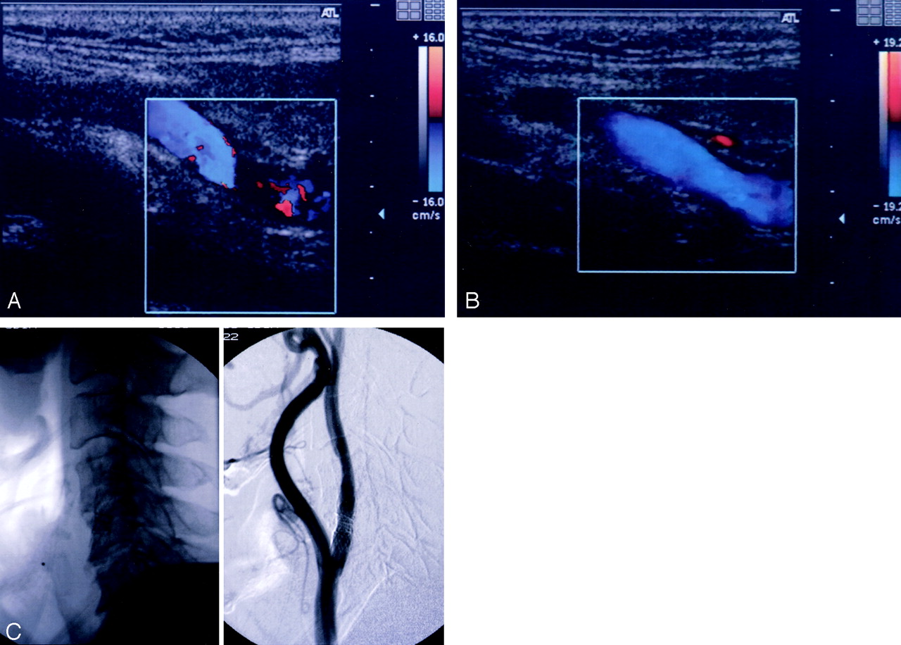
- Fig 1.
Benefit of CE-CCCD sonography in a patient with overlapped stents (patient 14).
A, Nonenhanced CCCD image did not allow visualization of blood flow in the overlapped stents.
B, CE-CCCD sonogram provides anatomic information almost equivalent to that on the DSA image.
C, Corresponding lateral radiograph (left) and DSA (right) images.
- Fig 2.
Benefit of CE-CCCD sonography in a patient with a high cervical lesion (patient 5).
A, Nonenhanced CCCD image allowed visualization of only the proximal half of the stented lumen.
B, After the application of the contrast agent, flow in the distally positioned stent could fully be detected by means of CE-CCCD imaging.
C, Corresponding lateral X-p (left) and DSA (right) images.


Tables
- TABLE 1:
Summary of CS procedures, follow-up findings, and depiction of the stented lumen at CCCD and CE-CCCD sonography
Patient Side Location and Type of Stent* Patency on DSA CCCD CE-CCCD 1 R C2, overlapped (Smart) No stenosis Partial Complete‡ 2 R C2–C3, single (Smart) No stenosis Partial Complete‡ 3 R C2–C3, single (Palmaz) No stenosis Partial Complete‡ 4 R C2–C4, single (Smart) Intimal hyperplasia§ Partial Partial 5 R C2–C4, single (Palmaz) No stenosis Partial Complete‡ 6 L C3, single (Palmaz) Not evaluated Partial Complete‡ 7 L C3–C4, single (Palmaz) No stenosis Complete Complete 8 L C3–C4, single (Smart) No stenosis Complete Complete 9 R C3–C4, single (Palmaz) No stenosis Complete Complete 10 R C3–C4, single (Palmaz) No stenosis Complete Complete 11 R C3–C4, single (Palmaz) No stenosis Complete Complete 12 R C3–C5, single (Palmaz) No stenosis Complete Complete 13 R C4–C5, single (Smart) No stenosis Complete Complete 14 L C4–C5, overlapped (Palmaz, Wiktor) No stenosis Partial Complete‡ 15 L C6–T1, single (Smart) No stenosis Complete Complete * C indicates cervical vertebra; T, thoracic vertebra. Smart, Smart stent; Palmaz, Palmaz stent; Wiktor, Wiktor stent.
† SA indicates digital subtraction angiography.
‡ In six patients who had overlapped stents or a high cervical lesion, CE-CCCD was helpful for visualization of the entire stented lumen. The location of the stent was confirmed on cervical radiograph, lateral views.
§ Asymptomatic intimal hyperplasia with luminal narrowing of approximately 50%.
In this issue


Jump to section
Related Articles
Cited By...
- No citing articles found.