Advertisement

Main menu

  • Home
  • Content
    • Current Issue
    • Accepted Manuscripts
    • Article Preview
    • Past Issue Archive
    • Video Articles
    • AJNR Case Collection
    • Case of the Week Archive
    • Case of the Month Archive
    • Classic Case Archive
  • Special Collections
    • AJNR Awards
    • Low-Field MRI
    • Alzheimer Disease
    • ASNR Foundation Special Collection
    • Photon-Counting CT
    • View All
  • Multimedia
    • AJNR Podcasts
    • AJNR SCANtastic
    • Trainee Corner
    • MRI Safety Corner
    • Imaging Protocols
  • For Authors
    • Submit a Manuscript
    • Submit a Video Article
    • Submit an eLetter to the Editor/Response
    • Manuscript Submission Guidelines
    • Statistical Tips
    • Fast Publishing of Accepted Manuscripts
    • Graphical Abstract Preparation
    • Imaging Protocol Submission
    • Author Policies
  • About Us
    • About AJNR
    • Editorial Board
    • Editorial Board Alumni
  • More
    • Become a Reviewer/Academy of Reviewers
    • Subscribers
    • Permissions
    • Alerts
    • Feedback
    • Advertisers
    • ASNR Home

User menu

  • Alerts
  • Log in

Search

  • Advanced search
American Journal of Neuroradiology

American Journal of Neuroradiology

ASHNR American Society of Functional Neuroradiology ASHNR American Society of Pediatric Neuroradiology ASSR
  • Alerts
  • Log in

Advanced Search

  • Home
  • Content
    • Current Issue
    • Accepted Manuscripts
    • Article Preview
    • Past Issue Archive
    • Video Articles
    • AJNR Case Collection
    • Case of the Week Archive
    • Case of the Month Archive
    • Classic Case Archive
  • Special Collections
    • AJNR Awards
    • Low-Field MRI
    • Alzheimer Disease
    • ASNR Foundation Special Collection
    • Photon-Counting CT
    • View All
  • Multimedia
    • AJNR Podcasts
    • AJNR SCANtastic
    • Trainee Corner
    • MRI Safety Corner
    • Imaging Protocols
  • For Authors
    • Submit a Manuscript
    • Submit a Video Article
    • Submit an eLetter to the Editor/Response
    • Manuscript Submission Guidelines
    • Statistical Tips
    • Fast Publishing of Accepted Manuscripts
    • Graphical Abstract Preparation
    • Imaging Protocol Submission
    • Author Policies
  • About Us
    • About AJNR
    • Editorial Board
    • Editorial Board Alumni
  • More
    • Become a Reviewer/Academy of Reviewers
    • Subscribers
    • Permissions
    • Alerts
    • Feedback
    • Advertisers
    • ASNR Home
  • Follow AJNR on Twitter
  • Visit AJNR on Facebook
  • Follow AJNR on Instagram
  • Join AJNR on LinkedIn
  • RSS Feeds

AJNR Awards, New Junior Editors, and more. Read the latest AJNR updates

Research ArticlePediatric Neuroimaging

Diffusion Tensor Brain Imaging Findings At Term-equivalent Age May Predict Neurologic Abnormalities in Low Birth Weight Preterm Infants

Y. Arzoumanian, M. Mirmiran, P.D. Barnes, K. Woolley, R.L. Ariagno, M.E. Moseley, B.E. Fleisher and S.W. Atlas
American Journal of Neuroradiology September 2003, 24 (8) 1646-1653;
Y. Arzoumanian
  • Find this author on Google Scholar
  • Find this author on PubMed
  • Search for this author on this site
M. Mirmiran
  • Find this author on Google Scholar
  • Find this author on PubMed
  • Search for this author on this site
P.D. Barnes
  • Find this author on Google Scholar
  • Find this author on PubMed
  • Search for this author on this site
K. Woolley
  • Find this author on Google Scholar
  • Find this author on PubMed
  • Search for this author on this site
R.L. Ariagno
  • Find this author on Google Scholar
  • Find this author on PubMed
  • Search for this author on this site
M.E. Moseley
  • Find this author on Google Scholar
  • Find this author on PubMed
  • Search for this author on this site
B.E. Fleisher
  • Find this author on Google Scholar
  • Find this author on PubMed
  • Search for this author on this site
S.W. Atlas
  • Find this author on Google Scholar
  • Find this author on PubMed
  • Search for this author on this site
  • Article
  • Figures & Data
  • Info & Metrics
  • Responses
  • References
  • PDF
Loading

Article Figures & Data

Figures

  • Tables
  • Fig 1
    • Download figure
    • Open in new tab
    • Download powerpoint
    Fig 1

    Images show region of interest placement for the posterior limb of the internal capsule. B0, image without diffusion weighting; DWI, diffusion-weighted image; FA, fractional anisotropy image; ADC, apparent diffusion coefficient map.

  • Fig 2
    • Download figure
    • Open in new tab
    • Download powerpoint
    Fig 2

    Average FA histograms by clinical subgroup. N, normal neurologic outcome; AN abnormal neurologic outcome.
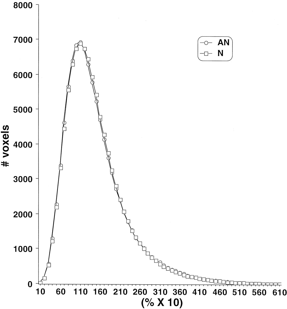
  • Fig 3
    • Download figure
    • Open in new tab
    • Download powerpoint
    Fig 3

    Average ADC histograms by clinical subgroup. N, normal neurologic outcome; AN abnormal neurologic outcome.

Tables

  • Figures
    • View popup
    TABLE 1:

    Study population MR imaging and neurologic follow-up

    GA ≤ 33 weeks Birthweight < 1800 gInitial populationn = 137
    Age at time of MR imaging, with 34–42 weeks
    30 with incomplete follow-upsOutcome analysisn = 107
    17 with category > 2DTI populationn = 90
    27 with incomplete DTIDTI study groupnc = 63
    • Note.—GA indicates gestational age; DTI, diffusion tensor imaging.

    • View popup
    TABLE 2:

    Neurologic outcome and conventional MR imaging category for 63 infants fully studied

    MR Imaging CategoryNormalAN
    13211
    2182
    • Note.—Normal indicates normal neurologic outcome; AN, abnormal neurologic outcome; Category 1, no abnormality; category 2, minimal subependymal hemorrhage or mineralization.

    • View popup
    TABLE 3:

    Region of interest fractional anisotropy values by pooled clinical subgroup

    FAMeanSDRangeP Value
    Corpus callosum genuN.449.082.255–.621.98
    AN.448.078.332–.580
    Corpus callosum spleniumN.451.079.273–.616.16
    AN.416.075.238–.519
    Internal capsule anterior LN.259.054.153–.381.14
    AN.230.026.197–.267
    Internal capsule anterior RN.267.054.148–.384.91
    AN.265.079.171–.411
    Internal capsule posterior LN.397.059.280–.539.08
    AN.365.049.305–.488
    Internal capsule posterior RN.411.046.284–.509.0002
    AN.351.061.259–.499
    Cerebral white matter LN.188.041.028–.277.13
    AN.169.030.113–.208
    Cerebral white matter RN.184.033.114–.290.16
    AN.170.031.120–.220
    • Note.—FA indicates fractional anisotropy; N, normal neurologic outcome; AN, abnormal neurologic outcome; L, left; R, right.

    • View popup
    TABLE 4:

    Region of interest apparent diffusion coefficient (μ mm2/s) values by pooled clinical subgroup

    ADCMeanSDRangeP Value
    Corpus callosum genuN1.312.1321.024–1.7030.84
    AN1.320.1221.100–1.499
    Corpus callosum spleniumN1.268.1451.050–2.0550.09
    AN1.340.0711.211–1.461
    Internal capsule anterior LN1.216.0921.039–1.4260.10
    AN1.122.3200.359–1.376
    Internal capsule anterior RN1.209.1081.001–1.4290.33
    AN1.248.0881.126–1.363
    Internal capsule posterior LN1.095.0710.970–1.3290.51
    AN1.080.0570.983–1.200
    Internal capsule posterior RN1.091.0750.893–1.2890.92
    AN1.093.0680.965–1.208
    Cerebral white matter LN1.413.1171.186–1.6870.23
    AN1.458.1301.287–1.741
    Cerebral white matter RN1.424.1341.205–1.8180.20
    AN1.478.1351.274–1.736
    • Note.—ADC indicates apparent diffusion coefficient; N, normal neurologic outcome; AN, abnormal neurologic outcome; L, left; R, right.

PreviousNext
Back to top

In this issue

American Journal of Neuroradiology: 24 (8)
American Journal of Neuroradiology
Vol. 24, Issue 8
1 Sep 2003
  • Table of Contents
  • Index by author
Advertisement
Print
Download PDF
Email Article

Thank you for your interest in spreading the word on American Journal of Neuroradiology.

NOTE: We only request your email address so that the person you are recommending the page to knows that you wanted them to see it, and that it is not junk mail. We do not capture any email address.

Enter multiple addresses on separate lines or separate them with commas.
Diffusion Tensor Brain Imaging Findings At Term-equivalent Age May Predict Neurologic Abnormalities in Low Birth Weight Preterm Infants
(Your Name) has sent you a message from American Journal of Neuroradiology
(Your Name) thought you would like to see the American Journal of Neuroradiology web site.
CAPTCHA
This question is for testing whether or not you are a human visitor and to prevent automated spam submissions.
Cite this article
Y. Arzoumanian, M. Mirmiran, P.D. Barnes, K. Woolley, R.L. Ariagno, M.E. Moseley, B.E. Fleisher, S.W. Atlas
Diffusion Tensor Brain Imaging Findings At Term-equivalent Age May Predict Neurologic Abnormalities in Low Birth Weight Preterm Infants
American Journal of Neuroradiology Sep 2003, 24 (8) 1646-1653;

Citation Manager Formats

  • BibTeX
  • Bookends
  • EasyBib
  • EndNote (tagged)
  • EndNote 8 (xml)
  • Medlars
  • Mendeley
  • Papers
  • RefWorks Tagged
  • Ref Manager
  • RIS
  • Zotero
0 Responses
Respond to this article
Share
Bookmark this article
Diffusion Tensor Brain Imaging Findings At Term-equivalent Age May Predict Neurologic Abnormalities in Low Birth Weight Preterm Infants
Y. Arzoumanian, M. Mirmiran, P.D. Barnes, K. Woolley, R.L. Ariagno, M.E. Moseley, B.E. Fleisher, S.W. Atlas
American Journal of Neuroradiology Sep 2003, 24 (8) 1646-1653;
del.icio.us logo Twitter logo Facebook logo Mendeley logo
  • Tweet Widget
  • Facebook Like
  • Google Plus One

Jump to section

  • Article
    • Abstract
    • Methods
    • Results
    • Discussion
    • Conclusion
    • Footnotes
    • References
  • Figures & Data
  • Info & Metrics
  • Responses
  • References
  • PDF

Related Articles

  • No related articles found.
  • PubMed
  • Google Scholar

Cited By...

  • Cyto/myeloarchitectural changes of cortical gray matter and superficial white matter in early neurodevelopment: Multimodal MRI study of preterm neonates
  • Regional brain volumes, microstructure and neurodevelopment in moderate-late preterm children
  • Optimal Timing of Cerebral MRI in Preterm Infants to Predict Long-Term Neurodevelopmental Outcome: A Systematic Review
  • Role of Diffusion Tensor Imaging as an Independent Predictor of Cognitive and Language Development in Extremely Low-Birth-Weight Infants
  • Predictive Value of Neonatal Magnetic Resonance Imaging in Preterm Infants
  • Quantitative Fiber Tracking of the Optic Radiation Is Correlated with Visual-Evoked Potential Amplitude in Preterm Infants
  • Crossref
  • Google Scholar

This article has not yet been cited by articles in journals that are participating in Crossref Cited-by Linking.

More in this TOC Section

  • FRACTURE MR in Congenital Vertebral Anomalies
  • Comparing MRI Perfusion in Pediatric Brain Tumors
  • Sodium MRI in Pediatric Brain Tumors
Show more Pediatric Neuroimaging

Similar Articles

Advertisement

Indexed Content

  • Current Issue
  • Accepted Manuscripts
  • Article Preview
  • Past Issues
  • Editorials
  • Editor's Choice
  • Fellows' Journal Club
  • Letters to the Editor
  • Video Articles

Cases

  • Case Collection
  • Archive - Case of the Week
  • Archive - Case of the Month
  • Archive - Classic Case

More from AJNR

  • Trainee Corner
  • Imaging Protocols
  • MRI Safety Corner
  • Book Reviews

Multimedia

  • AJNR Podcasts
  • AJNR Scantastics

Resources

  • Turnaround Time
  • Submit a Manuscript
  • Submit a Video Article
  • Submit an eLetter to the Editor/Response
  • Manuscript Submission Guidelines
  • Statistical Tips
  • Fast Publishing of Accepted Manuscripts
  • Graphical Abstract Preparation
  • Imaging Protocol Submission
  • Evidence-Based Medicine Level Guide
  • Publishing Checklists
  • Author Policies
  • Become a Reviewer/Academy of Reviewers
  • News and Updates

About Us

  • About AJNR
  • Editorial Board
  • Editorial Board Alumni
  • Alerts
  • Permissions
  • Not an AJNR Subscriber? Join Now
  • Advertise with Us
  • Librarian Resources
  • Feedback
  • Terms and Conditions
  • AJNR Editorial Board Alumni

American Society of Neuroradiology

  • Not an ASNR Member? Join Now

© 2025 by the American Society of Neuroradiology All rights, including for text and data mining, AI training, and similar technologies, are reserved.
Print ISSN: 0195-6108 Online ISSN: 1936-959X

Powered by HighWire