
Article Figures & Data
Figures
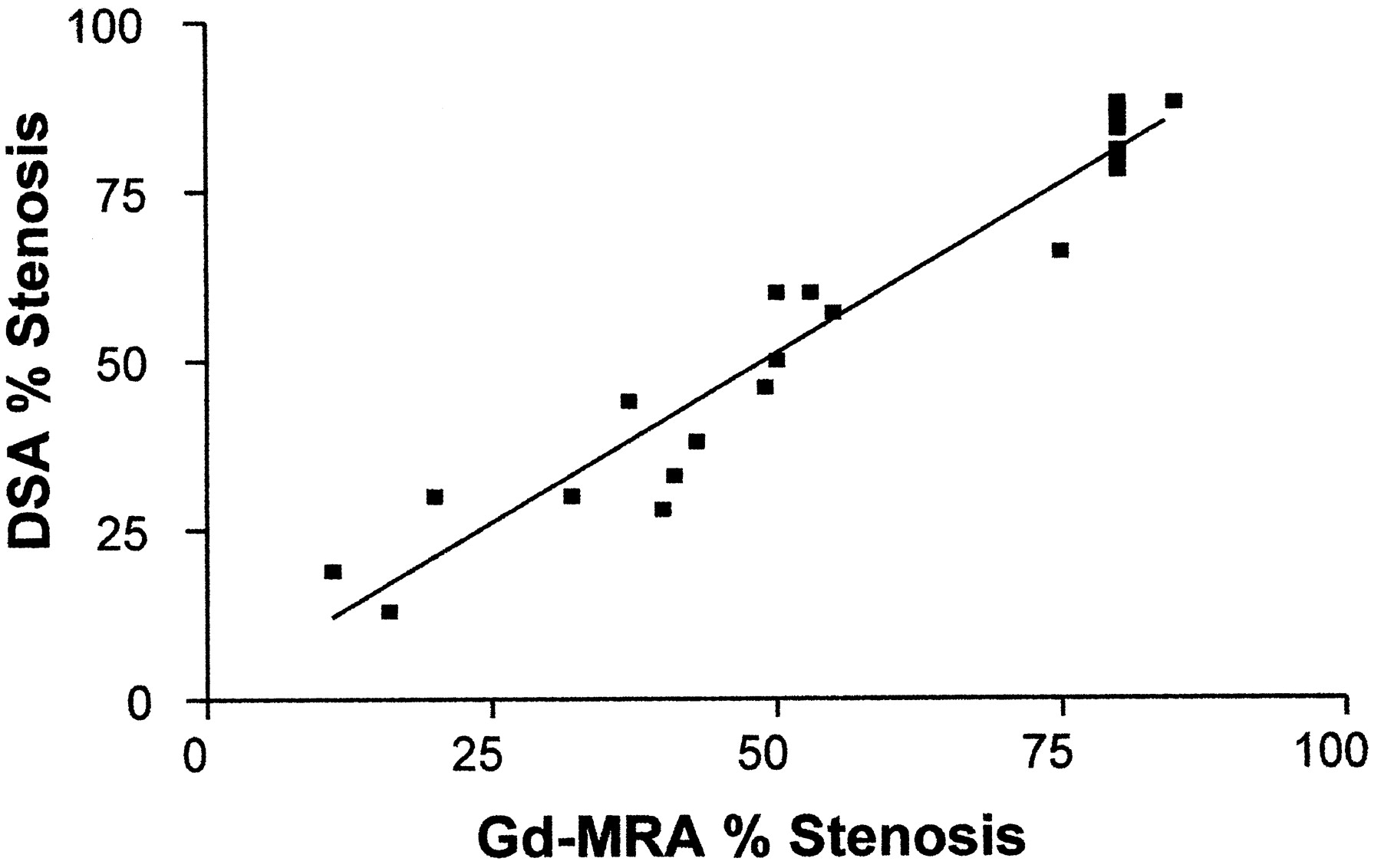
- Fig 1.
Scatter plot and regression line for digital subtraction angiographic measurement of stenosis versus contrast-enhanced MR angiographic measurement of stenosis. r = 0.967. The slope is very nearly 1, and the intercept is nearly 0, showing excellent linearity. DSA, digital subtraction angiography; Gd-MRA, contrast-enhanced MR angiography.
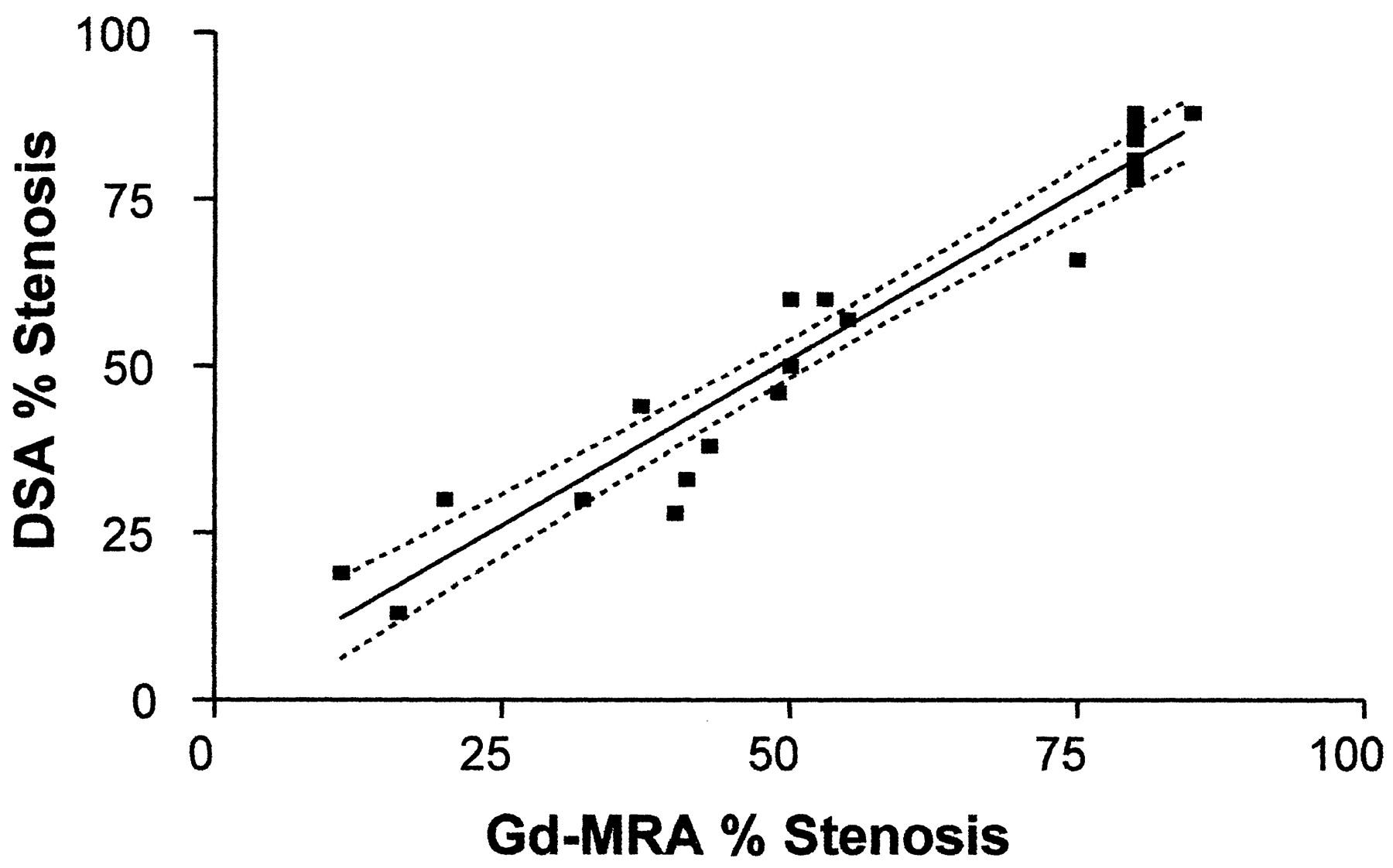
- Fig 2.
Comparison of digital subtraction angiographic measurement of stenosis versus contrast-enhanced MR angiographic measurement of stenosis shows regression line with 95% mean confidence intervals, which reach a minimum of ±2.8%, showing excellent correlation between digital subtraction angiography and contrast-enhanced MR angiography in the mean. DSA, digital subtraction angiography; Gd-MRA, contrast-enhanced MR angiography.
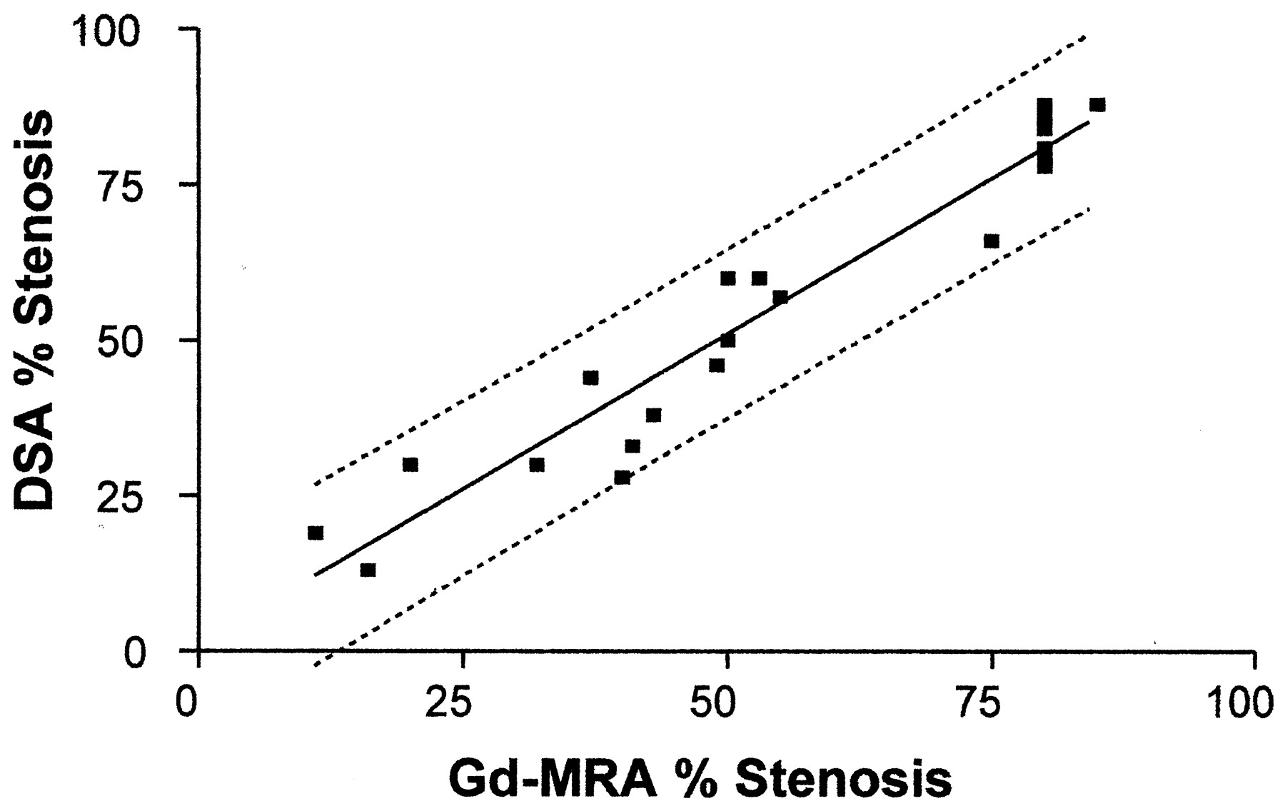
- Fig 3.
Comparison of digital subtraction angiographic measurement of stenosis versus contrast-enhanced MR angiographic measurement of stenosis shows regression line with 95% individual confidence intervals, which reach a minimum of ±13.6%. These intervals are significantly wider than the mean confidence intervals shown in Figure 2. DSA, digital subtraction angiography; Gd-MRA, contrast-enhanced MR angiography.
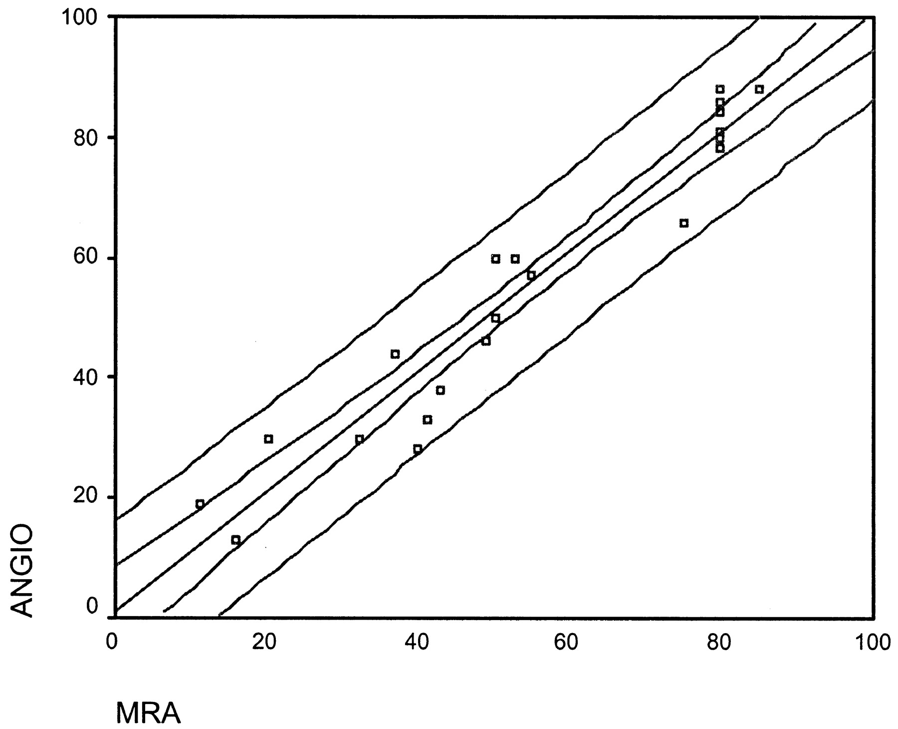
- Fig 4.
Comparison of digital subtraction angiographic measurement of stenosis versus contrast-enhanced MR angiographic measurement of stenosis shows regression line with both 95% mean confidence intervals and 95% individual confidence intervals overlaid on the same graph to depict the difference in relative width. DSA, digital subtraction angiography; Gd-MRA, contrast-enhanced MR angiography.
- Fig 5.
Comparison of digital subtraction angiographic measurement of stenosis versus ultrasound PSV shows regression line with 95% individual confidence intervals, which reach a minimum of ±27.3%. The fit is significantly less (r = 0.86) and the confidence intervals significantly wider than with contrast-enhanced MR angiography (Gd-MRA). DSA, digital subtraction angiography.


