
Article Figures & Data
Figures
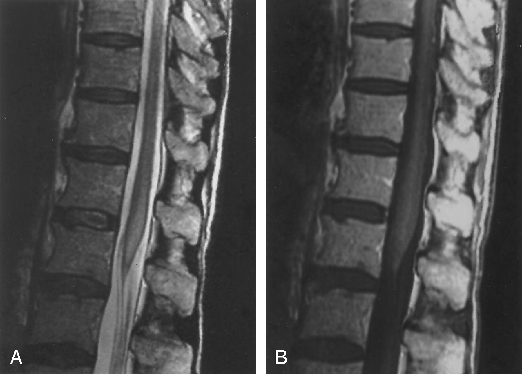
- Fig 1.
Imaging findings at initial imaging.
A, T2-weighted image shows a thick conus that is increased in signal intensity.
B, Contrast-enhanced T1-weighted image shows a thick conus without enhancement.
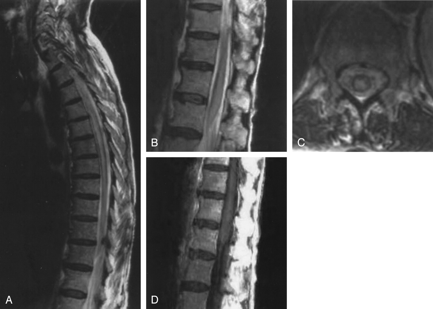
- Fig 2.
Imaging findings at 6-week follow-up.
A, MR image shows an enlargement of the thoracic cord associated with increased signal intensity in T2-weighted images.
B, The conus remains enlarged with signal intensity abnormality.
C, Axial T2-weighted MR image shows a central signal intensity abnormality within the cord at the T12 level.
D, There is still no enhancement of the conus following contrast medium injection.
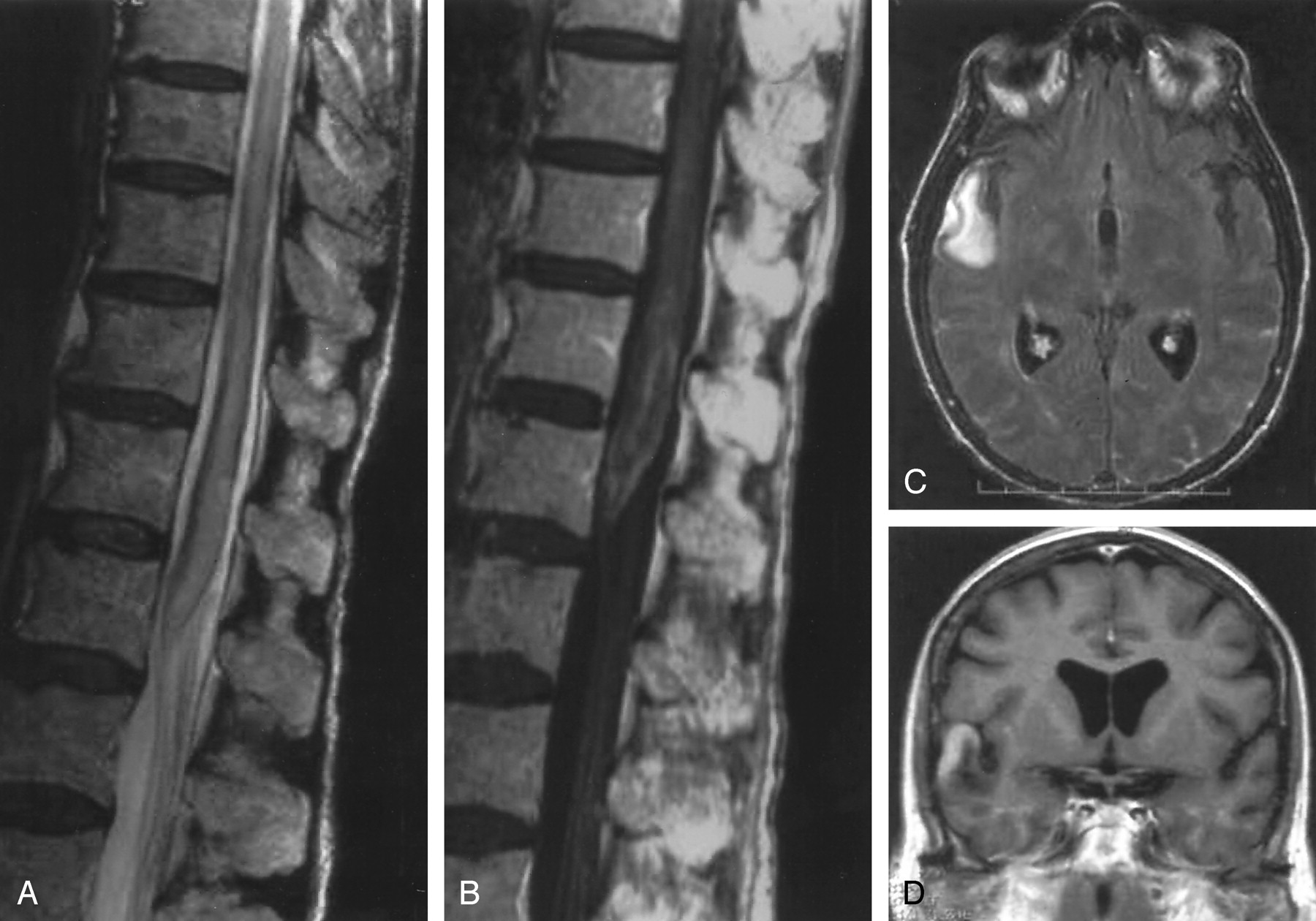
- Fig 3.
Imaging findings at 10-week follow-up.
A, There is an extension of spinal cord thickening on T2-weighted images.
B, Abnormal enhancement after Gd-DTPA injection on T1- weighted images.
C, T2-weighted FLAIR weighted image shows appearance of a right temporal lesion.
D, Postcontrast T1-weighted image shows lesion enhancement.
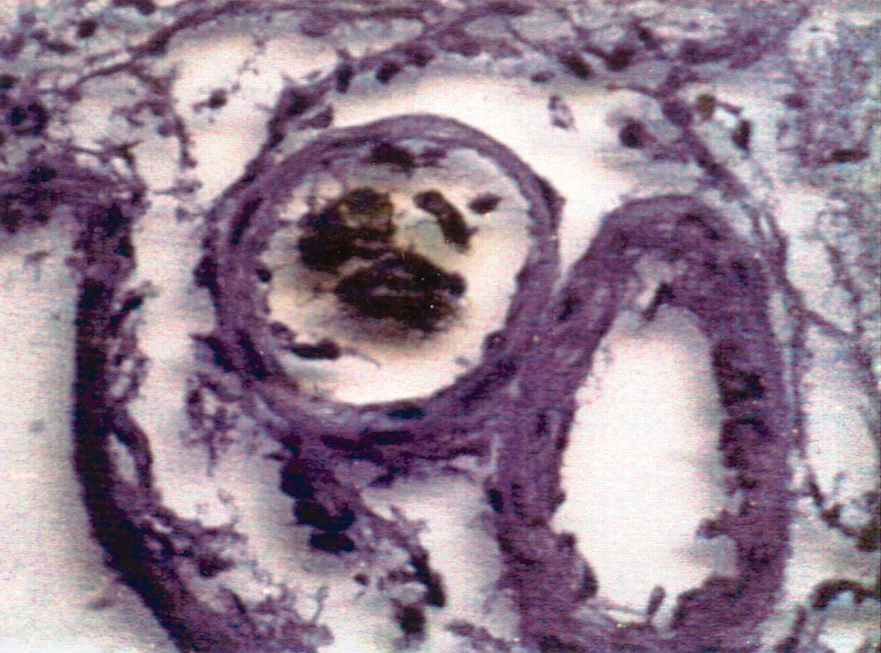
- Fig 4.
Large tumor cells in the lumen of small vessels of the brain (magnification ×400; hematoxylin and eosin). Biopsy fragments were taken from the right temporal lobe.
- Fig 5.
Large intravascular tumor cells express the cluster of differentiation 20,which is specific for B-cell lineage lymphocyte. (magnification ×400) (method with autoantibodies immunofixation). Biopsy fragments were taken from the right temporal lobe.
- Fig 6.
Imaging findings at 20-week follow-up.
A, T2-weighted FLAIR image following treatment shows disappearance of the previous signal intensity abnormality in the leptomeninges and in the right temporal lobe.
B, Contrast-enhanced T1-weighted image shows disappearance of prior enhancement.


Tables
Initial Status 6 Weeks Later 10 Weeks Later 20 Weeks Later Lumbar Cord Chemotherapy T2 Hyperintensity ++ ++ ++ − Enhancement − − ++ − Enlargement + + ++ + Thoracic Cord T2 Hyperintensity + ++ ++ − Enhancement − − − − Enlargement − + ++ + Cervical Cord T2 Hyperintensity + ++ ++ − Enhancement − − − − Enlargement − + ++ + Brain T2 Hyperintensity (not performed) − ++ + (toxic origin) Enhancement (not performed) − ++ −


