
Article Figures & Data
Figures
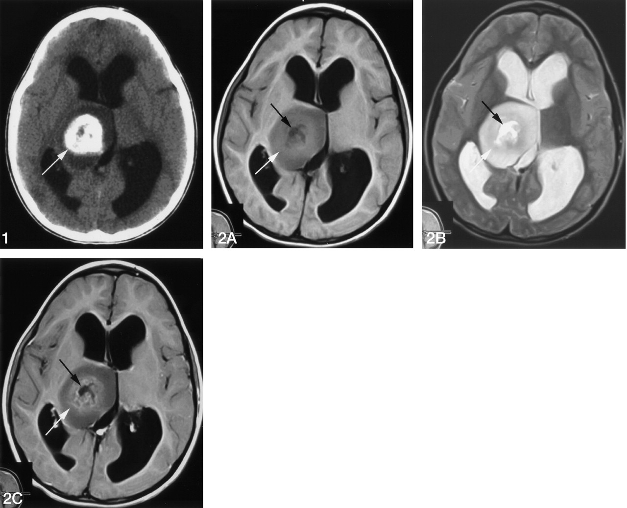
- Fig 1.
Noncontrast CT scan shows a low-attenuation mass in the right thalamus. Marked calcifications are seen in the center of the mass lesion (arrow).
- Fig 2.
Axial MR images. All images are obtained at same level.
A, T1-weighted image (500/20 [TR/TE]). B, T2-weighted image (5000/88 [TR/TE]). C, T1-weighted (500/20 [TR/TE]) image after gadolinium-DTPA administration.
The mass lesion shows lower signal intensity on the T1-weighted image (A) and higher signal intensity on the T2-weighted MR image (B) relative to gray matter. Calcified regions show slightly greater signal intensity on the T1-weighted image (A) and are enhanced after administration of gadolinium-DTPA (C, light arrows). CSF-like signal intensity exists in the core of the lesion (dark arrows).
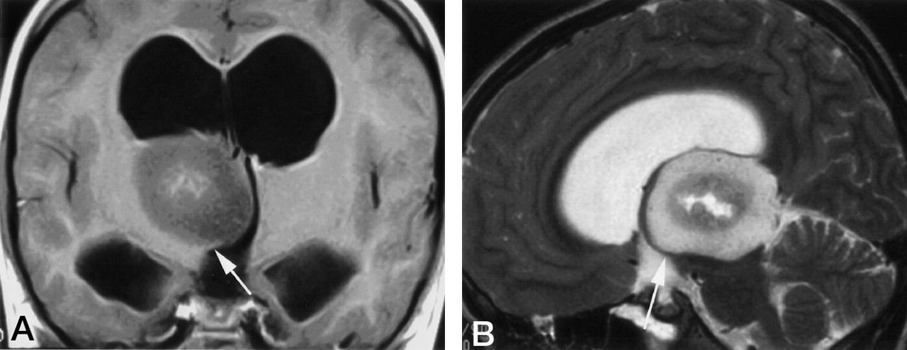
- Fig 3.
A, Coronal image after administration of gadolinium-DTPA (500/20 [TR/TE]). B, Sagittal T2-weighted image (4000/88.9 [TR/TE]). The margin of the mass lesion is covered with thalamus extending in a beaklike shape (arrows).
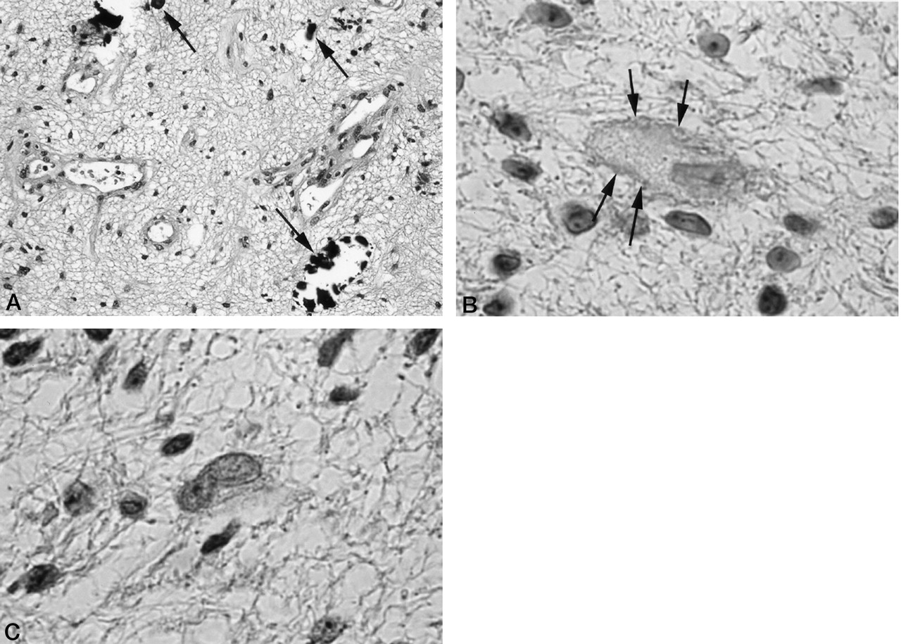
- Fig 4.
A, Hematoxylin and eosin stain (magnification ×100). B, N-CAM stain (magnification ×200). C, Polysialic acid stain (magnification ×200).
Proliferation of vessels and minute calcified corpuscles (dark arrows) are observed. There are no atypical cells or abnormal division of nucleus. Immunohistochemical staining is positive for N-CAM (dark arrows) and negative for polysialic acid.


