Advertisement

Main menu

  • Home
  • Content
    • Current Issue
    • Accepted Manuscripts
    • Article Preview
    • Past Issue Archive
    • Video Articles
    • AJNR Case Collection
    • Case of the Week Archive
    • Case of the Month Archive
    • Classic Case Archive
  • Special Collections
    • Advancing NeuroMRI with High-Relaxivity Contrast Agents
    • AJNR Awards
    • Low-Field MRI
    • Alzheimer Disease
    • ASNR Foundation Special Collection
    • Photon-Counting CT
    • View All
  • Multimedia
    • AJNR Podcasts
    • AJNR SCANtastic
    • Trainee Corner
    • MRI Safety Corner
    • Imaging Protocols
  • For Authors
    • Submit a Manuscript
    • Submit a Video Article
    • Submit an eLetter to the Editor/Response
    • Manuscript Submission Guidelines
    • Statistical Tips
    • Fast Publishing of Accepted Manuscripts
    • Graphical Abstract Preparation
    • Imaging Protocol Submission
    • Author Policies
  • About Us
    • About AJNR
    • Editorial Board
    • Editorial Board Alumni
  • More
    • Become a Reviewer/Academy of Reviewers
    • Subscribers
    • Permissions
    • Alerts
    • Feedback
    • Advertisers
    • ASNR Home

User menu

  • Alerts
  • Log in

Search

  • Advanced search
American Journal of Neuroradiology

American Journal of Neuroradiology

ASHNR American Society of Functional Neuroradiology ASHNR American Society of Pediatric Neuroradiology ASSR
  • Alerts
  • Log in

Advanced Search

  • Home
  • Content
    • Current Issue
    • Accepted Manuscripts
    • Article Preview
    • Past Issue Archive
    • Video Articles
    • AJNR Case Collection
    • Case of the Week Archive
    • Case of the Month Archive
    • Classic Case Archive
  • Special Collections
    • Advancing NeuroMRI with High-Relaxivity Contrast Agents
    • AJNR Awards
    • Low-Field MRI
    • Alzheimer Disease
    • ASNR Foundation Special Collection
    • Photon-Counting CT
    • View All
  • Multimedia
    • AJNR Podcasts
    • AJNR SCANtastic
    • Trainee Corner
    • MRI Safety Corner
    • Imaging Protocols
  • For Authors
    • Submit a Manuscript
    • Submit a Video Article
    • Submit an eLetter to the Editor/Response
    • Manuscript Submission Guidelines
    • Statistical Tips
    • Fast Publishing of Accepted Manuscripts
    • Graphical Abstract Preparation
    • Imaging Protocol Submission
    • Author Policies
  • About Us
    • About AJNR
    • Editorial Board
    • Editorial Board Alumni
  • More
    • Become a Reviewer/Academy of Reviewers
    • Subscribers
    • Permissions
    • Alerts
    • Feedback
    • Advertisers
    • ASNR Home
  • Follow AJNR on Twitter
  • Visit AJNR on Facebook
  • Follow AJNR on Instagram
  • Join AJNR on LinkedIn
  • RSS Feeds

AJNR Awards, New Junior Editors, and more. Read the latest AJNR updates


Improved Turnaround Times | Median time to first decision: 12 days

Research ArticleHead and Neck Imaging

Intraobserver Variability in the MR Determination of Tumor Volume in Squamous Cell Carcinoma of the Pharynx

Andrew R. Gordon, Laurie A. Loevner, Amita Shukla-Dave, Regina O. Redfern, Adina I. Sonners, Alex M. Kilger, Mark A. Elliott, Mitchell Machtay, Randal S. Weber, Jerry D. Glickson and David I. Rosenthal
American Journal of Neuroradiology June 2004, 25 (6) 1092-1098;
Andrew R. Gordon
  • Find this author on Google Scholar
  • Find this author on PubMed
  • Search for this author on this site
Laurie A. Loevner
  • Find this author on Google Scholar
  • Find this author on PubMed
  • Search for this author on this site
Amita Shukla-Dave
  • Find this author on Google Scholar
  • Find this author on PubMed
  • Search for this author on this site
Regina O. Redfern
  • Find this author on Google Scholar
  • Find this author on PubMed
  • Search for this author on this site
Adina I. Sonners
  • Find this author on Google Scholar
  • Find this author on PubMed
  • Search for this author on this site
Alex M. Kilger
  • Find this author on Google Scholar
  • Find this author on PubMed
  • Search for this author on this site
Mark A. Elliott
  • Find this author on Google Scholar
  • Find this author on PubMed
  • Search for this author on this site
Mitchell Machtay
  • Find this author on Google Scholar
  • Find this author on PubMed
  • Search for this author on this site
Randal S. Weber
  • Find this author on Google Scholar
  • Find this author on PubMed
  • Search for this author on this site
Jerry D. Glickson
  • Find this author on Google Scholar
  • Find this author on PubMed
  • Search for this author on this site
David I. Rosenthal
  • Find this author on Google Scholar
  • Find this author on PubMed
  • Search for this author on this site
  • Article
  • Figures & Data
  • Info & Metrics
  • Responses
  • References
  • PDF
Loading

Article Figures & Data

Figures

  • Tables
  • Fig 1.
    • Download figure
    • Open in new tab
    • Download powerpoint
    Fig 1.

    MR imaging–based volumetric analysis of a carcinoma on the left side of the base of the tongue in a 54-year-old man.

    A and B, Two sequential axial T2-weighted MR images (4000/100/2) were used to outline the primary tumor on each sequential axial image by using a mouse-controlled cursor. In this case, the neoplasm (outlined) is hypointense relative to the lymphoid tissue (L) of the lingual tonsil.

    C, Serial axial T2-weighted MR images (4000/100/2) show all images where a primary tumor is present, and the outlines of the tumor on each section. A computer program calculated the volume by counting voxels included within the outlined regions.
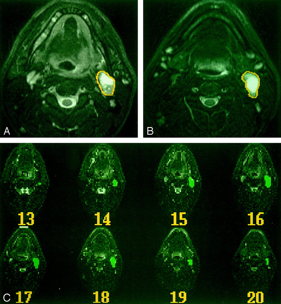
  • Fig 2.
    • Download figure
    • Open in new tab
    • Download powerpoint
    Fig 2.

    MR imaging–based volumetric analysis of a regional nodal metastasis in a patient with a primary cancer at the base of the tongue.

    A and B, Two sequential axial T2-weighted images show how the nodal mass was outlined within a mouse-controlled cursor.

    C, Serial axial T2-weighted MR images (4000/100/2) show all images on which nodal metastases are outlined and the outlines on each image.

Tables

  • Figures
    • View popup
    TABLE 1:

    Characteristics of primary cancers

    Patient (No.)Tumor LocationStageMR Imaging Time PointsPercentage Measurement Error, %
    1Base of tongueT4223*, 19
    2Pyriform sinusT4318, 20, 16
    3Base of tongueT355, 7, 12, 17, 2
    4Base of tongueT410
    5TonsilT431, 2, 10
    6TonsilT341, 3, 22†, 29†
    7Base of tongueT30‡None
    8TonsilT436, 15, 53†
    9Base of tongueT2413, 8, 37*, 11
    10Base of tongueT3216, 11
    11Base of tongueT231, 3, 12
    12Base of tongueT23§29*, 22*, 2
    13Base of tongueT334, 8, 19
    14Base of tongueT439, 3, 22†
    15Pyriform sinusT437, 2, 23†
    16TonsilT230, 13, 26†
    17Base of tongueT234, 4, 2
    • * Percentage measurement error was greater than 20% because of difficulty in differentiating the neoplasm from adjacent lymphoid tissue at the base of the tongue.

    • † Percentage measurement error was greater than 20% because of a small absolute differences in the volume measurements of small lesions.

    • ‡ The primary tumor was too large to measure.

    • § T1-weighted images were used because of artifact on T2-weighted images.

    • View popup
    TABLE 2:

    Characteristics of regional nodal metastases

    Patient (No.)StageNodal LevelMR Imaging Time PointsPercentage Measurement Error, %
    1N0None0*None
    2N2cL II/III414, 13, 11, 11
    3N2bR II52, 1, 3, 9, 31†
    4N2cR II/III32, 7, 3
    5N2cR II/III31, 2, 10
    6N2bL II-IV41, 1, 3, 8
    7N2cR II/III314, 26‡, 4
    8N2aR II35, 1, 4
    9N2bR II44, 12, 0, 11
    10N2bR II/III23, 30†
    11N2aL II34, 1, 1
    12N3R II317, 16, 27*
    13N2bR II33, 17, 17
    14N2cL II44, 16, 13, 19
    15N3R I-V33, 7, 20
    16N2bR II34, 9, 37†
    17N2cL II/III39, 6, 4
    • * No nodal metastases.

    • † Percentage measurement error was greater than 20% because of small absolute differences in the volume measurements of small lesions.

    • ‡ Percentage measurement error was greater than 20% because of inaccuracies in the volume measurements due to patient motion.

    • View popup
    TABLE 3:

    Percentage measurement error for primary pharyngeal cancer and regional nodal metastases

    Percent Measurement ErrorPrimary TumorNodal Mass
    Mean, %139
    Standard deviation, %119
    Median, %127
    95% CI, %10,167,12
    Range, %0,530,37
PreviousNext
Back to top

In this issue

American Journal of Neuroradiology: 25 (6)
American Journal of Neuroradiology
Vol. 25, Issue 6
1 Jun 2004
  • Table of Contents
  • Index by author
Advertisement
Print
Download PDF
Email Article

Thank you for your interest in spreading the word on American Journal of Neuroradiology.

NOTE: We only request your email address so that the person you are recommending the page to knows that you wanted them to see it, and that it is not junk mail. We do not capture any email address.

Enter multiple addresses on separate lines or separate them with commas.
Intraobserver Variability in the MR Determination of Tumor Volume in Squamous Cell Carcinoma of the Pharynx
(Your Name) has sent you a message from American Journal of Neuroradiology
(Your Name) thought you would like to see the American Journal of Neuroradiology web site.
CAPTCHA
This question is for testing whether or not you are a human visitor and to prevent automated spam submissions.
Cite this article
Andrew R. Gordon, Laurie A. Loevner, Amita Shukla-Dave, Regina O. Redfern, Adina I. Sonners, Alex M. Kilger, Mark A. Elliott, Mitchell Machtay, Randal S. Weber, Jerry D. Glickson, David I. Rosenthal
Intraobserver Variability in the MR Determination of Tumor Volume in Squamous Cell Carcinoma of the Pharynx
American Journal of Neuroradiology Jun 2004, 25 (6) 1092-1098;

Citation Manager Formats

  • BibTeX
  • Bookends
  • EasyBib
  • EndNote (tagged)
  • EndNote 8 (xml)
  • Medlars
  • Mendeley
  • Papers
  • RefWorks Tagged
  • Ref Manager
  • RIS
  • Zotero
0 Responses
Respond to this article
Share
Bookmark this article
Intraobserver Variability in the MR Determination of Tumor Volume in Squamous Cell Carcinoma of the Pharynx
Andrew R. Gordon, Laurie A. Loevner, Amita Shukla-Dave, Regina O. Redfern, Adina I. Sonners, Alex M. Kilger, Mark A. Elliott, Mitchell Machtay, Randal S. Weber, Jerry D. Glickson, David I. Rosenthal
American Journal of Neuroradiology Jun 2004, 25 (6) 1092-1098;
del.icio.us logo Twitter logo Facebook logo Mendeley logo
  • Tweet Widget
  • Facebook Like
  • Google Plus One

Jump to section

  • Article
    • Abstract
    • Methods
    • Results
    • Discussion
    • Conclusions
    • Footnotes
    • References
  • Figures & Data
  • Info & Metrics
  • Responses
  • References
  • PDF

Related Articles

  • No related articles found.
  • PubMed
  • Google Scholar

Cited By...

  • Efficacy of Diffusion-Weighted Imaging for the Differentiation between Lymphomas and Carcinomas of the Nasopharynx and Oropharynx: Correlations of Apparent Diffusion Coefficients and Histologic Features
  • Evaluating lingual carcinoma for surgical management: what does volumetric measurement with MRI offer?
  • Specific Growth Rate versus Doubling Time for Quantitative Characterization of Tumor Growth Rate
  • Crossref
  • Google Scholar

This article has not yet been cited by articles in journals that are participating in Crossref Cited-by Linking.

More in this TOC Section

  • Diagnostic Performance of US in Neck Node NIRADS 2
  • Hydrops Herniation into the Semicircular Canals
  • ASL Sensitivity for Head and Neck Paraganglioma
Show more HEAD AND NECK IMAGING

Similar Articles

Advertisement

Indexed Content

  • Current Issue
  • Accepted Manuscripts
  • Article Preview
  • Past Issues
  • Editorials
  • Editor's Choice
  • Fellows' Journal Club
  • Letters to the Editor
  • Video Articles

Cases

  • Case Collection
  • Archive - Case of the Week
  • Archive - Case of the Month
  • Archive - Classic Case

More from AJNR

  • Trainee Corner
  • Imaging Protocols
  • MRI Safety Corner
  • Book Reviews

Multimedia

  • AJNR Podcasts
  • AJNR Scantastics

Resources

  • Turnaround Time
  • Submit a Manuscript
  • Submit a Video Article
  • Submit an eLetter to the Editor/Response
  • Manuscript Submission Guidelines
  • Statistical Tips
  • Fast Publishing of Accepted Manuscripts
  • Graphical Abstract Preparation
  • Imaging Protocol Submission
  • Evidence-Based Medicine Level Guide
  • Publishing Checklists
  • Author Policies
  • Become a Reviewer/Academy of Reviewers
  • News and Updates

About Us

  • About AJNR
  • Editorial Board
  • Editorial Board Alumni
  • Alerts
  • Permissions
  • Not an AJNR Subscriber? Join Now
  • Advertise with Us
  • Librarian Resources
  • Feedback
  • Terms and Conditions
  • AJNR Editorial Board Alumni

American Society of Neuroradiology

  • Not an ASNR Member? Join Now

© 2025 by the American Society of Neuroradiology All rights, including for text and data mining, AI training, and similar technologies, are reserved.
Print ISSN: 0195-6108 Online ISSN: 1936-959X

Powered by HighWire