
Article Figures & Data
Figures
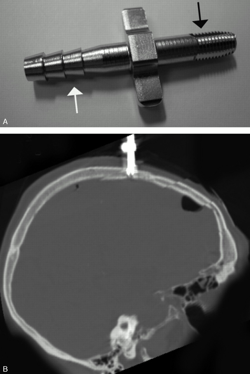
- Fig 1.
A, This image shows the metal device that is screwed into the burr hole. The threaded end (black arrow) is self-tapping. The notched tubing (white arrow) is then attached to the silicone tubing.
B, This reformat of the CT data shows the metal insertion portion of the device orientation perpendicular to the skull.
- Fig 2.
Oblique (A) and A-P (B) views, reformatted from CT data on a 3D workstation (Vitrea), demonstrate the winged canula and its relationship to the skull.
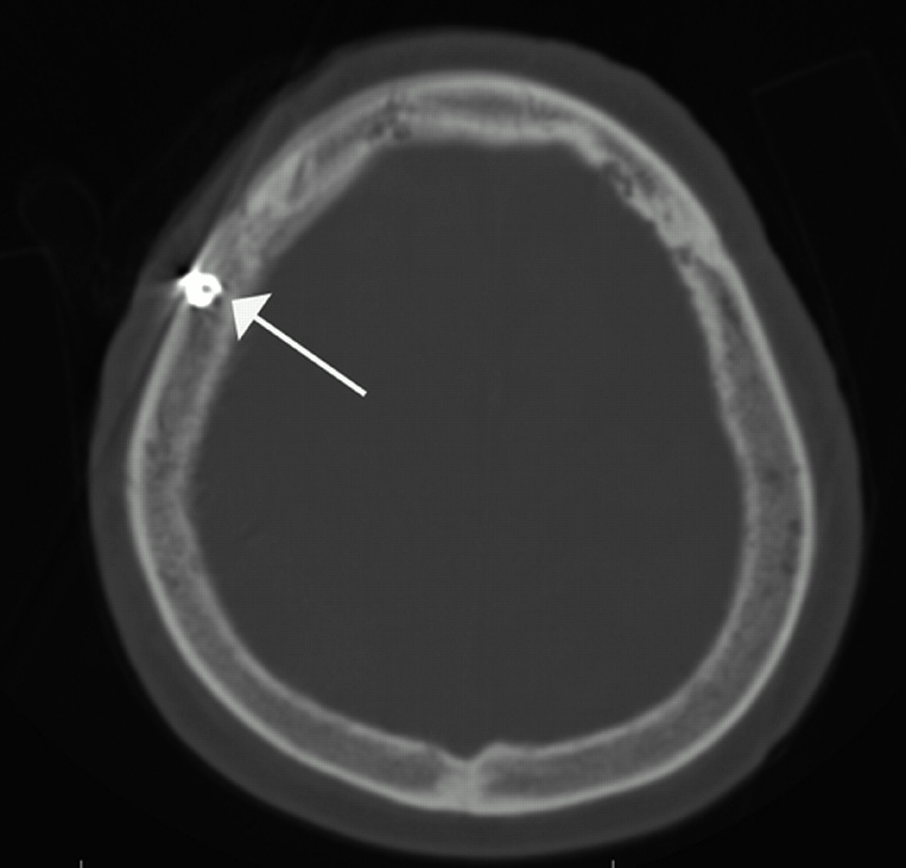
- Fig 3.
The bone image from that same study shows the tip of the device positioned in the diploic space of the right calvaria (arrow).
- Fig 4.
The reformatted 2D image demonstrates the relationships of the subdural hematoma (short arrows) and the device positioned in the skull (long arrow).


In this issue


Jump to section
Related Articles
Cited By...
- No citing articles found.