
Article Figures & Data
Figures
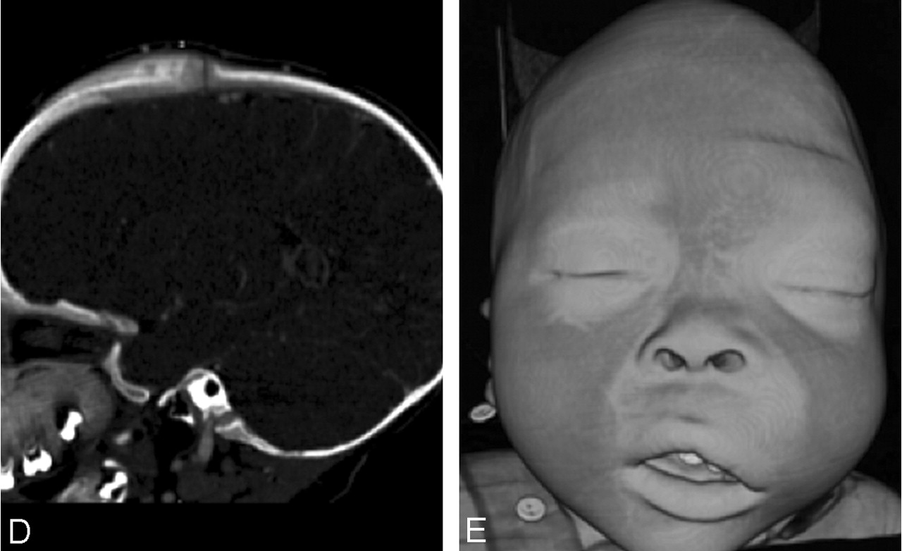
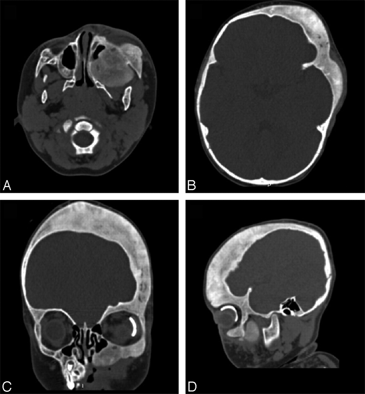
- Fig 1.
Axial (A–C) and reformatted sagittal (D) images from CTA obtained after intravenous injection of 1 mL/kg of Omnipaque 350 showing osseous expansion and periosteal elevation involving the left maxilla, orbital rim, and left frontal-temporal skull. Note a left orbit prosthetic device from previous surgery for congenital glaucoma. Prominent focal meningeal enhancement is noted beneath the left frontal calvaria (C), and there is narrowing of the left coronal suture (D). These changes cause oromaxillofacial asymmetry depicted on the surface-rendered 3D image (E).
- Fig 2.
Surface-rendered 3D images in vascular algorithm (by using a 3D image workstation Vitrea II, Vital Imaging) demonstrate capillary blush in the left vertex crossing the midline, left temporal-parietal skull, periorbital, and maxillary regions corresponding to the known facial port-wine stain (V1 and V2 distribution), as well as sites of osseous hypertrophy.
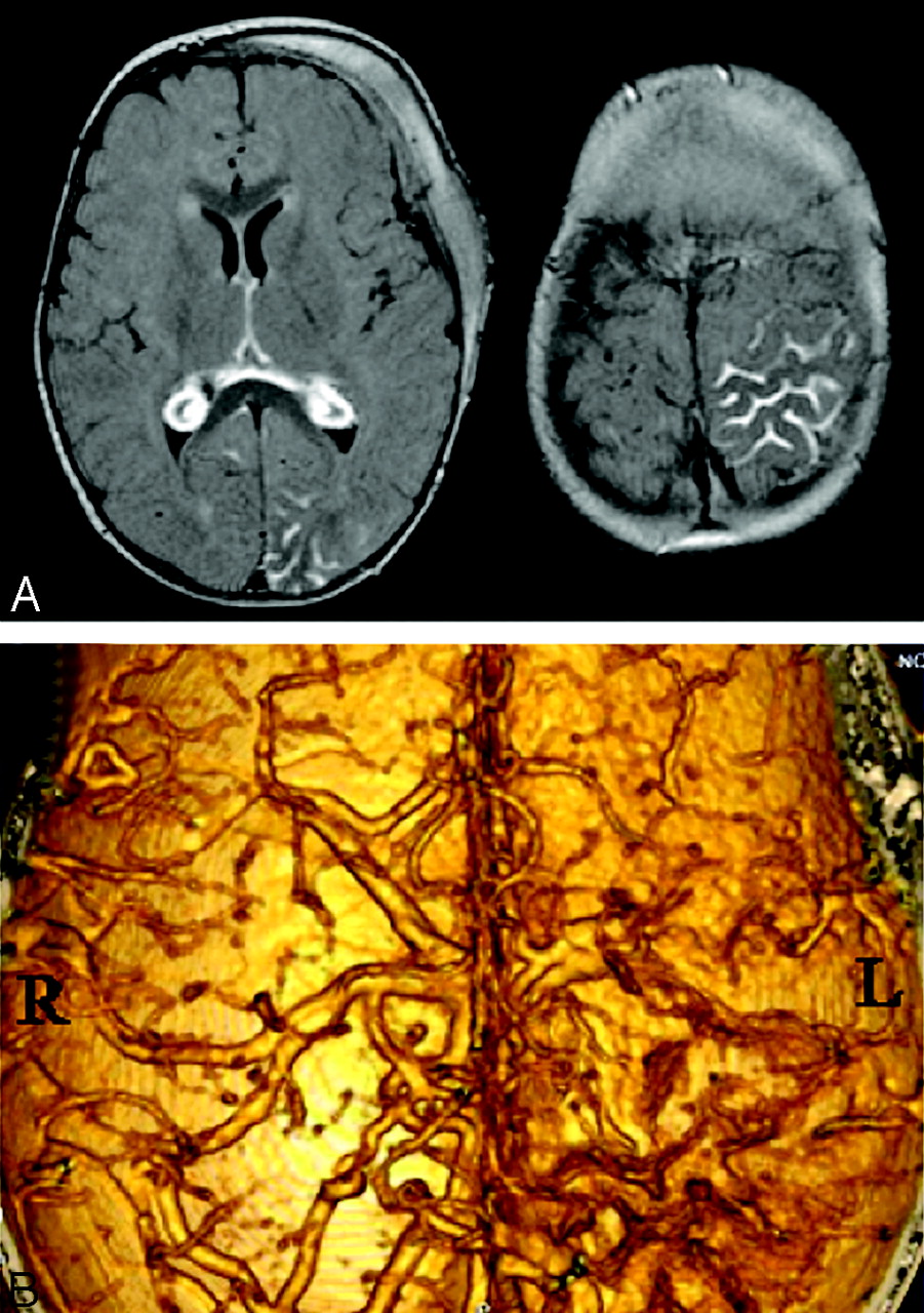
- Fig 3.
A, Axial contrast-enhanced FLAIR (TR/TE/TI, 8802/133/2200) images show leptomeningeal enhancement in the left parietal and occipital regions, and bilateral choroid glomus. B, Surface-rendered 3D image of the interior of the skull depicts dysplastic, irregular vascular structures over the surface of the left cerebrum most prominent in the parietal regions. This is in sharp contrast to a normal appearance of the cortical veins on the right side.
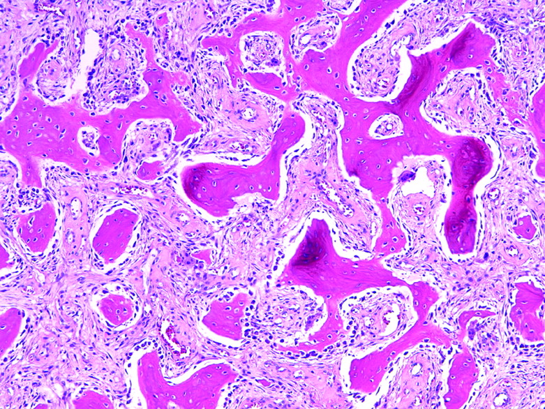
- Fig 4.
Low-powered photomicrograph (H&E ×20) showing woven bone distributed randomly throughout a fibrovascular stroma.
- Fig 5.
Follow-up CTA 8 months after the initial scan and 2 months after surgery shows extended maxillectomy with reconstruction of the left maxillary sinus. There is marked thickening and ground-glass appearance to the remaining left frontal and temporal skull, orbital rim, and maxillary sinus wall. Active periostitis has resolved.


