
Article Figures & Data
Figures
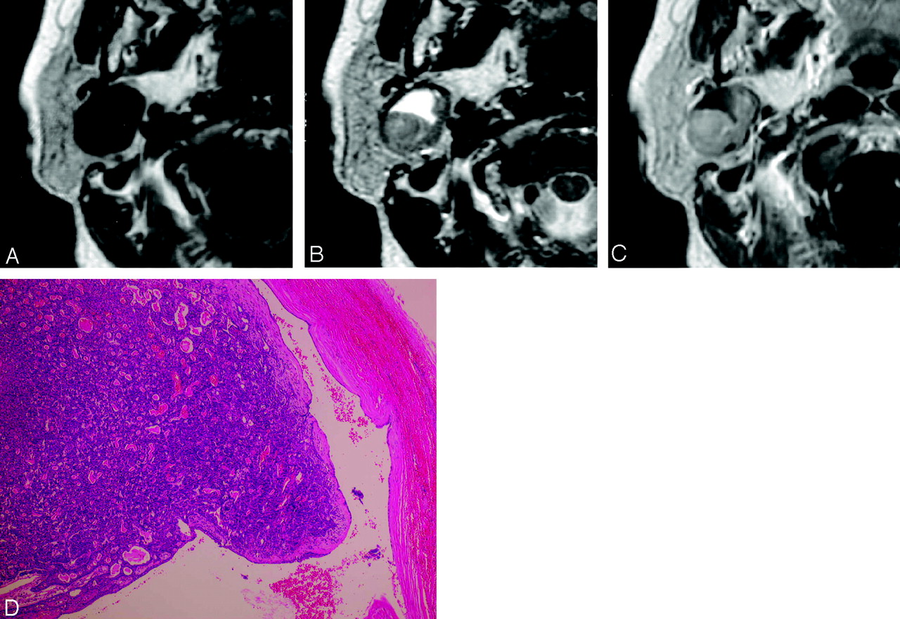
- Fig 1.
A 82-year-old man with basal cell adenoma (BCA) (membranous type, marked enhancement)
A, A T1-weighted image (T1WI) shows a homogeneously low-SI mass with a well-defined, smooth margin in the superficial lobe of the left parotid gland.
B, On T2WI, the tumor shows slightly lower SI than the normal parotid gland.
C, On Gd-enhanced fat-suppressed T1WI, the tumor shows homogeneous and strong enhancement.
D, On dynamic study, the tumor shows a rapid and prolonged enhancement pattern.
E, Microscopic examination (hematoxylin-eosin stain [H&E]) shows conspicuous hyaline matrix surrounding epithelial cells. Relatively large vessels are seen in the tumor (arrows).
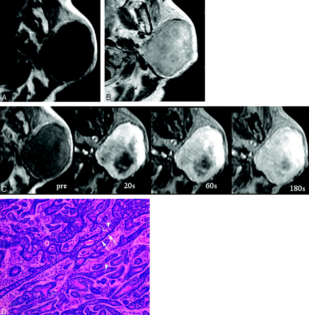
- Fig 2.
A 68-year-old woman with basal cell adenoma (BCA) (solid type, large cystic change)
A, A T1-weighted image (T1WI) shows a homogeneously low-SI mass with well-defined and smooth margin in the deep lobe of the right parotid gland.
B, On T2WI, the tumor shows slightly lower SI than the normal parotid gland with very high SI (cystic change) at the anterior half of the tumor.
C, On Gd-enhanced T1WI, the tumor shows homogeneous moderate enhancement of the solid component. On T2WI, a very-high-SI area is not enhanced.
D, Microscopic examination (hematoxylin-eosin stain [H&E]) shows a solid pattern with many microcystic changes around a large cystic change. Dilated vascular channels are prominent.
- Fig 3.
A 69-year-old woman with basal cell adenoma (BCA) (trabecular type)
A, A T1-weighted image (T1WI) shows a homogeneously low-SI mass with a well-defined, smooth margin in the left parotid gland.
B, On Gd-enhanced T1WI, the tumor shows inhomogeneous moderate enhancement.
C, On dynamic study, the tumor shows rapid enhancement at the periphery of the tumor and is gradually enhanced from the periphery to central part of the tumor.
D, Microscopic examination (hematoxylin-eosin stain [H&E]) shows a trabecular pattern of a BCA. The tumor is composed of small uniform basaloid cells (arrowheads) arranged in solid or trabecular pattern. The stroma is loose, scantily collagenous tissue. There is characteristic palisading (arrows) in the peripheral portion of the tumor cell nests.


Tables
Summary of results
MR findings Size (cm) 1.0–5.8 (avg 2.8) Marginal morphology Margin Well defined, 8 Contour Smooth, 8 Capsule 2 Signal intensity T1WI Homogeneous low signal intensity, 7 T2WI Slightly lower signal intensity than parotid gland, 8 Gd-T1WI (n = 7) Moderate:marked, 6:1 Dynamic study (n = 4) Rapid and prolonged enhancement, 4 Cystic change 4 Pathologic findings Pathologic subtype Solid:trabecular:tubular:membranous, 5:2:0:1
In this issue


Jump to section
Related Articles
Cited By...
- No citing articles found.