
Article Figures & Data
Figures
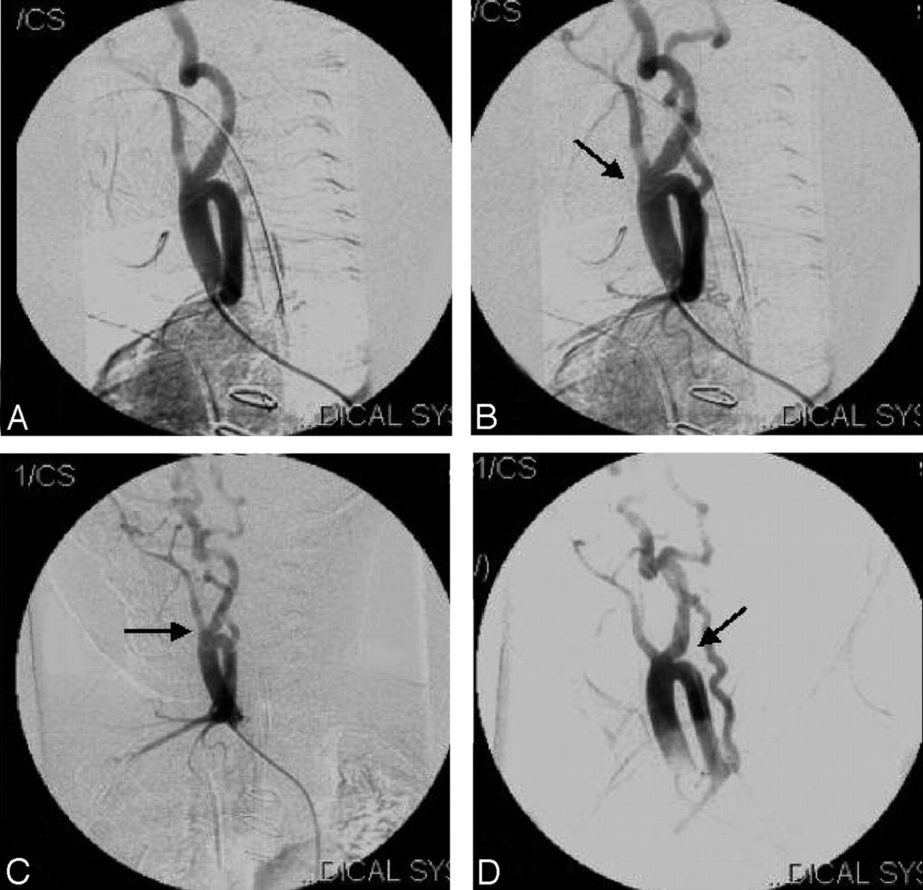
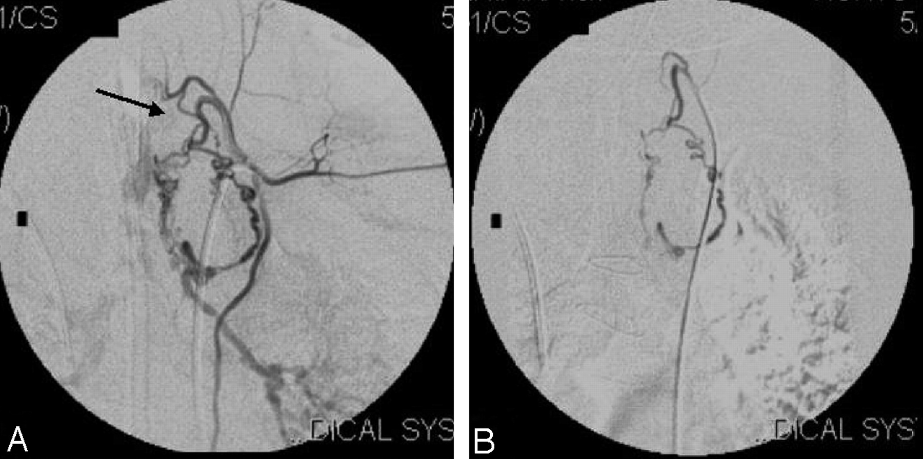
- Fig 1.
A, Arch aortogram shows the origin of brachiocephalic artery and other vessels. B, Little distal injection in the arch shows the separate origins of external carotid artery, internal carotid artery and left subclavian artery. Note the loops at the origin of both the external and internal carotid arteries (thick arrow).
- Fig 2.
A–D, Right brachiocephalic artery injection with early (A) and late arterial phases (B–D) on different angulations show the trifurcation of brachiocephalic artery into right subclavian, right internal and external carotid arteries (thick arrows, B–D). Note the CORSA and the vessel coursing downward and laterally into the arm (B and D).
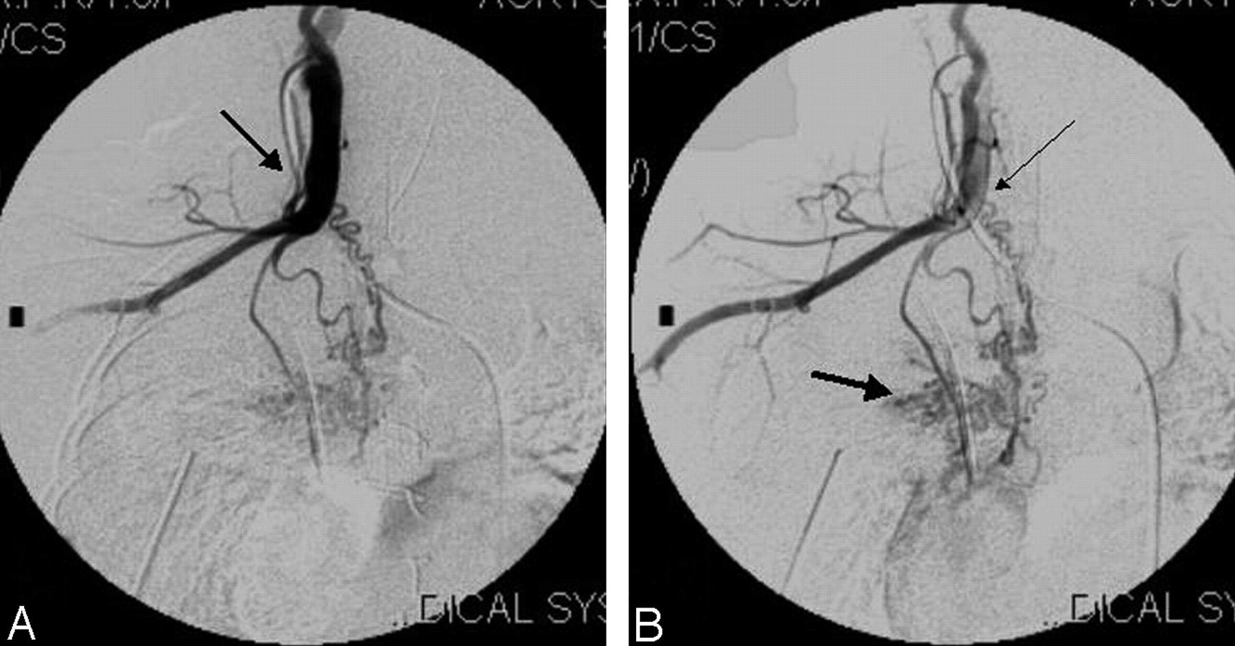
- Fig 3.
A, Selective injection of the right subclavian artery shows its cervical origin and the vessel coursing inferolaterally (arrow) in the neck and then to the right arm.
B, The right thyrocervical artery (thin arrow) is seen to supply major pulmonary collaterals (MAPCA). Note the abnormal blush in lung parenchyma (thick arrow).
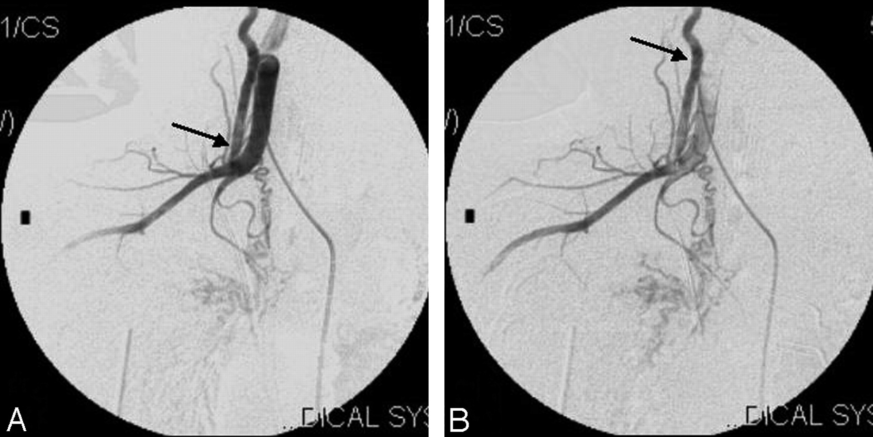
- Fig 4.
Right subclavian artery injection, (A) early and (B) late phases, showing the normal origin of the right vertebral artery (arrows).
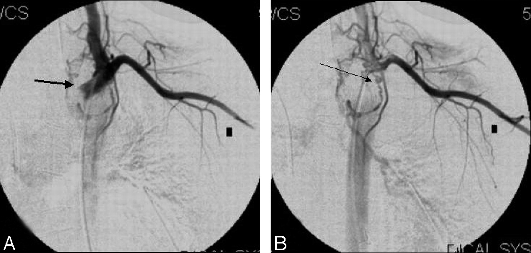
- Fig 5.
A, Injection at the origin of the left subclavian artery shows its normal origin and course to the left arm (arrow).
B, Late phase of the same injection shows the MAPCA from right thyrocervical artery (thin arrow).
- Fig 6.
A, Right bronchial artery injection shows the abnormal blush (arrow).
B, Postembolization angiogram of right bronchial artery (arrow) shows the complete disappearance of the abnormal blush.
- Fig 7.
Pre-embolization (A, arrow) and postembolization (B) angiograms of left thyrocervical trunk supplying the pulmonary circulation.
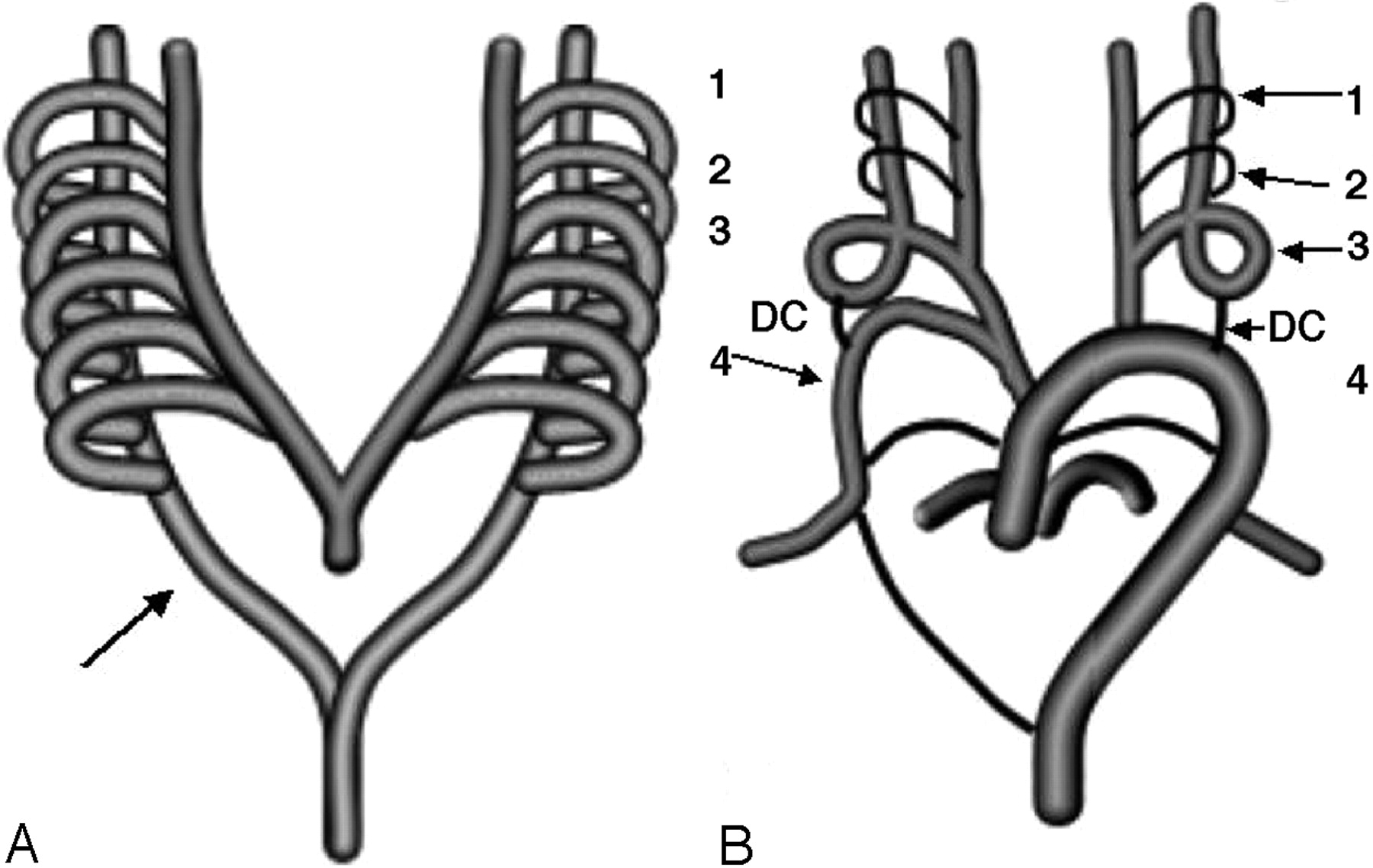
- Fig 8.
A, Normal development of aortic arch. Six aortic arches connecting the ventral to the dorsal aortas are seen (arrow).
B, Normal evolution of the arches: DC, ductus caroticus; 1 to 4, aortic arches (arrows). Fourth arch and the dorsal aorta on the right side form the normal subclavian artery (arrow). Third arch along with parts of the ventral and dorsal aorta form the internal and external carotid arteries. Fifth arches involute, and the sixth form the pulmonary arteries.
- Fig 9.
The right ductus caroticus (arrow), the right seventh intersegmental artery and the right dorsal aorta below the level of the third arch form the right subclavian artery arising from right common carotid artery. Persistence of ductus caroticus with regression of third arch or fourth arch causes the internal and external carotid arteries to arise from the aortic arch on the left side. DC, ductus caroticus; 1 to 4, aortic arches (arrows).


