
Article Figures & Data
Figures
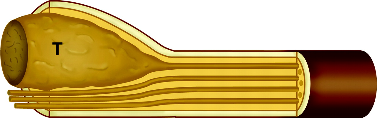
- Fig 1.
Diagram of a schwannoma. The tumor (T) originates within a Schwann cell cylinder that surrounds an axon. The tumor subsequently grows, eccentrically compressing the normal adjacent axons. Schwann cell cylinders surrounding axons (light brown), endoneurium (dark yellow), and perineurium (tan and dark brown).
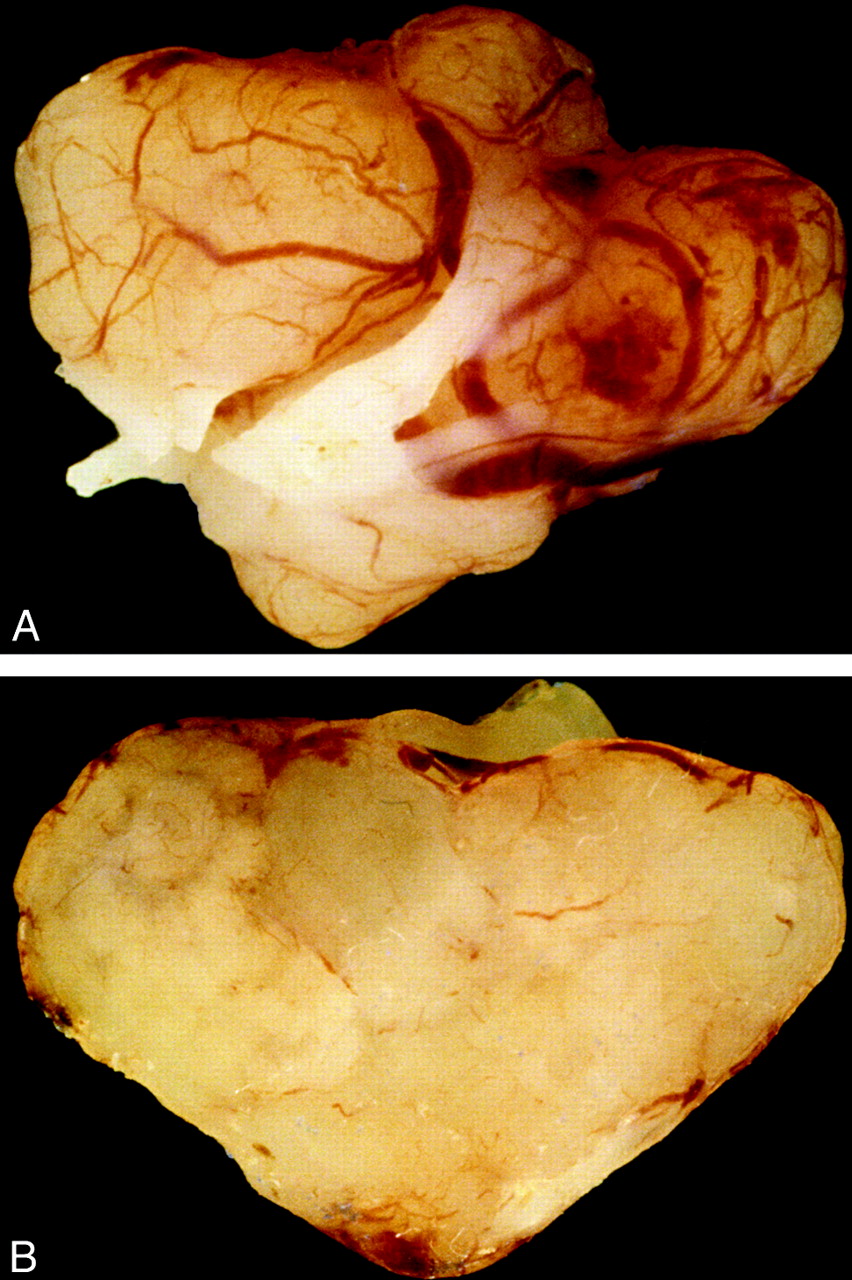
- Fig 2.
Photograph of surgical resection specimen of an intradural schwannoma arising in a 53-year-old woman. A, The yellowish tan gross specimen exhibits a thin capsule. B, Cut section of the tumor reveals a uniform, solid parenchyma. Architecture of schwannomas may vary and also exhibit cystic or nodular consistency.
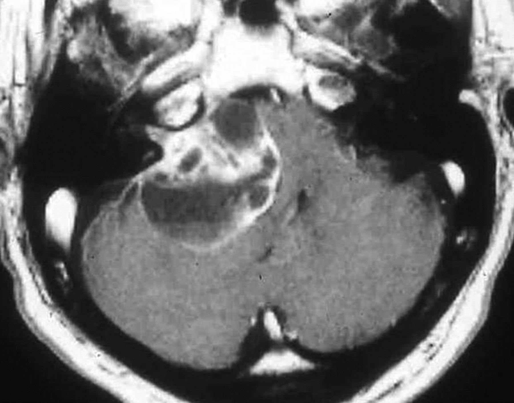
- Fig 3.
Vestibular schwannoma. Enhanced T1-weighted MR image demonstrating a large predominantly cystic schwannoma arising from cranial nerve VIII within the internal auditory canal. The tumor fills the cerebellopontine angle, compresses the pons, and displaces the fourth ventricle. Debris settles within the dependent portion of the tumor.
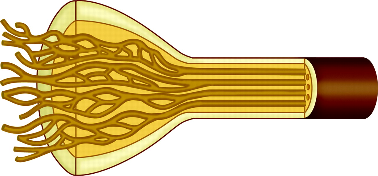
- Fig 4.
Diagram of a neurofibroma. The endoneurium (dark yellow) increases in volume. Schwann cell cylinders (light brown) separate from one another and become dolichoectatic. The mature tumor consists of a complex of Schwann cells, axons, and fibrous material within a myxomatous matrix surrounded by a thickened perineurium (tan and dark brown).
- Fig 5.
Solitary neurofibroma in a 34-year-old woman. A, Enhanced fat suppressed T1-weighted MR axial image at the L4–5 level demonstrates an enhancing tumor arising from the left L4 root. The neuroforamen is expanded. B, Photograph of cut sections of the gross specimen in the same patient shows a solid heterogenous-appearing tumor, which has grown within and expanded multiple fascicles of the nerve.
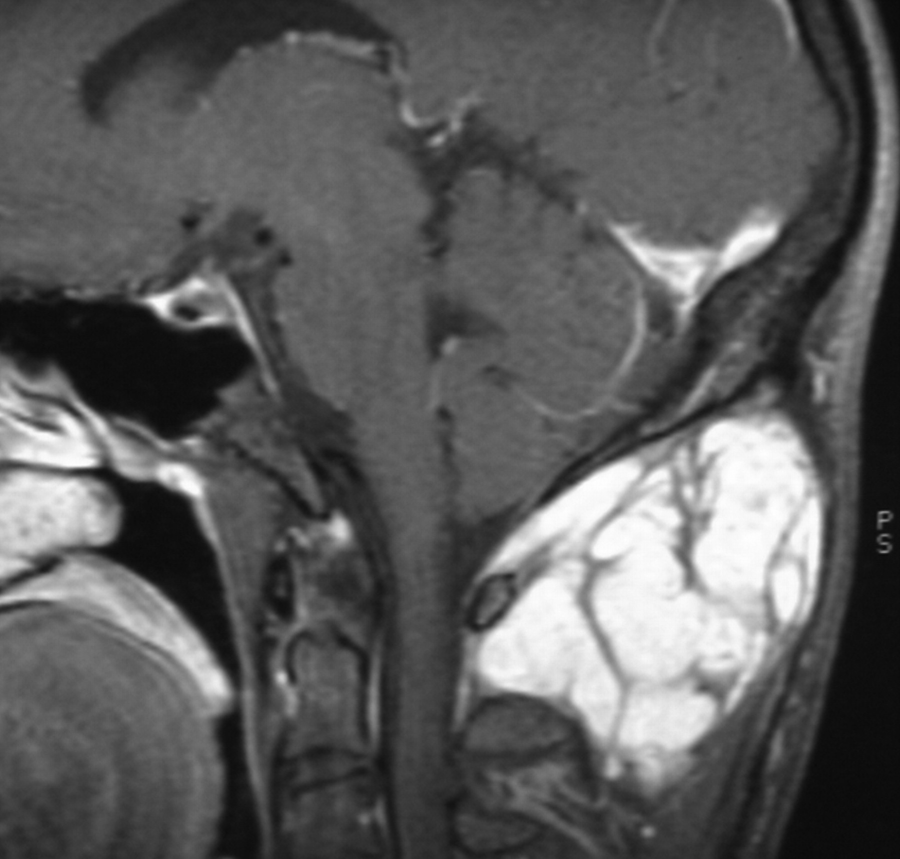
- Fig 6.
Plexiform neurofibroma in a 21-year-old man with no other signs of NF1. Sagittal enhanced T1-weighted MR image demonstrates an enhancing serpentine mass in the pontine soft tissues of the neck.
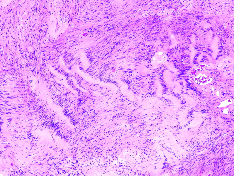
- Fig 7.
Photomicrograph of Antoni A tissue within a schwannoma. Wavy, tightly organized nuclear palisades known as Verocay bodies occupy the center of a highly cellular field (hematoxylin-eosin, original magnification ×400).
- Fig 8.
Photomicrograph of Antoni A tissue and Antoni B tissue within a schwannoma. The highly cellular Antoni A region on the right of the field is contrasted with the loosely organized hypocellular Antoni B region on left of the field (hematoxylin-eosin, original magnification ×400).


In this issue


Jump to section
Related Articles
Cited By...
- Extratemporal intraparotid facial nerve schwannoma
- Cellular mechanisms of heterogeneity in NF2-mutant schwannoma
- Structural Origin and Surgical Complications of Peripheral Schwannomas
- Diffusivity Measurements Differentiate Benign from Malignant Lesions in Patients with Peripheral Neuropathy or Plexopathy