
Article Figures & Data
Figures
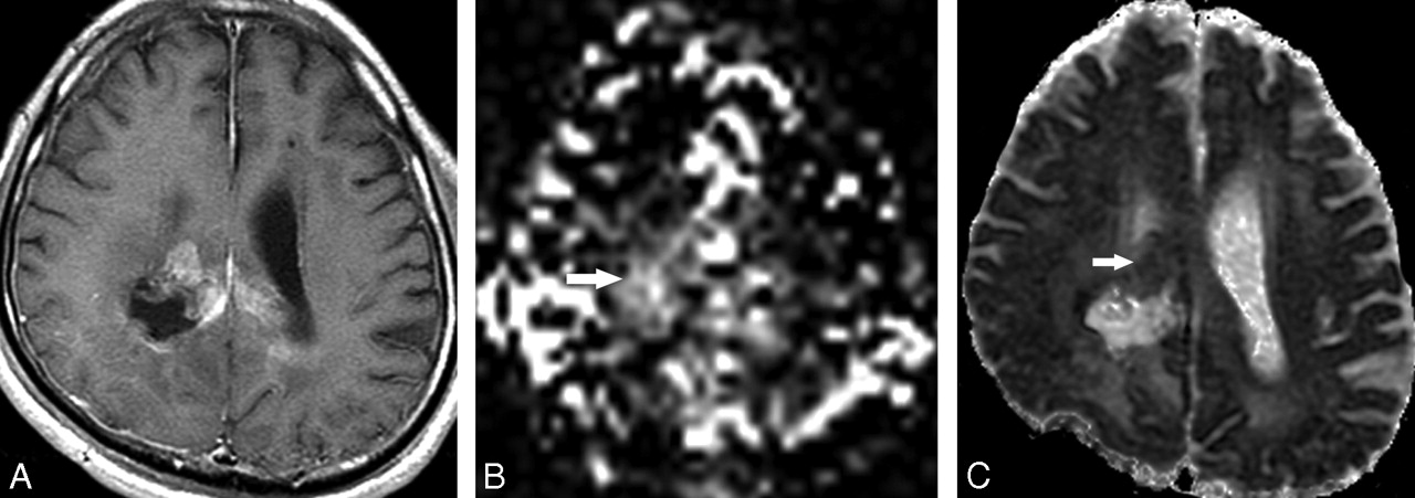
- Fig 1.
A 41-year-old man with a suspected recurred tumor after surgery. There is a contrast-enhancing lesion around the previous tumor resection site on contrast-enhanced T1-weighted axial image (A). Tumor perfusion score (sTP) on pulsed arterial spin-labeling map is 4 (B, arrow). Visual apparent diffusion coefficient score (sADC) is 3 (C, arrow); therefore, the combined sTP and sADC is 7. Initial imaging diagnosis was glioblastoma multiforme (grade 4); however, on pathologic examination this tumor was confirmed as an anaplastic astrocytoma (grade 3).
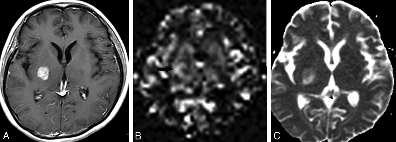
- Fig 2.
A 45-year-old woman with a suspected brain tumor in the right basal ganglia. Contrast-enhanced T1-weighted axial image shows enhancing mass in the right basal ganglia (A). Tumoral perfusion score (sTP) is 3 (B, arrow). The visual apparent diffusion coefficient score (sADC) is 2 (C); therefore, the combined sTP and sADC is 5. On pathologic examination, this tumor was confirmed as an anaplastic astrocytoma (grade 3).
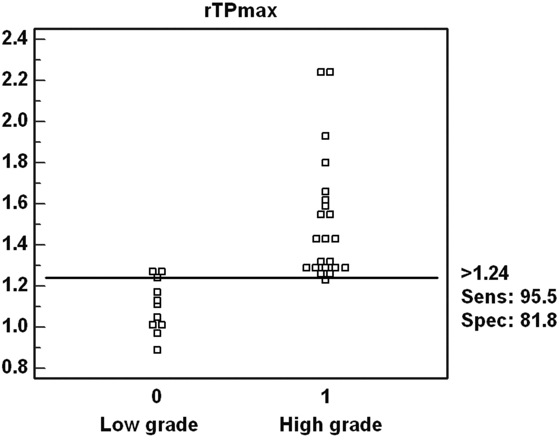
- Fig 3.
Interactive dot diagram. The sensitivity and specificity for the determination of a glioma grade with the ratio of maximum tumoral perfusion signal intensity (rTPmax) were 95.1 and 81.8, with a threshold value of 1.24.
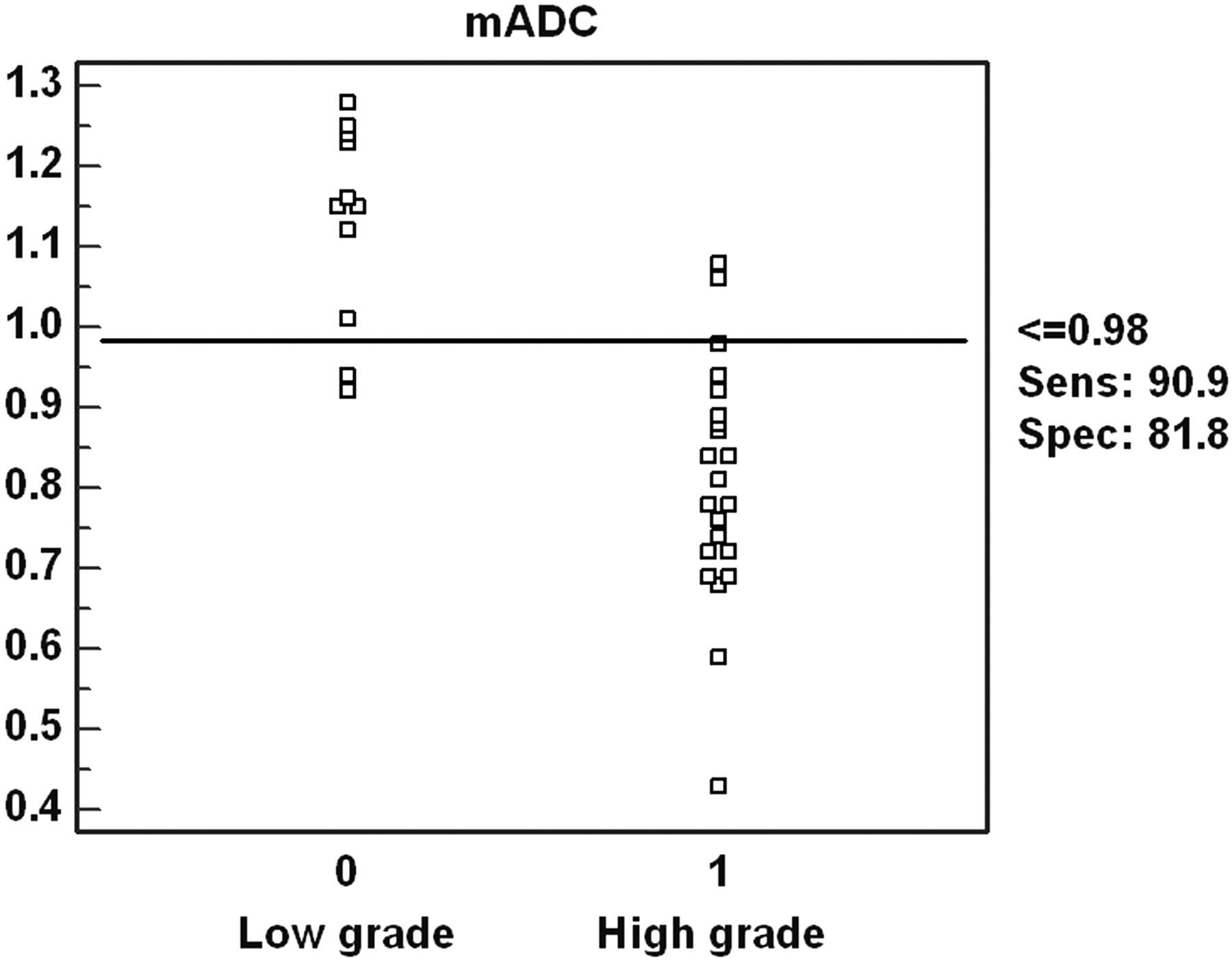
- Fig 4.
Interactive dot diagram. The sensitivity and specificity for the determination of a glioma grade with a minimum apparent diffusion coefficient value (mADC) were 90.9 and 81.8, with a threshold value of 0.98 × 10⁻³ mm/s2.
- Fig 5.
The ROC curve of 2 quantitative (rTPmax, mADC) and 3 qualitative (sTP, sADC, sTP and sADC) parameters for glioma grading. The areas under the ROC curve for the 5 parameters are as follows: rTPmax, 0.97; sTP and sADC, 0.96; mADC, 0.93; sTP, 0.90; and sADC, 0.80.


Tables
Patient No./Age (y)/Sex Pathologic Diagnosis (Grade of Gliomas, 1–4) Diagnostic Method rTPmax mADC ×10−3 mm/s2 1/41/M 2 Biopsy 1.01 0.92 2/51/M 4 Surgery 1.55 0.89 3/29/M 2 Surgery 1.05 1.23 4/30/F 2 Biopsy 1.27 1.16 5/42/F 4 Surgery 1.59 0.71 6/41/M 3 Surgery 1.29 0.98 7/45/M 4 Biopsy 1.62 0.43 8/43/F 3 Surgery 1.32 0.68 9/30/F 2 Surgery 0.97 0.94 10/32/M 2 Surgery 1.13 1.24 11/35/F 4 Surgery 2.25 0.92 12/41/F 3 Surgery 1.23 0.78 13/42/M 4 Biopsy 1.93 0.72 14/40/M 3 Surgery 1.29 0.84 15/32/M 2 Surgery 1.28 1.45 16/46/F 4 Surgery 1.80 1.06 17/39/F 4 Surgery 2.24 0.88 18/52/M 3 Biopsy 1.29 0.87 19/42/M 4 Surgery 1.43 0.74 20/35/F 2 Surgery 0.89 1.22 21/39/M 4 Surgery 1.56 0.76 22/30/F 3 Surgery 1.26 0.78 23/40/F 4 Surgery 1.43 0.59 24/42/M 2 Surgery 1.24 1.15 25/52/M 4 Biopsy 1.66 0.88 26/45/F 3 Biopsy 1.26 0.69 27/42/M 4 Surgery 1.29 0.69 28/37/F 2 Surgery 1.11 1.28 29/50/M 2 Surgery 1.01 1.01 30/51/M 4 Biopsy 1.44 0.72 31/29/M 2 Surgery 1.17 1.15 32/51/F 4 Surgery 1.30 0.84 33/67/M 4 Surgery 1.32 0.94 Note:—rTPmax indicates ratio of maximum tumor perfusion signal intensity; mADC, minimum apparent diffusion coefficient value.
- Table 2:
Sensitivity, specificity, PPV, and NPV for conventional imaging and the five qualitative and quantitative methods of PASL and ADC for distinguishing high-grade from low-grade gliomas
CI rTPmax mADC sTP sADC sTP and sADC Sensitivity (%) 77.3 95.5 90.9 86.4 86.4 90.9 Specificity (%) 72.7 81.8 81.8 90.9 72.7 90.9 PPV (%) 85.0 91.3 90.9 95.0 86.4 95.2 NPV (%) 66.7 90.1 81.8 76.9 72.7 83.3 Note:—CI indicates conventional image; rTPmax, ratio of maximum tumor perfusion signal intensity; mADC, minimum ADC value; sTP, qualitative scoring of tumor perfusion signal intensity; sADC, qualitative scoring of ADC value; PPV, positive predictive value; NPV, negative predictive value.
In this issue


Jump to section
Related Articles
Cited By...
- Arterial Spin-Labeling in Children with Brain Tumor: A Meta-Analysis
- 3D Pseudocontinuous Arterial Spin-Labeling MR Imaging in the Preoperative Evaluation of Gliomas
- MR Imaging-Based Analysis of Glioblastoma Multiforme: Estimation of IDH1 Mutation Status
- Arterial Spin-Labeling Perfusion MRI Stratifies Progression-Free Survival and Correlates with Epidermal Growth Factor Receptor Status in Glioblastoma
- MRI Grading versus Histology: Predicting Survival of World Health Organization Grade II-IV Astrocytomas
- Comparison of Multiple Parameters Obtained on 3T Pulsed Arterial Spin-Labeling, Diffusion Tensor Imaging, and MRS and the Ki-67 Labeling Index in Evaluating Glioma Grading
- The Added Value of Apparent Diffusion Coefficient to Cerebral Blood Volume in the Preoperative Grading of Diffuse Gliomas
- Does MR Perfusion Imaging Impact Management Decisions for Patients with Brain Tumors? A Prospective Study
- Quantitative Blood Flow Measurements in Gliomas Using Arterial Spin-Labeling at 3T: Intermodality Agreement and Inter- and Intraobserver Reproducibility Study
- Isolated Diffusion Restriction Precedes the Development of Enhancing Tumor in a Subset of Patients with Glioblastoma
- Apparent Diffusion Coefficient of Glial Neoplasms: Correlation with Fluorodeoxyglucose-Positron-Emission Tomography and Gadolinium-Enhanced MR Imaging