
Article Figures & Data
Figures
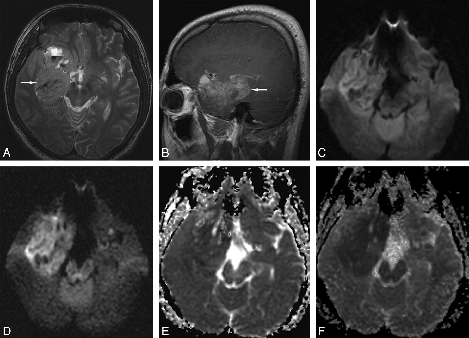
- Fig 1.
Grade IV glioblastoma in a 27-year-old man. A, Transverse T2-weighted image shows a slightly hyperintense main mass (arrow) in the right medial temporal lobe. B, Sagittal contrast-enhanced T1-weighted image shows diffuse tumor enhancement (arrow). C, Transverse DWI at b = 1000 s/mm2 shows slight tumor hyperintensity with some hypointense foci. D, Transverse DWI at b = 3000 s/mm2 shows more conspicuous main mass hyperintensity. E, Transverse ADC map at b = 1000 s/mm2 shows subtle or slight tumor hypointensity. F, Transverse ADC map at b = 3000 s/mm2 shows more conspicuous tumor hypointensity.
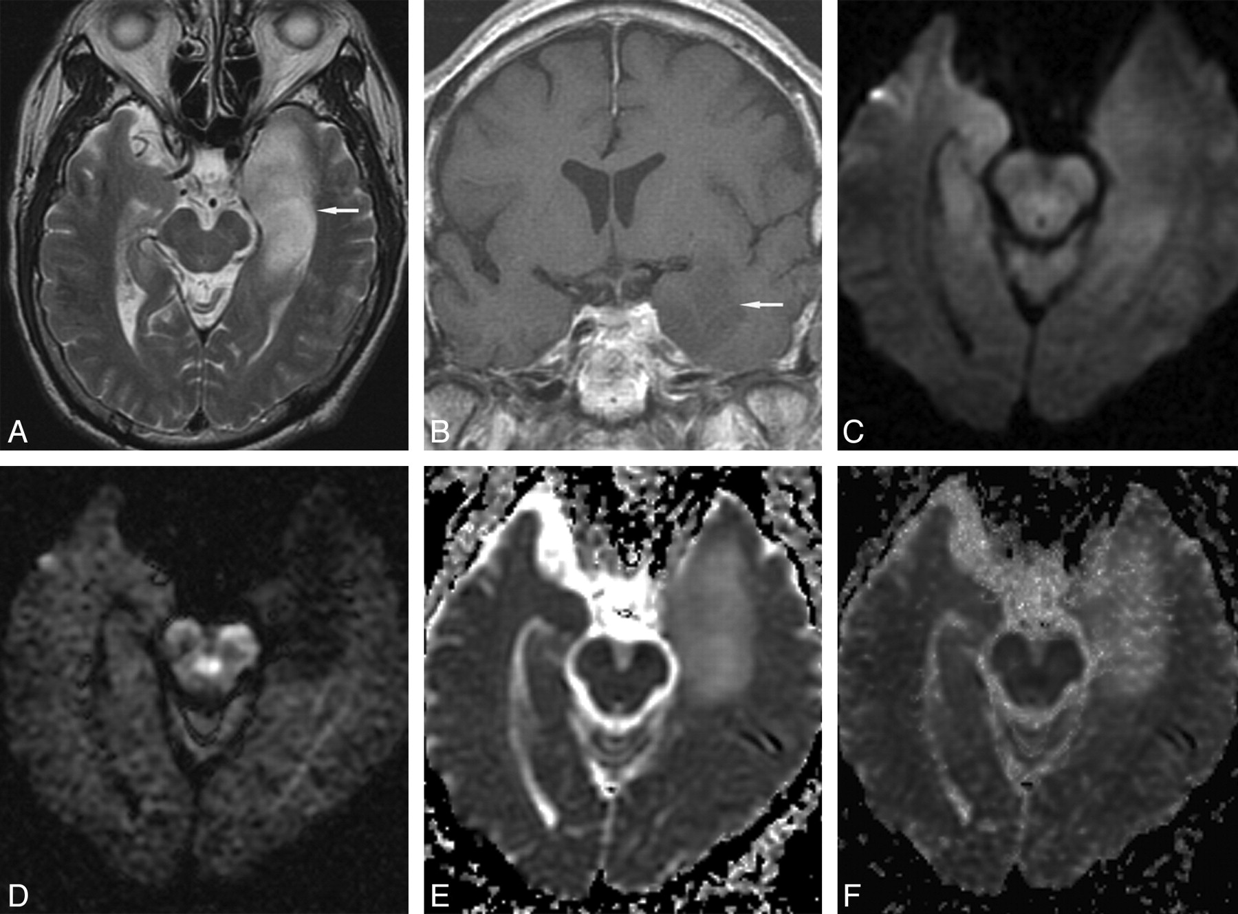
- Fig 2.
Grade II diffuse astrocytoma in a 66-year-old man. A, Transverse T2-weighted image shows a hyperintense mass (arrow) in the left medial temporal lobe. B, Coronal contrast-enhanced T1-weighted image shows slight tumor hypointensity without enhancement (arrow). C, Transverse DWI at b = 1000 s/mm2 shows tumor isointensity. D, Transverse DWI at b = 3000 s/mm2 shows marked tumor hypointensity. Transverse ADC maps at b = 1000 s/mm2 (E) and b = 3000 s/mm2 (F) show tumor hyperintensity.
- Fig 3.
ROC curves derived from 5-point scale visual assessment scores at b = 1000 and 3000 s/mm2. The Az value of DWI at b = 3000 s/mm2 was higher than at b = 1000 s/mm2 but this was not statistically significant (0.864 vs 0.790; P =.08).
- Fig 4.
Box plots of DWI SI ratios (A) in high-grade and low-grade gliomas, and ADC values (B) of high-grade and low-grade gliomas and normal subcortical white matter at b = 1000 and 3000 s/mm2. The horizontal line is the median, and the upper and lower ends of the boxes are the upper and lower quartiles, respectively. The vertical lines represent data ranges. A, The mean SI ratio of high-grade gliomas was significantly higher than that of low-grade gliomas at both b-values (P < .01). Note that the difference between the mean SI ratios of high-grade and low-grade gliomas at b = 3000 s/mm2 was significantly greater than at b = 1000 s/mm2 (P < .05). B, The mean ADC value of high-grade gliomas was significantly lower than that of low-grade gliomas at both b-values (P < .01). The mean ADC values of high-grade and low-grade gliomas and normal subcortical white matter at b = 3000 s/mm2 were significantly lower than at b = 1000 s/mm2, respectively (P < .01).


Tables
Tumor Type No. of Patients Age (y) Gender Mean Range Male Female High-grade glioma WHO grade III 20 44.7 23–72 14 6 Anaplastic astrocytoma 10 42.9 23–63 6 4 Anaplastic oligodendroglioma 5 45.2 29–63 4 1 Anaplastic oligoastrocytoma 3 42.7 37–53 2 1 Gliomatosis cerebri 2 55 38–72 2 0 WHO grade IV 29 50.9 6–77 18 11 Glioblastoma 28 50.9 6–77 17 11 Gliosarcoma 1 52 52 1 0 Low-grade glioma WHO grade II 13 40.9 29–65 10 3 Oligodendroglioma 7 42.9 30–65 6 1 Diffuse astrocytoma 6 38.5 29–53 4 2 Note:—WHO indicates World Health Organization.
- Table 2:
Mean scores and ranges of DWI signal intensities (SIs) in cerebral gliomas according to the five-point visual assessment scale used in this study
b = 1000 s/mm2 b = 3000 s/mm2 Mean ± SD Range Mean ± SD Range High-grade glioma (n = 49) 4.7 ± 0.5 4–5 4.6 ± 0.8 2–5 WHO grade III (n = 20) 4.7 ± 0.5 4–5 4.4 ± 1.0 2–5 WHO grade IV (n = 29) 4.7 ± 0.4 4–5 4.7 ± 0.7 3–5 Low-grade (II) glioma (n = 13) 3.7 ± 1.0 2–5 2.7 ± 1.3 1–5 Note:—WHO indicates World Health Organization; SD, standard deviation.
DWI Az* Cutoff Score† Sensitivity (%) Specificity (%) PPV‡ (%) NPV‡ (%) κ-value§ b = 1000 s/mm2 0.790 5 69.4 76.9 94.4 40.0 0.56 b = 3000 s/mm2 0.864 4 83.7 84.6 95.3 57.9 0.77 Note:—ROC indicates receiver operating characteristic; DWI, diffusion-weighted imaging.
* Az refers to area under the ROC curve; these were not significantly different with the 2 b-values (P = .08).
† Cutoff score refers to the optimal cutoff level for high-grade glioma with highest accuracy for obtaining a positive test for high-grade glioma with a minimum C1 error by ROC analysis, where C1 = 1 − (sensitivity+specificity)/2.
‡ PPV and NPV are positive and negative predictive values, respectively, for high-grade glioma.
§ κ-value is the kappa value for interobserver agreement.
- Table 4:
Comparison of mean DWI signal intensity (SI) ratios and standard deviations between b = 1000 s/mm2 and b = 3000 s/mm2 in high-grade and low-grade gliomas
SI Ratio b = 1000 s/mm2 SI Ratio b = 3000 s/mm2 High-grade glioma (n = 49) 1.96 ± 0.57 2.13 ± 1.05 WHO grade III (n = 20) 1.91 ± 0.56 2.12 ± 1.17 WHO grade IV (n = 29) 1.99 ± 0.59 2.14 ± 0.97 Low-grade (II) glioma (n = 13) 1.39 ± 0.43 0.83 ± 0.50 Note:—WHO indicates World Health Organization.
- Table 5:
Comparison of mean ADC values between b = 1000 s/mm2 (ADC1) and b = 3000 s/mm2 (ADC3) in high-grade and low-grade gliomas and normal subcortical white matter
ADC1(μm2/s) ADC3(μm2/s) High-grade glioma (n = 49) 914.6 ± 278.9 597.6 ± 195.9 WHO grade III (n = 20) 970.8 ± 333.6 627.3 ± 248.2 WHO grade IV (n = 29) 875.9 ± 232.4 577.2 ± 151.3 Low-grade (II) glioma (n = 13) 1379.2 ± 428.2 910.8 ± 227.1 Normal subcortical WM (n = 62) 779.1 ± 70.7 625.0 ± 68.9 Note:—WM indicates white matter; ADC, apparent diffusion coefficient.
In this issue


Jump to section
Related Articles
Cited By...
- Differentiation of Recurrent Tumor and Posttreatment Changes in Head and Neck Squamous Cell Carcinoma: Application of High b-Value Diffusion-Weighted Imaging
- Apparent Diffusion Coefficient with Higher b-Value Correlates Better with Viable Cell Count Quantified from the Cavity of Brain Abscess
- High-b-Value Diffusion MR Imaging and Basal Nuclei Apparent Diffusion Coefficient Measurements in Variant and Sporadic Creutzfeldt-Jakob Disease