Advertisement

Main menu

  • Home
  • Content
    • Current Issue
    • Accepted Manuscripts
    • Article Preview
    • Past Issue Archive
    • AJNR Case Collection
    • Case of the Week Archive
    • Classic Case Archive
    • Case of the Month Archive
  • Special Collections
    • Spinal CSF Leak Articles (Jan 2020-June 2024)
    • 2024 AJNR Journal Awards
    • Most Impactful AJNR Articles
  • Multimedia
    • AJNR Podcast
    • AJNR Scantastics
    • Video Articles
  • For Authors
    • Submit a Manuscript
    • Author Policies
    • Fast publishing of Accepted Manuscripts
    • Graphical Abstract Preparation
    • Manuscript Submission Guidelines
    • Imaging Protocol Submission
    • Submit a Case for the Case Collection
  • About Us
    • About AJNR
    • Editorial Board
  • More
    • Become a Reviewer/Academy of Reviewers
    • Subscribers
    • Permissions
    • Alerts
    • Feedback
    • Advertisers
    • ASNR Home
  • Other Publications
    • ajnr

User menu

  • Alerts
  • Log in

Search

  • Advanced search
American Journal of Neuroradiology

American Journal of Neuroradiology

ASHNR American Society of Functional Neuroradiology ASHNR American Society of Pediatric Neuroradiology ASSR
  • Alerts
  • Log in

Advanced Search

  • Home
  • Content
    • Current Issue
    • Accepted Manuscripts
    • Article Preview
    • Past Issue Archive
    • AJNR Case Collection
    • Case of the Week Archive
    • Classic Case Archive
    • Case of the Month Archive
  • Special Collections
    • Spinal CSF Leak Articles (Jan 2020-June 2024)
    • 2024 AJNR Journal Awards
    • Most Impactful AJNR Articles
  • Multimedia
    • AJNR Podcast
    • AJNR Scantastics
    • Video Articles
  • For Authors
    • Submit a Manuscript
    • Author Policies
    • Fast publishing of Accepted Manuscripts
    • Graphical Abstract Preparation
    • Manuscript Submission Guidelines
    • Imaging Protocol Submission
    • Submit a Case for the Case Collection
  • About Us
    • About AJNR
    • Editorial Board
  • More
    • Become a Reviewer/Academy of Reviewers
    • Subscribers
    • Permissions
    • Alerts
    • Feedback
    • Advertisers
    • ASNR Home
  • Follow AJNR on Twitter
  • Visit AJNR on Facebook
  • Follow AJNR on Instagram
  • Join AJNR on LinkedIn
  • RSS Feeds

Welcome to the new AJNR, Updated Hall of Fame, and more. Read the full announcements.


AJNR is seeking candidates for the position of Associate Section Editor, AJNR Case Collection. Read the full announcement.


OtherPEDIATRICS

Imaging Findings of Congenital Glaucoma in Opitz Syndrome

A.K. Choudhary and B. Jha
American Journal of Neuroradiology May 2008, 29 (5) 1003-1005; DOI: https://doi.org/10.3174/ajnr.A1020
A.K. Choudhary
  • Find this author on Google Scholar
  • Find this author on PubMed
  • Search for this author on this site
B. Jha
  • Find this author on Google Scholar
  • Find this author on PubMed
  • Search for this author on this site
  • Article
  • Figures & Data
  • Info & Metrics
  • Responses
  • References
  • PDF
Loading

Abstract

SUMMARY: Opitz syndrome is a rare autosomal recessive disorder of cholesterol metabolism associated with mental retardation and multiple congenital malformations. It is also uncommonly associated with congenital glaucoma. We describe the orbital findings on CT in this rare case of a patient with Opitz syndrome who presented with congenital glaucoma, with a review of the literature. The CT findings of congenital glaucoma, which have not been described before in the literature, are also discussed. It is important for the radiologist to be aware of this rare association. It is also important to be aware of the findings of congenital glaucoma on CT because patients with Opitz syndrome and other syndromes associated with learning difficulties may not present with typical clinical features of glaucoma. A high index of suspicion will lead to a correct diagnosis and earlier intervention.

Opitz syndrome is a rare autosomal recessive disorder caused by a defect of cholesterol metabolism. It is associated with mental retardation and multiple congenital malformations. Multiple midline neurologic abnormalities have been described in the literature. There have been rare reports of congenital glaucoma in association with Opitz syndrome. To the best of our knowledge, radiologic findings on CT of congenital glaucoma have not been described before in the literature. We describe the association as well as radiologic findings on CT of congenital glaucoma.

Case Report

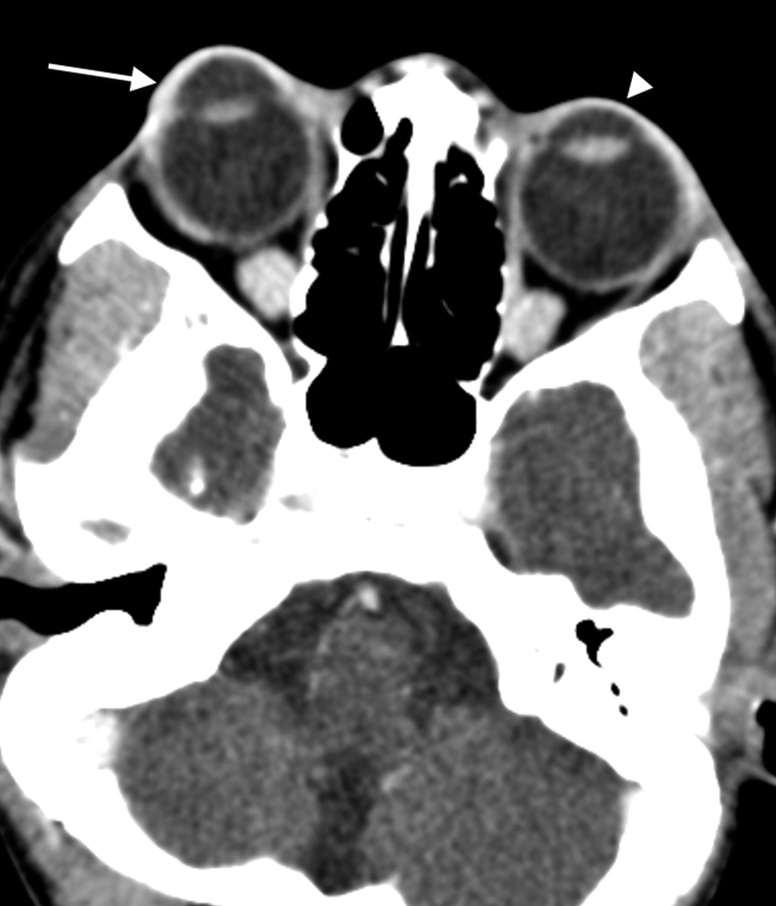
A 12-year-old female patient with Opitz syndrome came to the emergency department with a history of increasing prominence of right eye. She had profound mental retardation; cleft palate; nasal, forehead, finger, and foot deformities; and micrognathia. On previous barium swallow study, her epiglottis was described as short and stubby. A CT study was obtained to exclude a retro-orbital mass and showed a markedly enlarged anterior chamber of the right eye, which measured 6.7 mm in the anteroposterior dimension compared with 4.0 mm on the left side, which is also enlarged (Figs 1 and 2). Trigonocephaly was noted (Fig 3). Bilateral optic nerve atrophy (Fig 4) was also noted. On ophthalmologic examination, she was found to have large cornea with a pressure of more than 40 bilaterally, worse on the right side. She subsequently was given a topical beta-blocker, systemic carbonic anhydrase inhibitor, and brimonidine. One week later, she had trabeculotomy and trabeculectomy of her right eye and, a few weeks later, trabeculotomy and trabeculectomy of her left eye.

Fig 1.
  • Download figure
  • Open in new tab
  • Download powerpoint
Fig 1.

Axial CT image through the orbits after administration of intravenous (IV) contrast medium, which demonstrates a markedly enlarged anterior chamber of the right globe (arrow) and mildly enlarged anterior chamber of the left globe (arrowhead).

Fig 2.
  • Download figure
  • Open in new tab
  • Download powerpoint
Fig 2.

Sagittal reconstructed CT images through the orbit after IV contrast medium, which demonstrates a markedly enlarged anterior chamber of the right globe (arrow) and a mildly enlarged anterior chamber of the left globe (arrowhead).

Fig 3.
  • Download figure
  • Open in new tab
  • Download powerpoint
Fig 3.

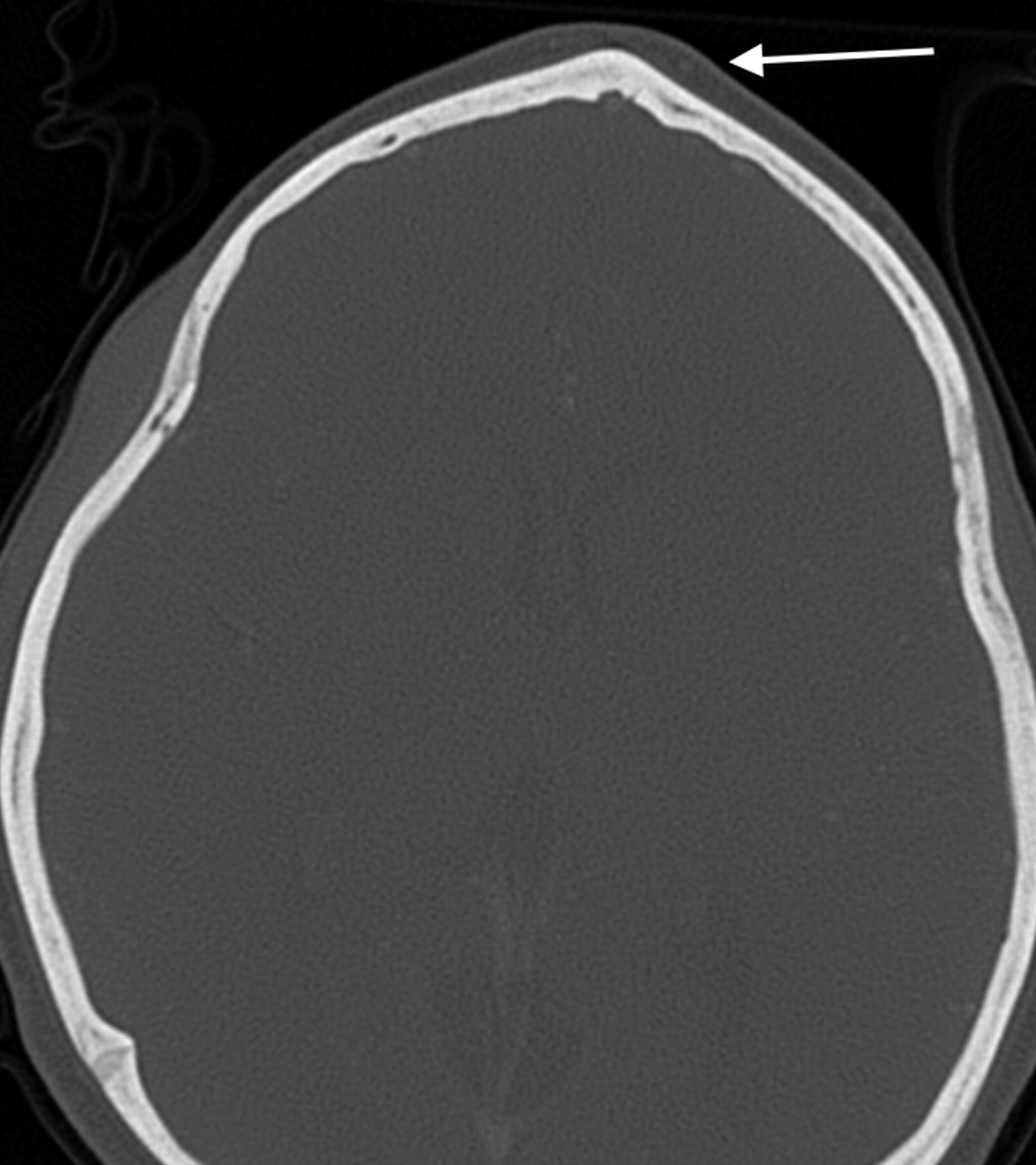
Axial CT image through the frontal bone in bony windows demonstrating trigonocephaly (arrow).

Fig 4.
  • Download figure
  • Open in new tab
  • Download powerpoint
Fig 4.

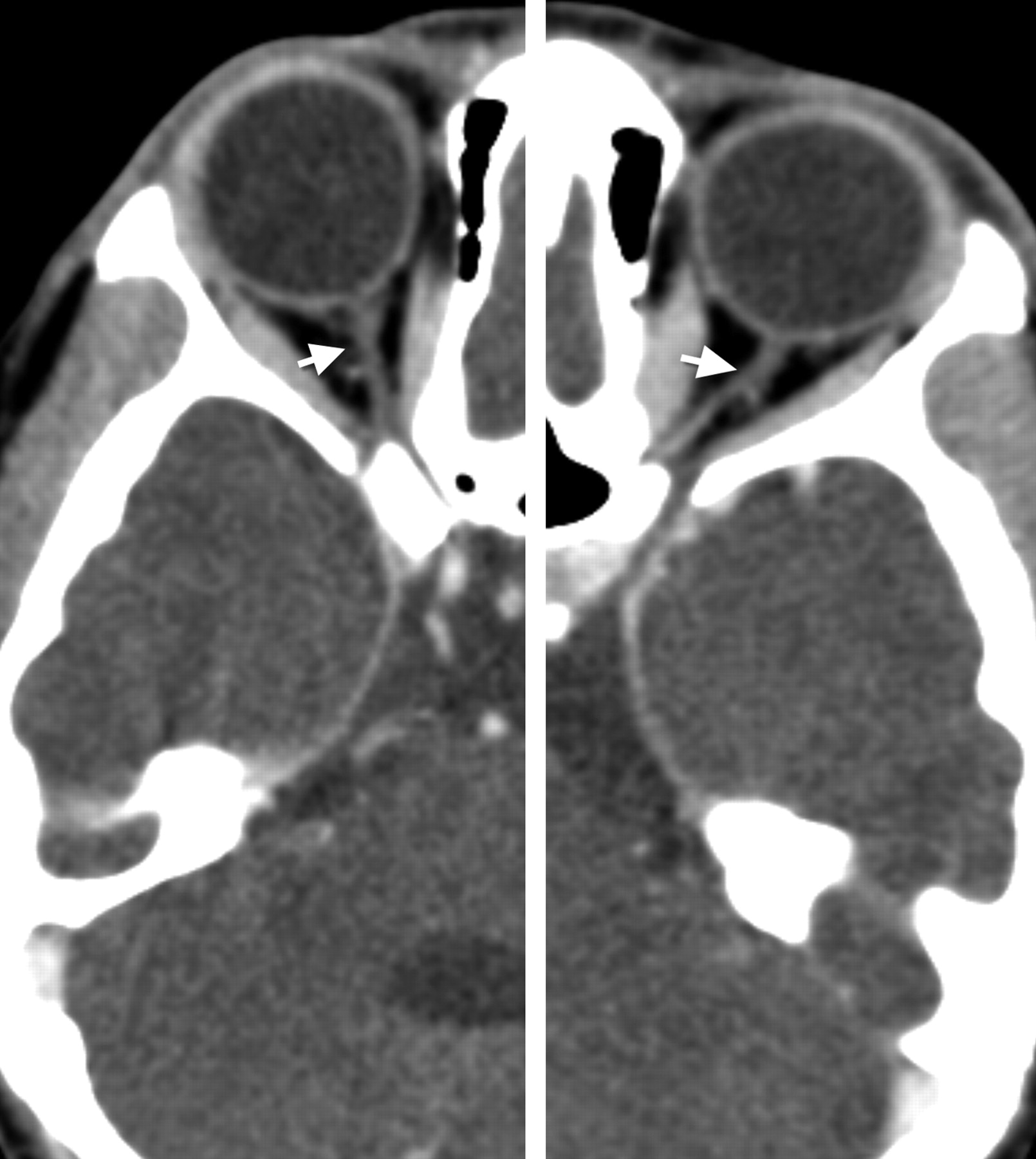
Axial CT images through the optic nerve after IV contrast medium showing bilateral optic nerve atrophy (arrows).

Discussion

Opitz syndrome is a rare autosomal disorder caused by a defect in cholesterol metabolism. It is a heterogeneous disorder ranging from lethality to minor abnormalities. The abnormalities involve multiple systems including mental retardation; structural brain abnormalities; craniofacial abnormalities; and abnormalities of the heart, bowel, liver, lung, extremities, and genitalia. The ophthalmologic findings described have included blepharoptosis, congenital cataracts, optic atrophy, nystagmus, blepharoptosis, epicanthal folds, convergent strabismus, downslanting small palpebral fissures, mild exophthalmos, retinal pigment epithelial defects, pale disks, nystagmus, microcornea, aniridia, congenital cataracts, postlenticular membrane, Duane retraction syndrome, absence of lacrimal punctaopsoclonus-like eye movements, lack of visual-following responses and opticokinetic reflexes, corneal enlargement, abnormal iris insertion, bilateral optic pits, sclerocornea, and optic nerve demyelination.1 Congenital glaucoma is uncommonly seen with Opitz syndrome.2,3 Glaucoma can also be associated with other congenital disorders including Sturge-Weber syndrome, neurofibromatosis, Lowe syndrome, Pierre Robin syndrome/sequence, Marfan syndrome, homocystinuria, aniridia, Axenfeld anomaly, and Reiger syndrome.4 Secondary causes of infantile glaucoma causing similar appearance include maternal rubella, syphilis, retinopathy of prematurity, infantile xanthogranuloma, trauma, and retinoblastoma.5

The dimensions of the globe are relatively constant, varying within a millimeter or so with the average sagittal diameter measuring approximately 24.15 mm (outer) and 22.12 mm (inner). In boys, the diameter is approximately 0.5 mm more than that in girls. At birth, the sagittal diameter of the eyes varies from 16 to 17 mm and then increases to an average value of 22.5 to 23 mm at the age of 3 years. It further increases by approximately 1 mm between the ages of 3 and 13 years and then stays stable. The overall incidence of congenital glaucoma is 1:5000 to 1:10,000 live births. It is more common in boys (60%–70%) and is bilateral in 64% of 88% of patients.4 In congenital glaucoma, developmental abnormalities cause obstruction to the flow of the aqueous humor from the anterior chamber, resulting in increased intraocular pressure. The different theories to explain obstruction include the Barkan membrane covering the trabecular meshwork, congenital absence of the Schlemm canal, and an embryonic anterior chamber angle.4 In young children, this results in enlargement, stretching, and thinning of the coats of the eye, as the eyes are of sufficient plasticity. This is especially marked in the anterior chamber, resulting in a deep anterior chamber. The appearance is said to resemble the eyes of an ox (buphthalmos). In the adult, a similar degree of raised pressure will only cause stretching in the region of the lamina cribrosa.5 The normal mean anterior chamber depth is approximately 3 mm, which varies with the measuring method and the age and sex of the patient.6,7 In congenital glaucoma, the mean anterior chamber depth is 6.3 mm.5 In our patient, the sagittal diameter of the globe on the right side measured 27 mm, with the anterior chamber measuring 6.7 mm. Therefore, it has been predominantly an enlargement of the anterior chamber. These eyes may develop subluxated lens. Complications include atrophy of the optic nerve, which was noted on our study. The optic nerve should measure approximately 3 to 4 mm in the orbit.6 On our patient, it measured approximately 2 mm. It is also important to note that the primary closed-angle glaucoma is associated with a narrow anterior chamber.8 The condition tends to stabilize in early youth, possibly because of a stretching process opening up the trabeculae of the angle of the anterior chamber. In the absence of effective treatment, the end result is almost invariably blindness.5

On the axial sections, it is important to measure the depth of the anterior chamber at the equator because a globe rotated superiorly or inferiorly may give an erroneous measurement. The best way to assess the anterior chamber is on sagittal images, which on CT examination can be easily reconstructed from the raw data of most of the modern multidetector CT studies. A significant difference in the size of the 2 globes or an absolute measurement of more than 5 mm may also be considered abnormal.9 An enlarged anterior chamber can also be seen in other causes of glaucoma, including posttrauma. Hampton10 has compiled a comprehensive classification of pseudodevelopmental glaucoma with its differentiating features. It has also been reported in cases of posterior scleral rupture.9 However, the diagnosis may be suspected by the clinical history of trauma as well as other signs of globe trauma including loss of volume, significant change in globe contour, or vitreous hemorrhage.11 Subluxation of the lens may also cause an enlarged anterior chamber, but the lens is usually rotated and the diagnosis is obvious clinically. Other differential diagnoses would include megalocornea, high congenital myopia, congenital staphyloma, and also congenital hereditary dystrophies of the cornea. Megalocornea may appear similar with enlarged anterior chamber but is because of a primary overgrowth rather than secondary to distension and, as such, does not have raised intraocular pressure or optic nerve atrophy.5 The other differential diagnoses listed above are usually clinically obvious.

Conclusion

It is important for the radiologist to be aware of the association of Opitz syndrome with congenital glaucoma. It is also important to be aware of the findings of congenital glaucoma on CT because patients with Opitz and other syndromes associated with learning difficulties may not present with typical clinical complaints of glaucoma. An enlarged anterior chamber sign in the absence of trauma may be a sign of congenital glaucoma, particularly in the presence of optic nerve atrophy, and additional clinical assessment to evaluate for raised intraocular pressure would be required. A high index of suspicion will lead to a correct diagnosis and earlier intervention.

References

  1. ↵
    Atchaneeyasakul LO, Linck LM, Connor WE, et al. Eye findings in 8 children and a spontaneously aborted fetus with RSH/Smith-Lemli-Opitz syndrome. Am J Med Genet 1998;80:501–05
    CrossRefPubMed
  2. ↵
    Kelley RI, Hennekam RC. The Smith-Lemli-Opitz syndrome. J Med Genet 2000;37:321–35
    Abstract/FREE Full Text
  3. ↵
    Curry CJ, Carey JC, Holland JS, et al. Smith-Lemli-Opitz syndrome-type II: multiple congenital anomalies with male pseudohermaphroditism and frequent early lethality. Am J Med Genet 1987;26:45–57
    CrossRefPubMed
  4. ↵
    Yanoff M, Fine BS. Ocular Pathology, 4th ed. St Louis: Mosby-Year Book;1996 :580–83
  5. ↵
    Duke-Elder S. System of Ophthalmology. Vol III: Congenital Deformities. St Louis: Mosby;1961 :489–565
  6. ↵
    Duke-Elder S. System of Ophthalmology. Vol II: The Anatomy of the Visual System. St Louis: Mosby;1961 :187–277
  7. ↵
    Lavanya R, Teo L, Friedman DS, et al. Comparison of anterior chamber depth measurements using the IOLMaster, scanning peripheral anterior chamber depth analyser, and anterior segment optical coherence tomography. Br J Ophthalmol 2007;91:1023–26
    Abstract/FREE Full Text
  8. ↵
    Duke-Elder S. System of Ophthalmology. Vol XI: Diseases of the Lens and Vitreous, Glaucoma and Hypotony. St Louis: Mosby;1961 :571
  9. ↵
    Weissman JL, Beatty RL, Hirsch WL, et al. Enlarged anterior chamber: CT finding of a ruptured globe. AJNR Am J Neuroradiol 1995;16 (4 Suppl):936–38
    Abstract
  10. ↵
    Hampton RF. Pseudodevelopmental glaucoma. Appearance of developmental glaucoma. Ann Ophthalmol 2005;37:245–51
    CrossRef
  11. ↵
    Arey ML, Mootha VV, Whittemore AR, et al. Computed tomography in the diagnosis of occult open-globe injuries. Ophthalmology 2007;14:1448–52
  • Received November 14, 2007.
  • Accepted after revision December 16, 2007.
  • Copyright © American Society of Neuroradiology
View Abstract
PreviousNext
Back to top

In this issue

American Journal of Neuroradiology: 29 (5)
American Journal of Neuroradiology
Vol. 29, Issue 5
May 2008
  • Table of Contents
  • Index by author
Advertisement
Print
Download PDF
Email Article

Thank you for your interest in spreading the word on American Journal of Neuroradiology.

NOTE: We only request your email address so that the person you are recommending the page to knows that you wanted them to see it, and that it is not junk mail. We do not capture any email address.

Enter multiple addresses on separate lines or separate them with commas.
Imaging Findings of Congenital Glaucoma in Opitz Syndrome
(Your Name) has sent you a message from American Journal of Neuroradiology
(Your Name) thought you would like to see the American Journal of Neuroradiology web site.
CAPTCHA
This question is for testing whether or not you are a human visitor and to prevent automated spam submissions.
Cite this article
A.K. Choudhary, B. Jha
Imaging Findings of Congenital Glaucoma in Opitz Syndrome
American Journal of Neuroradiology May 2008, 29 (5) 1003-1005; DOI: 10.3174/ajnr.A1020

Citation Manager Formats

  • BibTeX
  • Bookends
  • EasyBib
  • EndNote (tagged)
  • EndNote 8 (xml)
  • Medlars
  • Mendeley
  • Papers
  • RefWorks Tagged
  • Ref Manager
  • RIS
  • Zotero
0 Responses
Respond to this article
Share
Bookmark this article
Imaging Findings of Congenital Glaucoma in Opitz Syndrome
A.K. Choudhary, B. Jha
American Journal of Neuroradiology May 2008, 29 (5) 1003-1005; DOI: 10.3174/ajnr.A1020
del.icio.us logo Twitter logo Facebook logo Mendeley logo
  • Tweet Widget
  • Facebook Like
  • Google Plus One
Purchase

Jump to section

  • Article
    • Abstract
    • Case Report
    • Discussion
    • Conclusion
    • References
  • Figures & Data
  • Info & Metrics
  • Responses
  • References
  • PDF

Related Articles

  • No related articles found.
  • PubMed
  • Google Scholar

Cited By...

  • The CT Appearance of a Corneal Melt: Report of 2 Cases
  • Crossref (3)
  • Google Scholar

This article has been cited by the following articles in journals that are participating in Crossref Cited-by Linking.

  • Capsular Tension Ring in the Management of Occult Lens Zonular Dehiscence in Infantile Glaucoma
    Suqin Guo, Rudolph Wagner, Brian Forbes, Bradford Tannen, Anthony Caputo
    Journal of Pediatric Ophthalmology & Strabismus 2010 47 1
  • The CT Appearance of a Corneal Melt: Report of 2 Cases
    A. Bluestone, J. Ackert, P.M. Som
    American Journal of Neuroradiology 2012 33 5
  • Congenital Malformations of the Eye: A Pictorial Review and Clinico-Radiological Correlations
    Alessia Guarnera, Paola Valente, Luca Pasquini, Giulia Moltoni, Francesco Randisi, Chiara Carducci, Alessia Carboni, Giulia Lucignani, Antonio Napolitano, Antonino Romanzo, Daniela Longo, Carlo Gandolfo, Maria Camilla Rossi-Espagnet, Arturo Carta
    Journal of Ophthalmology 2024 2024

More in this TOC Section

  • Comparison of Image Quality and Radiation Dose in Pediatric Temporal Bone CT Using Photon-Counting Detector CT and Energy-Integrating Detector CT
  • SyMRI & MR Fingerprinting in Brainstem Myelination
  • Pons&Vermis Localization on Fetal MRI Using U-Net
Show more Pediatrics

Similar Articles

Advertisement

Indexed Content

  • Current Issue
  • Accepted Manuscripts
  • Article Preview
  • Past Issues
  • Editorials
  • Editors Choice
  • Fellow Journal Club
  • Letters to the Editor

Cases

  • Case Collection
  • Archive - Case of the Week
  • Archive - Case of the Month
  • Archive - Classic Case

Special Collections

  • Special Collections

Resources

  • News and Updates
  • Turn around Times
  • Submit a Manuscript
  • Author Policies
  • Manuscript Submission Guidelines
  • Evidence-Based Medicine Level Guide
  • Publishing Checklists
  • Graphical Abstract Preparation
  • Imaging Protocol Submission
  • Submit a Case
  • Become a Reviewer/Academy of Reviewers
  • Get Peer Review Credit from Publons

Multimedia

  • AJNR Podcast
  • AJNR SCANtastic
  • Video Articles

About Us

  • About AJNR
  • Editorial Board
  • Not an AJNR Subscriber? Join Now
  • Alerts
  • Feedback
  • Advertise with us
  • Librarian Resources
  • Permissions
  • Terms and Conditions

American Society of Neuroradiology

  • Not an ASNR Member? Join Now

© 2025 by the American Society of Neuroradiology All rights, including for text and data mining, AI training, and similar technologies, are reserved.
Print ISSN: 0195-6108 Online ISSN: 1936-959X

Powered by HighWire