
Article Figures & Data
Figures
- Fig 1.
A and B, Head CT and T2-weighted MR images of CSDH, respectively. C and D, Regions of interest in the bilateral pyramidal tract of the cerebral peduncle. C, FA map. D, Apparent diffusion coefficient map.
- Fig 2.
Region-of-interest analysis of FA values in the cerebral peduncle for lesion sides and contralateral sides. The Wilcoxon signed rank test was used for analysis of the differences. A, In patients with CSDH, FA values of the affected sides are significantly lower than those of intact sides (P < .0001). B, In healthy volunteers, there is no significant difference between bilateral sides (P > .5). Lt. indicates left; Rt, right.
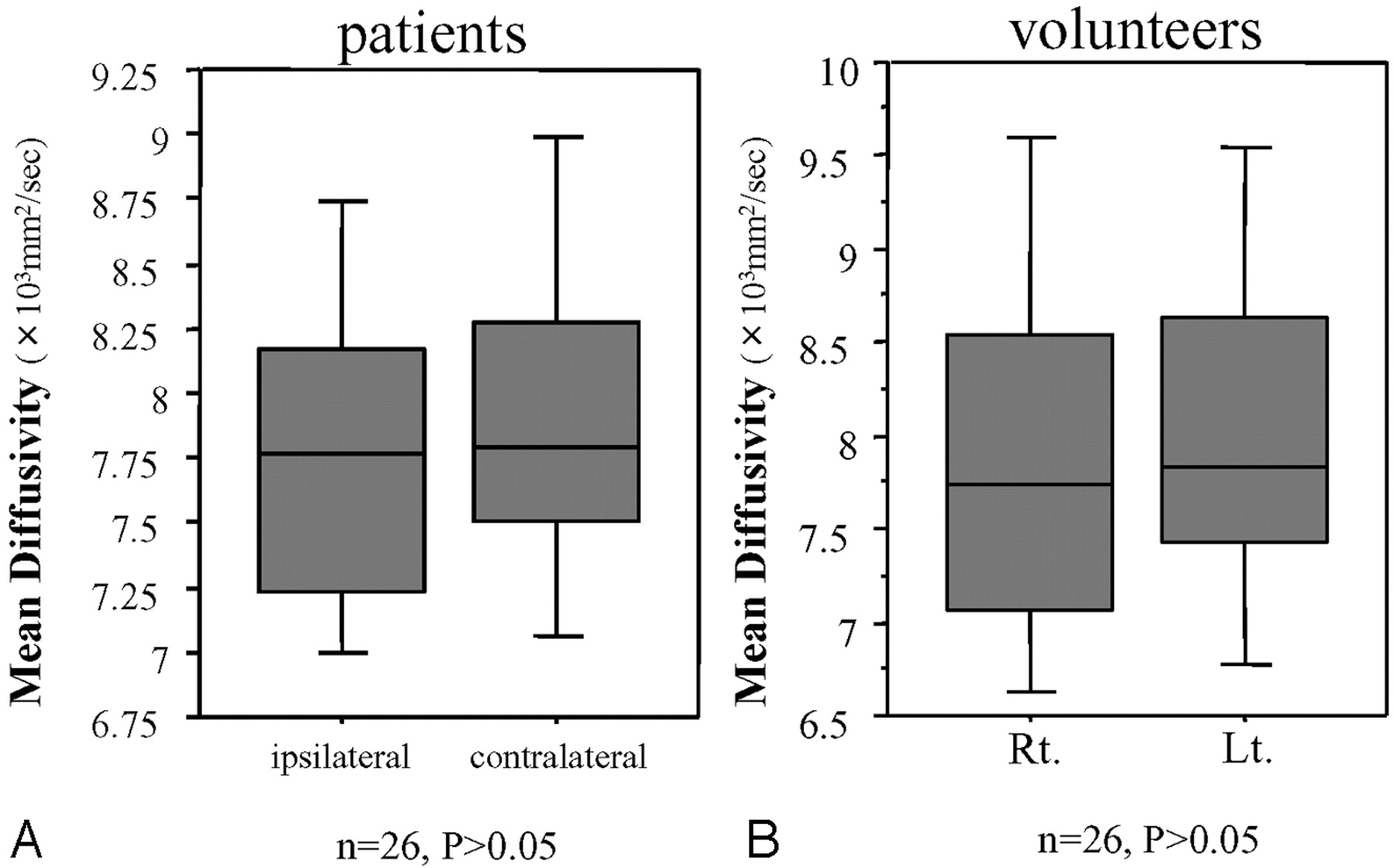
- Fig 3.
Region-of-interest analysis of MD values in the cerebral peduncle for lesion sides and contralateral sides. The Wilcoxon signed rank test was used for analysis of the differences. A, In patients with CSDH, there is no significant difference in MD values between the affected sides and the contralateral sides (P > .5). B, In healthy volunteers, there is no significant difference between bilateral sides (P > .5). Lt. indicates left; Rt, right.
- Fig 4.
The correlation between the initial FA ratio and motor weakness. Initial FA ratios are significantly correlated with motor weakness (R2 = 0.32, P = .002) by linear regression analysis. Preope. indicates preoperative.
- Fig 5.
The comparison FA ratios before and after burr-hole craniotomy in 23 patients with CSDH. FA ratios are significantly increased after surgery (Wilcoxon signed rank test, P = .0004). Pre-ope indicates preoperative; Post-ope, postoperative.


Tables
Case No./Age (yr)/Sex Side Motor Fun. (MMT) Preop. FA (affect.) Preop. FA Ratio Preop. MD Ratio Postop. FA Ratio* 1/83/F Rt 4 0.685 0.92 0.93 0.99 2/88/M Rt 4 0.645 0.95 0.97 – 3/82/M Lt 5 0.68 0.9 1.07 – 4/89/M Lt 4 0.627 0.84 1.09 – 5/73/M Rt 2 0.625 0.84 1.16 0.94 6/78/M Rt 4 0.595 0.78 0.91 1.02 7/67/M Rt 3 0.699 0.88 0.86 1 8/59/M Rt 5 0.71 0.96 1.15 0.97 9/80/M Lt 5 0.796 1 0.98 1.05 10/90/M Rt 3 0.536 0.79 0.95 0.96 11/73/M Rt 5 0.687 0.92 0.97 0.97 12/74/F Lt 4 0.489 0.79 0.95 0.96 13/64/M Rt 4 0.674 0.86 1.14 1.12 14/78/M Lt 5 0.709 0.93 0.97 0.89 15/82/F Lt 5 0.694 0.91 1.05 1.04 16/67/F Rt 4 0.673 0.94 0.84 0.92 17/77/M Rt 4 0.763 0.96 0.96 0.88 18/81/F Lt 3 0.498 0.7 1.25 0.72 19/44/M Lt 4 0.73 0.92 0.76 1 20/77/M Lt 5 0.719 0.91 1.06 0.97 21/88/M Lt 4 0.615 0.77 1.08 0.91 22/76/M Rt 4 0.636 0.88 1.03 0.94 23/76/F Rt 5 0.655 0.97 0.79 0.99 24/76/F Rt 4 0.658 0.91 0.97 0.97 25/69/M Rt 5 0.628 1 1.08 1.02 26/65/F Lt 4 0.661 0.84 1.04 0.93 Note:—Fun. indicates function; affect., affected side; Preop, preoperative; Postop, postoperative; Rt, right; Lt, left; FA, fractional anisotropy; MD, mean diffusivity; MMT, Manual Muscle Test.
* Patients 2, 3, and 4 could not undergo postoperative DTI. They hoped to leave hospital within 5 days after surgery.
Factor Patient No. FA Ratio (mean ± SD) P Value* Age (yrs) <75 10 0.895 ± 0.064 .792 >75 16 0.883 ± 0.084 Sex Male 18 0.894 ± 0.070 .718 Female 8 0.873 ± 0.090 Hematoma thickness (mm)† <22 13 0.895 ± 0.078 .573 >22 13 0.879 ± 0.075 Midline shift (mm)‡ <9.0 14 0.914 ± 0.068 .037 >9.0 12 0.856 ± 0.074 Interval from trauma (days) <40 9 0.839 ± 0.071 .0008 >40 10 0.944 ± 0.041 CT Mixed density 15 0.906 ± 0.071 .078 Non-mixed density 11 0.862 ± 0.077 * The Mann-Whitney U test was used for analysis of the differences.
† Calculated on CT as the maximal width of the hematoma.
‡ Calculated on CT as the distance from the center of the Monro foramen to a perpendicular line connecting the anterior and posterior insertions of the falx cerebri.


