
Article Figures & Data
Figures
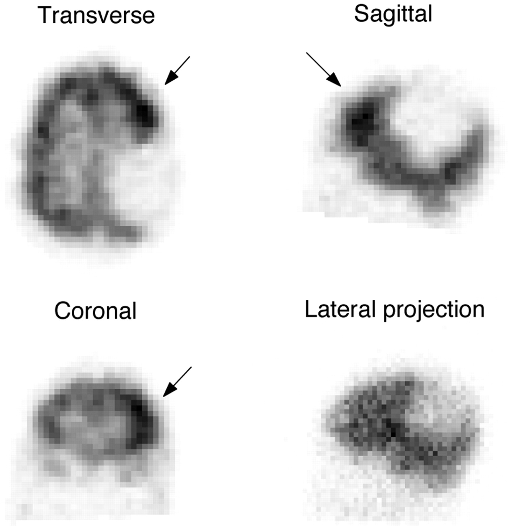
- Fig 1.
Cerebral perfusion in a 13-year-old boy with a history of seizures. Ictal study following injection of 1.258 GBq (34 mCi) Tc99m-ECD. The image shows a focus of increased uptake in the left frontal lobe (arrows) suggestive of a seizure focus. The photopenic area in the left posterior parietal lobe corresponds to a large cyst previously identified on MR imaging. The increased uptake focus is hardly recognizable in the projection data (bottom right).
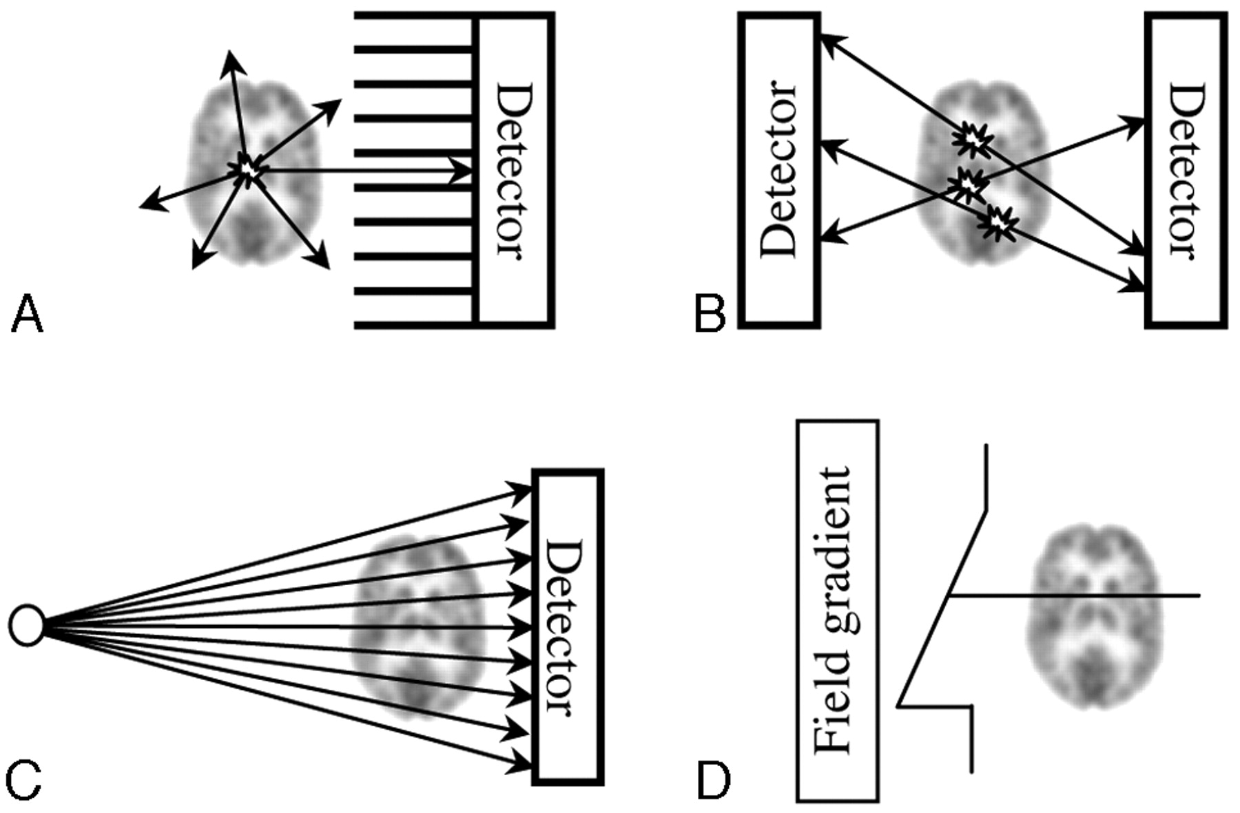
- Fig 2.
Pictorial view of photon emission and determination of LOR (drawings are not to scale). A, Parallel-hole collimator SPECT. At each point, photons are emitted isotropically. Only those emitted along lines parallel to the channels of the collimator reach the detector. Thus, the direction of the channels of the collimator identifies the LOR. B, In PET, LORs are identified by connecting the location of 2 photons arriving in temporal coincidence at opposing detectors. C, In CT, LORs are determined by the location of detection and the known location of the x-ray beam spot. D, In MR imaging, all spins subject to the same magnetic field have the same precession frequency: Their signal intensity is summed along the lines seeing the same field, just as photons originating along the same line in A, B, or C contribute to the same LOR. Just as in other 3D radiologic techniques, the LOR, or the total signal intensity coming from a line traversing the patient, is the basic piece of information acquired in SPECT from which 3D reconstruction starts.
- Fig 3.
Pictorial view of collimation (drawings are not to scale). A, Parallel-hole collimator. The object is projected onto the detector without magnification. The projection does not use the whole detector. Ideally, photons reach the detector only traveling along lines (LORs, dashed lines) parallel to the collimator channels. B, The finite width of the collimator channels allows lines nearly parallel to the collimator also to reach the detector. C, Longer channels restrict the acceptance angle and resolution improves (see how a more restricted region of space than that in B is seen from each channel). Resolution worsens with distance. D, A fan-beam collimator magnifies the object, projecting it on the whole detector. LORs converge to the focal line (a point in the transverse section shown).
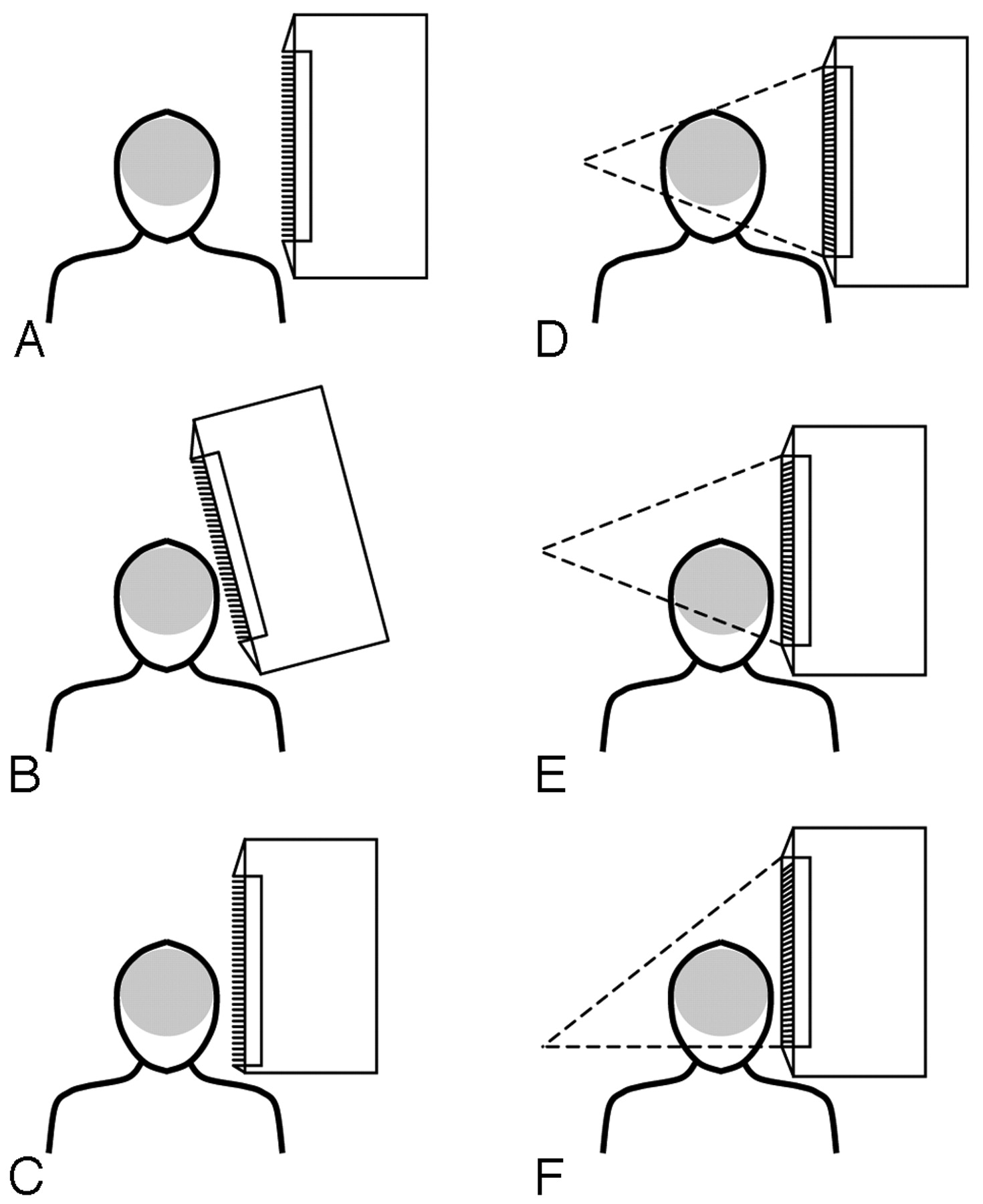
- Fig 4.
Pictorial representation of the problem of clearing the shoulders of the patient and proposed solutions. A, Due to side shielding and dead area at the borders of the FOV of the detector, collimators do not reach the side of the detector and prevent positioning the camera head next to the patient's head. B, If a parallel-hole with slant holes is used, the camera can be tilted inwards to allow clearance. C, The camera head can be redesigned to minimize side dead space. D and E, The same problem is present for cone-beam collimation, even with minimal side shielding. A possible solution is to tilt the collimator as in B. F, An alternative is to shift caudally the focal point of the collimator. The case in which the shift equals half the size of the collimator, resulting in a half-cone-beam collimator, is shown.
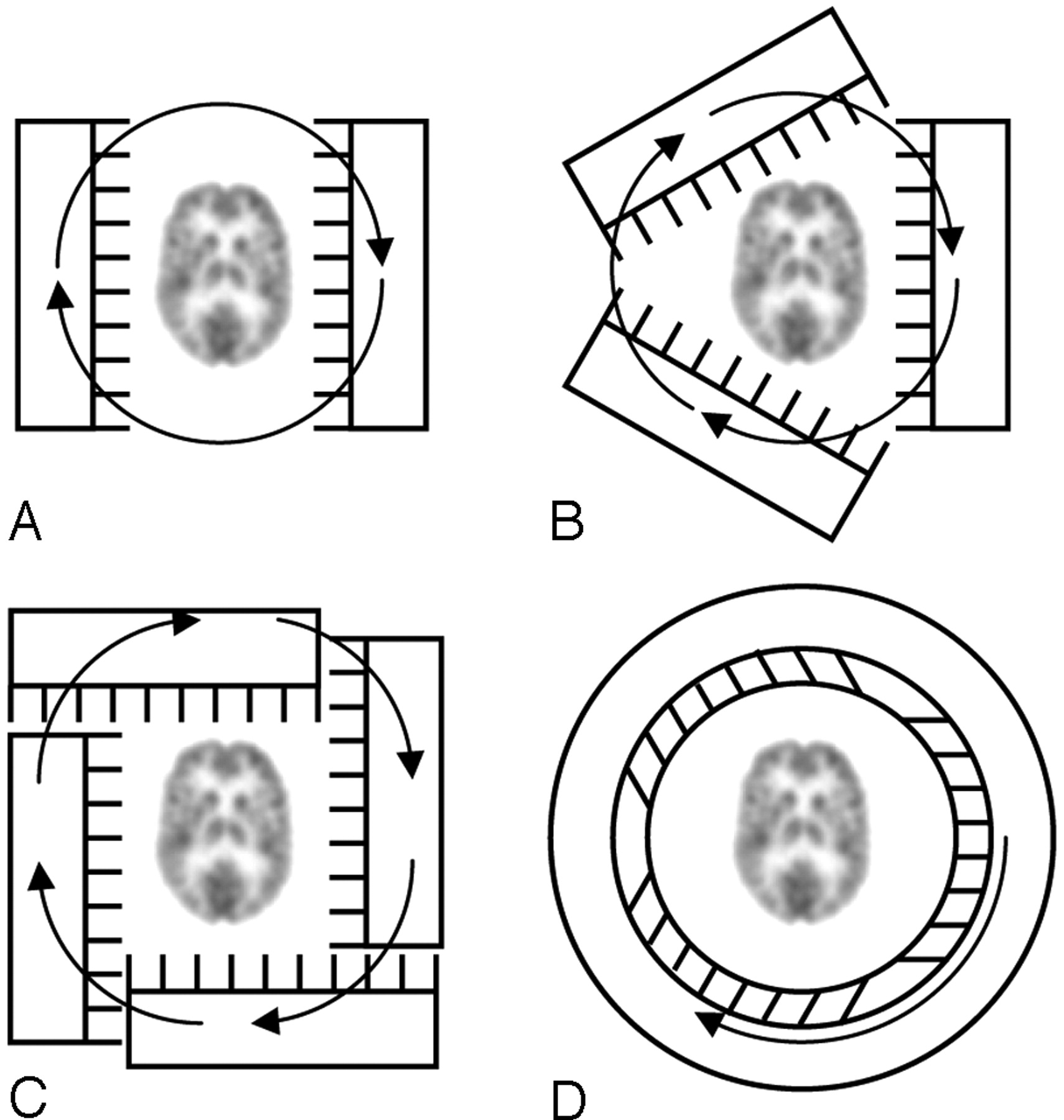
- Fig 5.
Brain SPECT designs. A, With a dual-head system, the acquisition time can be half that for a single-head camera. B, With a 3-head system, the acquisition time is reduced by a factor of 3. C, Four heads can be offset to allow large-FOV cameras to image close to the head. D, In a ring (helmet) detector, it is possible to rotate only the collimator inside the crystal and shielding.


Tables
Sample brain SPECT compounds and applications
Nuclide Half-Life Compound Acronym Measurement 133Xe 5.24 days 133Xenon 133Xe rCBF 123I 13.2 hours 123I-isopropyliodoamphetamine 123I-IMP rCBF Tc99m 6.02 hours Tc99m-hexamethylpropyleneamineoxime Tc99m-HMPAO rCBF Tc99m 6.02 hours Tc99m-ethyl cysteinate dimer (bicisate) Tc99m-ECD rCBF Tc99m 6.02 hours Tc99m-diethylenetriaminepentaacetic acid Tc99m-DTPA CSF, brain death Tc99m 6.02 hours Tc99m-pertechnetate Tc99m-TcO4 CSF, brain death 111In 2.83 days 111In-diethylenetriaminepentaacetic acid 111In-DTPA CSF, brain death 67Ga 78.3 hours 67Ga-ethylenediaminetetraacetic acid 67Ga-EDTA Blood-brain barrier permeability 123I 13.2 hours 123Iomazenil 123I-IMZ Central type benzodiazepine-receptor binding20 123I 13.2 hours 123I-2-((2-((dimethylamino)methyl)phenyl)thio)-5-iodophenylamine 123I-ADAM Serotonin transporter imaging21 123I 13.2 hours 123I-ß-carbomethoxy-3-ß-(4-iodophenyl)-tropane 123I-CIT Dopamine and serotonin transporters22 123I 13.2 hours 123I-iodobenzofuran 123I-IBF Dopamine D-2 receptor ligand23 123I 13.2 hours 123I-iodobenzamide 123I-IBZM Dopamine D-2 receptor ligand24,25 Tc99m 6.02 hours Tc99m-TRODAT Tc99m-TRODAT Dopamine transporter sites26 Tc99m 6.02 hours Tc99m-pyrophosphate Tc99m-PPi Bone scanning, TMJ27,28 Tc99m 6.02 hours Tc99m-methylene diphosphonate Tc99m-MDP Bone scanning, TMJ28,29 111In 2.83 days [111In-DOTA0,D-Phe1,Tyr3] octreotide 111In-DOTA-TOC Somatostatin receptor imaging30 Tc99m 6.02 hours Tc99m-hydrazinonicotinyl-Tyr3-octreotide Tc99m-HYNIC-TOC Somatostatin receptor imaging 111In 2.83 days 111In-pentetreotide 111In-pentetreotide Somatostatin receptor imaging31 123I 13.2 hours [123I]5-iodo-3-[2(S)-2-azetidinylmethoxy]pyridine 123I-5IA Nicotinic acetylcholine receptors32 123I 13.2 hours 6-iodo-2-(4′-dimethylamino-)phenylimidazo[1,2-a]pyridine 123I-IMPY β-amyloid plaque imaging33 Note:—TMJ indicates temporomandibular joint; CBF, relative cerebral blood flow; TRODAT, [2-[[2-[[[3-(4-chlorophenyl)-8-methyl-8-azabicyclo[3,2,1]oct-2-yl]methyl](2-mercaptoethyl)amino]ethyl]amino]ethanethiolato(3-)-N2,N2′,S2,S2′]oxo-[1R-(exo-exo)].


