Advertisement

Main menu

  • Home
  • Content
    • Current Issue
    • Accepted Manuscripts
    • Article Preview
    • Past Issue Archive
    • AJNR Case Collection
    • Case of the Week Archive
    • Classic Case Archive
    • Case of the Month Archive
  • Special Collections
    • Spinal CSF Leak Articles (Jan 2020-June 2024)
    • 2024 AJNR Journal Awards
    • Most Impactful AJNR Articles
  • Multimedia
    • AJNR Podcast
    • AJNR Scantastics
    • Video Articles
  • For Authors
    • Submit a Manuscript
    • Author Policies
    • Fast publishing of Accepted Manuscripts
    • Graphical Abstract Preparation
    • Manuscript Submission Guidelines
    • Imaging Protocol Submission
    • Submit a Case for the Case Collection
  • About Us
    • About AJNR
    • Editorial Board
  • More
    • Become a Reviewer/Academy of Reviewers
    • Subscribers
    • Permissions
    • Alerts
    • Feedback
    • Advertisers
    • ASNR Home
  • Other Publications
    • ajnr

User menu

  • Alerts
  • Log in

Search

  • Advanced search
American Journal of Neuroradiology

American Journal of Neuroradiology

ASHNR American Society of Functional Neuroradiology ASHNR American Society of Pediatric Neuroradiology ASSR
  • Alerts
  • Log in

Advanced Search

  • Home
  • Content
    • Current Issue
    • Accepted Manuscripts
    • Article Preview
    • Past Issue Archive
    • AJNR Case Collection
    • Case of the Week Archive
    • Classic Case Archive
    • Case of the Month Archive
  • Special Collections
    • Spinal CSF Leak Articles (Jan 2020-June 2024)
    • 2024 AJNR Journal Awards
    • Most Impactful AJNR Articles
  • Multimedia
    • AJNR Podcast
    • AJNR Scantastics
    • Video Articles
  • For Authors
    • Submit a Manuscript
    • Author Policies
    • Fast publishing of Accepted Manuscripts
    • Graphical Abstract Preparation
    • Manuscript Submission Guidelines
    • Imaging Protocol Submission
    • Submit a Case for the Case Collection
  • About Us
    • About AJNR
    • Editorial Board
  • More
    • Become a Reviewer/Academy of Reviewers
    • Subscribers
    • Permissions
    • Alerts
    • Feedback
    • Advertisers
    • ASNR Home
  • Follow AJNR on Twitter
  • Visit AJNR on Facebook
  • Follow AJNR on Instagram
  • Join AJNR on LinkedIn
  • RSS Feeds

Welcome to the new AJNR, Updated Hall of Fame, and more. Read the full announcements.


AJNR is seeking candidates for the position of Associate Section Editor, AJNR Case Collection. Read the full announcement.


Getting new auth cookie, if you see this message a lot, tell someone!
Research ArticleHEAD & NECK

Crista Galli Pneumatization Is an Extension of the Adjacent Frontal Sinuses

P.M. Som, E.E. Park, T.P. Naidich and W. Lawson
American Journal of Neuroradiology January 2009, 30 (1) 31-33; DOI: https://doi.org/10.3174/ajnr.A1291
P.M. Som
  • Find this author on Google Scholar
  • Find this author on PubMed
  • Search for this author on this site
E.E. Park
  • Find this author on Google Scholar
  • Find this author on PubMed
  • Search for this author on this site
T.P. Naidich
  • Find this author on Google Scholar
  • Find this author on PubMed
  • Search for this author on this site
W. Lawson
  • Find this author on Google Scholar
  • Find this author on PubMed
  • Search for this author on this site
  • Article
  • Figures & Data
  • Info & Metrics
  • Responses
  • References
  • PDF
Loading

Abstract

BACKGROUND AND PURPOSE: The crista galli is part of the ethmoid bone and, as such, it could be expected that aeration of the crista would come from ethmoid cells. After observing crista pneumatization from the frontal sinuses in several cases, we undertook this study to establish how often crista galli pneumatization came from the frontal sinuses rather than from the ethmoid complex.

MATERIALS AND METHODS: Two hundred consecutive CT scans of the paranasal sinuses were studied in adult patients to obtain the incidence of crista galli pneumatization and the cell of origin for this phenomenon. A second group of 132 children, 0–7 years of age, was studied to see if any crista galli pneumatization occurred before frontal sinus development. A third group of 79 children, 7–12 years of age, was also studied to see when crista pneumatization occurred in children whose frontal sinuses had already extended into the squamosal portion of the frontal bone.

RESULTS: Of the 200 adult cases, there were 26 patients (13%) with crista galli pneumatization, all from either the left or right frontal sinuses. In the second group of children 0–7 years of age, there were no cases of crista pneumatization. In the third group of children 7–12 years of age, there were 4 cases of crista galli pneumatization, all from well-developed frontal sinuses.

CONCLUSIONS: Our study indicates that crista galli pneumatization is virtually exclusively from either the left or right frontal sinuses and not from displaced ethmoid complex cells in the frontal recess. This finding may have surgical implications when disease is present in the crista galli.

The crista galli sits in the midline above the cribriform plate. The falx cerebri attaches to its thin and slightly curved posterior border, whereas its shorter thicker anterior border is joined to the frontal bone by 2 small alae, completing the margins of the foramen caecum. Embryologically, the crista galli is derived from the ethmoid bone, and as such, it would seem reasonable that any eventual pneumatization of the crista galli would come from the ethmoid complex. In fact, the conventional thinking has been that the pneumatization occurs from displaced ethmoid cells from the frontal recess region.1,2 However, supraorbital ethmoid cells are known to extend from the ethmoid complex into the frontal bone, thus crossing from one bone to the other. Because sinus pneumatization is known to cross from one bone to another and with the recent observation that most frontal sinus interseptal cells (immediately adjacent to the crista galli) come from the frontal sinuses and not from the ethmoid complex as previously thought, the possibility exists that pneumatization of the crista galli could also come primarily from the adjacent frontal sinuses.3 To examine the pathway of crista galli pneumatization, we studied axial and coronal CT scans of the paranasal sinuses on 200 consecutive adult patients to obtain the incidence of crista galli pneumatization and then, if possible, the origin of the pneumatizing cell. In addition, 211 CT scans of the brain, orbits, and/or sinuses were studied in children 0–12 years of age to see if any crista galli pneumatization occurred before pneumatization of the main frontal sinus cavity into the squamosal portion of the frontal bone.

Materials and Methods

A prospective study of 200 consecutive axial and coronal CT scans of the paranasal sinuses in adult patients was made to assess specifically if the crista galli was pneumatized. If it was pneumatized, an assessment was made as to whether the bony margins of the crista galli were completely intact except at its caudal margin or if there was an extension of either frontal sinus into the crista galli. In the former case, the pneumatizing cell was said to be from the ethmoid complex. In the latter case, the pneumatizing cell was said to be from the frontal sinus. The patients ranged from 21 to 74 years of age, and there were 127 women and 73 men. The images were reviewed by a head and neck radiologist, a neuroradiologist, and an otolaryngologist. Any differing opinions were resolved by consensus.

A second group of 132 CT scans was also studied. These included brain, paranasal sinus, and orbital CT scans on patients 0–7 years of age, performed in the past year at our institution. These CT scans were assessed for any pneumatization in the crista galli. The number of children in each year of life in this study is shown in the Table. There were 79 boys and 53 girls.

View this table:
  • View inline
  • View popup

Age range of children whose CT studies were examined for the presence of crista galli pneumatization

A third group of 79 CT scans was studied in patients who were 7–12 years of age. These included brain, paranasal sinus, and orbital CT scans obtained at our institution in the past year. The number of children in each year of life in this study is shown in the Table as is the number of cases of crista galli aeration. There was 1 girl 8 years of age, 1 boy 10 years of age, and 2 boys 11 years of age who had pneumatization of the crista galli, all from the frontal sinuses.

The study was performed with the approval of the internal review board (number 06-0254).

Results

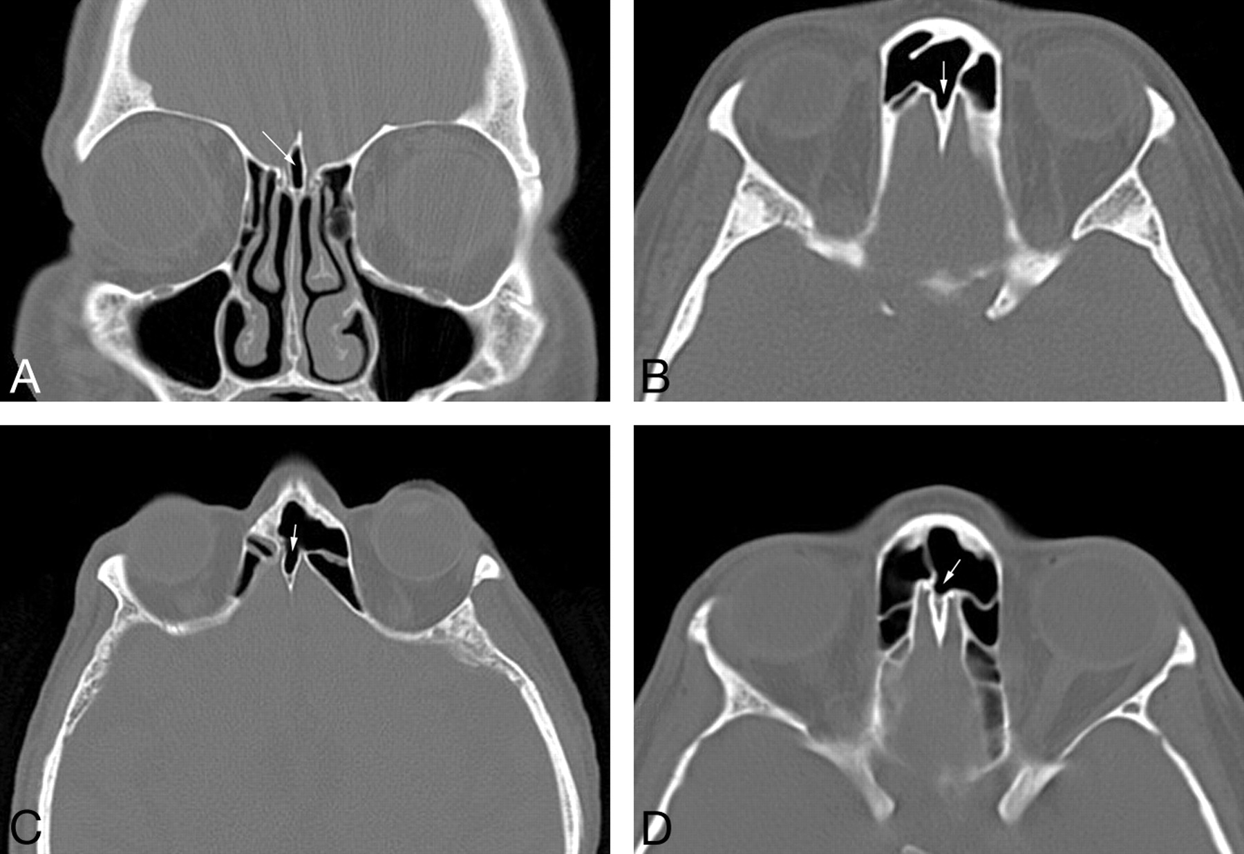
Of the 200 CT scans in adults, there were 26 with some degree of pneumatization of the crista galli (13%). There were 4 cases with only minimal pneumatization and 22 cases with extensive pneumatization (Fig 1). In all cases, the pneumatizing air cell was either the left (11 cases) or right (15 cases) frontal sinus and the crista galli was pneumatized from its anteriormost surface. In 2 cases, inflammatory disease in a frontal sinus extended directly into the pneumatized crista galli (Fig 2).

Fig 1.
  • Download figure
  • Open in new tab
  • Download powerpoint
Fig 1.

A, Coronal CT scan of the paranasal sinuses in a 45-year-old women with difficulty breathing shows the typical appearance of crista galli pneumatization (arrow). B, Axial CT scan of the paranasal sinuses in a 51-year-old man with sinus pain shows extensive pneumatization of the crista galli from the right frontal sinus (arrow). C, Axial CT scan of the paranasal sinuses in a 63-year-old man with difficulty breathing shows extensive pneumatization of the crista galli from the left frontal sinus (arrow). D, Axial CT scan of the paranasal sinuses in a 33-year-old woman with difficulty breathing shows minimal pneumatization of the anterior crista galli from the left frontal sinus (arrow).

Fig 2.
  • Download figure
  • Open in new tab
  • Download powerpoint
Fig 2.

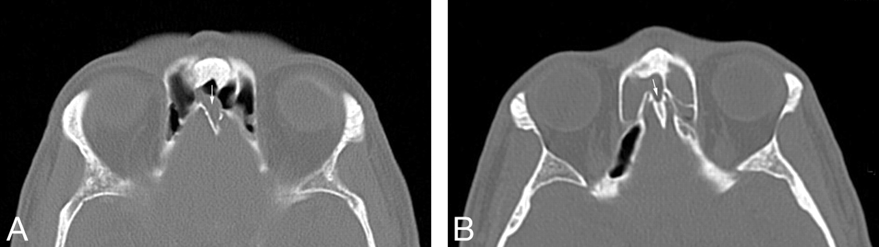
A, Axial CT scan of the paranasal sinuses in a 27-year-old man with sinus pain shows polypoid mucosal thickening extending from the right frontal sinus into a well-pneumatized crista galli (arrow). B, Axial CT scan of the paranasal sinuses in a 49-year-old woman with sinus pain shows extensive mucosal thickening extending from the right frontal sinus into a well-pneumatized crista galli (arrow). There is also mucosal disease in the left frontal sinus.

Of the 132 CT scans in patients ≤7 years of age, none had any findings of pneumatization of the crista galli. In the 13 cases with the beginning of frontal sinus pneumatization, the developing frontal sinuses were all anterior and lateral to the crista galli, with no extension toward the crista galli.

Of the 79 CT scans in children 7–12 years of age, there were 4 cases of crista galli pneumatization. In these 4 children, all had well-developed frontal sinuses and the aerating cell was from the frontal sinuses.

There were no discrepancies between observers in interpreting the images.

Discussion

In the embryo, the ethmoidal cartilage consists of both a mesial mass (the mesethmoid), which extends from the sphenoid to the tip of the nasal process, and a pair of lateral masses developed from the lateral nasal processes (the ectethmoid), lateral to the olfactory sacs. The terminal portion of the mesial mass persists as the cartilaginous nasal septum, whereas ossification of the upper portion becomes the perpendicular plate and crista galli.4,5 Although some ossification has been reported in the fetus by the 12th week, most ossification of the cartilaginous crista galli starts at approximately 2 months of postnatal life, steadily increases in ossification to 14 months of age, and then slowly progresses until it finishes by 24 months of age.6,7

According to Dodd and Jing8 and Netter9, on average, the frontal sinuses reach the level of the superior orbital rims by 6–7 years of age. It was because of this observation that we examined CT scans in children younger than 7 years of age to see if any pneumatization of the crista galli could be found. Because the crista is from the ethmoid bone and the ethmoid complexes are pneumatized at birth, it seems reasonable that some crista galli pneumatization would be present if the pneumatizing cells were from the ethmoid complex. In fact, we found no crista pneumatization in this group of children.

We next examined CT scans in children 7–12 years of age to see if any crista pneumatization could be found. In this group of 79 cases, there were 4 children (5% of this group) with crista pneumatization and all 4 of these children had well-developed frontal sinuses, which were the source of the crista aeration.

Using CT scans, Basic et al10 in a series of 212 patients noted pneumatization of the crista galli in 2.4%. These authors also found that crista pneumatization came primarily from the frontal sinuses, though they did not reveal how many of their 5 patients had suggested pneumatization from the ethmoid sinuses.

Using anatomic material, McLaughlin et al11 also noted that when pneumatized, the crista galli can drain into either the ethmoid complex or the frontal sinuses, but these authors gave no statistics or case numbers. Traditionally, it had been thought that aeration of the crista galli came from the frontal recess and migrated ethmoid cells.1,2,12 Our series supports the concept that crista galli pneumatization is virtually exclusively from the frontal sinus as a diverticular-like extension into the crista from either the left or right frontal sinus cavity. In our series, we had 13% of adults with crista galli pneumatization.

Our findings are not surprising in view of our earlier observation that frontal intersinus septal cells also come primarily from the adjacent frontal sinuses rather than from ethmoid cells, as previously thought.

Conclusions

As previously noted, the derivation of the crista galli from the ethmoid bone has led, in the past, to the belief that its pneumatization would proceed from the ethmoid labyrinth. Accordingly, disease in a crista galli cell would represent extension of ethmoid sinus pathology, and surgical management should be directed at the ethmoid labyrinth. However, evidence that the primary origin of pneumatization of the crista galli is from the frontal sinus and demonstration of a persistent connection with it should now direct the clinician to focus therapy on the frontal sinus as the source of the disease.

References

  1. ↵
    Zinreich S, Albayram S, Benson M, et al. The ostiomeatal complex and functional endoscopic surgery. In: Som P, Curtin H, eds. Head and Neck Imaging. Vol 1. 4th ed. St. Louis: Mosby;2003 :165
  2. ↵
    Standring S, Berkovitz B, Hackney C, et al. Head and neck. In: Standring S, ed. Gray's Anatomy: The Anatomical Basis of Clinical Practice. 39th ed. Edinburgh, UK: Elsevier;2005 :474
  3. ↵
    Som P, Lawson W. The frontal intersinus septal air cell: a new hypothesis of its origin. AJNR Am J Neuroradiol 2008;29:1215–17. Epub 2008 Apr 3
    Abstract/FREE Full Text
  4. ↵
    Moss-Salentijn L. Anatomy and Embryology. In: Blitzer A, Lawson W, Friedman W, eds. Surgery of the Paranasal Sinuses. Philadelphia: WB Saunders;1985 :1–22
  5. ↵
    Arey L. Developmental Anatomy. 6th ed. Philadelphia: WB Saunders;1962 :411–19
  6. ↵
    Kier E. Fetal skull. In: Newton T, Potts D, eds. Radiology of the Skull and Brain: The Skull. Vol 1. Saint Louis: Mosby;1971 :100
  7. ↵
    Belden CJ, Mancuso AA, Kotzur IM. The developing anterior skull base: CT appearance from birth to 2 years of age. AJNR Am J Neuroradiol 1997;18:811–18
    Abstract/FREE Full Text
  8. ↵
    Dodd G, Jing B. Radiology of the Nose, Paranasal Sinuses, and Nasopharynx. Baltimore: Williams & Wilkins;1977 :60
  9. ↵
    Netter F. Atlas of Human Anatomy. Summit, NJ: Ciba-Geigy;1989 :44
  10. ↵
    Basic N, Basic V, Jukic T, et al. Computed tomographic imaging to determine the frequency of anatomical variations in pneumatization of the ethmoid bone. Eur Arch Otorhinolaryngol 1999;256:69–71
    CrossRefPubMed
  11. ↵
    McLaughlin RB Jr, Rehl RM, Lanza DC. Clinically relevant frontal sinus anatomy and physiology. Otolaryngol Clin North Am 2001;34:1–22
    CrossRefPubMed
  12. ↵
    Naidich T, Zimmerman R, Bauer B, et al. Midface: embryology and congenital lesions. In: Som P, Curtin H, eds. Head and Neck Imaging. Vol 1. 3rd ed. St. Louis: Mosby;1996 :3–60
  • Received May 9, 2008.
  • Accepted after revision July 14, 2008.
  • Copyright © American Society of Neuroradiology
View Abstract
PreviousNext
Back to top

In this issue

American Journal of Neuroradiology: 30 (1)
American Journal of Neuroradiology
Vol. 30, Issue 1
January 2009
  • Table of Contents
  • Index by author
Advertisement
Print
Download PDF
Email Article

Thank you for your interest in spreading the word on American Journal of Neuroradiology.

NOTE: We only request your email address so that the person you are recommending the page to knows that you wanted them to see it, and that it is not junk mail. We do not capture any email address.

Enter multiple addresses on separate lines or separate them with commas.
Crista Galli Pneumatization Is an Extension of the Adjacent Frontal Sinuses
(Your Name) has sent you a message from American Journal of Neuroradiology
(Your Name) thought you would like to see the American Journal of Neuroradiology web site.
CAPTCHA
This question is for testing whether or not you are a human visitor and to prevent automated spam submissions.
Cite this article
P.M. Som, E.E. Park, T.P. Naidich, W. Lawson
Crista Galli Pneumatization Is an Extension of the Adjacent Frontal Sinuses
American Journal of Neuroradiology Jan 2009, 30 (1) 31-33; DOI: 10.3174/ajnr.A1291

Citation Manager Formats

  • BibTeX
  • Bookends
  • EasyBib
  • EndNote (tagged)
  • EndNote 8 (xml)
  • Medlars
  • Mendeley
  • Papers
  • RefWorks Tagged
  • Ref Manager
  • RIS
  • Zotero
0 Responses
Respond to this article
Share
Bookmark this article
Crista Galli Pneumatization Is an Extension of the Adjacent Frontal Sinuses
P.M. Som, E.E. Park, T.P. Naidich, W. Lawson
American Journal of Neuroradiology Jan 2009, 30 (1) 31-33; DOI: 10.3174/ajnr.A1291
del.icio.us logo Twitter logo Facebook logo Mendeley logo
  • Tweet Widget
  • Facebook Like
  • Google Plus One
Purchase

Jump to section

  • Article
    • Abstract
    • Materials and Methods
    • Results
    • Discussion
    • Conclusions
    • References
  • Figures & Data
  • Info & Metrics
  • Responses
  • References
  • PDF

Related Articles

  • No related articles found.
  • PubMed
  • Google Scholar

Cited By...

  • No citing articles found.
  • Crossref (35)
  • Google Scholar

This article has been cited by the following articles in journals that are participating in Crossref Cited-by Linking.

  • CT of Anatomic Variants of the Paranasal Sinuses and Nasal Cavity: Poor Correlation With Radiologically Significant Rhinosinusitis but Importance in Surgical Planning
    Katya A. Shpilberg, Simon C. Daniel, Amish H. Doshi, William Lawson, Peter M. Som
    American Journal of Roentgenology 2015 204 6
  • Computed tomography anatomy of the paranasal sinuses and anatomical variants of clinical relevants in Nigerian adults
    Regina Chinwe Onwuchekwa, Nengi Alazigha
    Egyptian Journal of Ear, Nose, Throat and Allied Sciences 2017 18 1
  • An anatomical study of pneumatized crista galli
    Ranko Mladina, Romano Antunović, Cemal Cingi, Nuray Bayar Muluk, Neven Skitarelić, Mislav Malić
    Neurosurgical Review 2017 40 4
  • Anatomical variants of ethmoid bone on multidetector CT
    Daniele Gibelli, Michaela Cellina, Stefano Gibelli, Annalisa Cappella, Antonio Giancarlo Oliva, Giovanni Termine, Chiarella Sforza
    Surgical and Radiologic Anatomy 2018 40 11
  • A comprehensive morphometric analysis of crista galli for sex determination with a novel morphological classification on computed tomography images
    Erdal Komut, Murat Golpinar
    Surgical and Radiologic Anatomy 2021 43 12
  • Computed tomographic study of remarkable anatomic variations in paranasal sinus region and their clinical importance - A retrospective study
    Amit Nautiyal, Arun Narayanan, Deepanjan Mitra, ThittamarnahalliMuguregowda Honnegowda, Sivakumar
    Annals of Maxillofacial Surgery 2020 10 2
  • Crista galli mucocele: endoscopic marsupialization via frontoethmoid approach
    Sergio Santino Cervantes, Devyani Lal
    International Forum of Allergy & Rhinology 2014 4 7
  • Paranasal Sinuses
    Abdulmalik S. Alsaied
    2017
  • Anatomic Variations of Paranasal Region in Migraine
    Özlem Elvan, Kaan Esen, Havva Didem Çelikcan, Mesut Sabri Tezer, Anil Özgür
    Journal of Craniofacial Surgery 2019 30 6
  • The Radiological Evaluation of the Crista Galli and Its Clinical Implications for Anterior Skull Base Surgery
    Hadice Uçar, Ilhan Bahşi, Mustafa Orhan, Eda Didem Yalçin
    Journal of Craniofacial Surgery 2021 32 5

More in this TOC Section

  • WHO Classification Update: Nasal&Skull Base Tumors
  • Peritumoral Signal in Vestibular Schwannomas
  • Chondrosarcoma vs Synovial Chondromatosis: Imaging
Show more HEAD & NECK

Similar Articles

Advertisement

Indexed Content

  • Current Issue
  • Accepted Manuscripts
  • Article Preview
  • Past Issues
  • Editorials
  • Editors Choice
  • Fellow Journal Club
  • Letters to the Editor

Cases

  • Case Collection
  • Archive - Case of the Week
  • Archive - Case of the Month
  • Archive - Classic Case

Special Collections

  • Special Collections

Resources

  • News and Updates
  • Turn around Times
  • Submit a Manuscript
  • Author Policies
  • Manuscript Submission Guidelines
  • Evidence-Based Medicine Level Guide
  • Publishing Checklists
  • Graphical Abstract Preparation
  • Imaging Protocol Submission
  • Submit a Case
  • Become a Reviewer/Academy of Reviewers
  • Get Peer Review Credit from Publons

Multimedia

  • AJNR Podcast
  • AJNR SCANtastic
  • Video Articles

About Us

  • About AJNR
  • Editorial Board
  • Not an AJNR Subscriber? Join Now
  • Alerts
  • Feedback
  • Advertise with us
  • Librarian Resources
  • Permissions
  • Terms and Conditions

American Society of Neuroradiology

  • Not an ASNR Member? Join Now

© 2025 by the American Society of Neuroradiology All rights, including for text and data mining, AI training, and similar technologies, are reserved.
Print ISSN: 0195-6108 Online ISSN: 1936-959X

Powered by HighWire