Advertisement

Main menu

  • Home
  • Content
    • Current Issue
    • Accepted Manuscripts
    • Article Preview
    • Past Issue Archive
    • AJNR Case Collection
    • Case of the Week Archive
    • Classic Case Archive
    • Case of the Month Archive
  • Special Collections
    • Spinal CSF Leak Articles (Jan 2020-June 2024)
    • 2024 AJNR Journal Awards
    • Most Impactful AJNR Articles
  • Multimedia
    • AJNR Podcast
    • AJNR Scantastics
    • Video Articles
  • For Authors
    • Submit a Manuscript
    • Author Policies
    • Fast publishing of Accepted Manuscripts
    • Graphical Abstract Preparation
    • Manuscript Submission Guidelines
    • Imaging Protocol Submission
    • Submit a Case for the Case Collection
  • About Us
    • About AJNR
    • Editorial Board
  • More
    • Become a Reviewer/Academy of Reviewers
    • Subscribers
    • Permissions
    • Alerts
    • Feedback
    • Advertisers
    • ASNR Home
  • Other Publications
    • ajnr

User menu

  • Alerts
  • Log in

Search

  • Advanced search
American Journal of Neuroradiology

American Journal of Neuroradiology

ASHNR American Society of Functional Neuroradiology ASHNR American Society of Pediatric Neuroradiology ASSR
  • Alerts
  • Log in

Advanced Search

  • Home
  • Content
    • Current Issue
    • Accepted Manuscripts
    • Article Preview
    • Past Issue Archive
    • AJNR Case Collection
    • Case of the Week Archive
    • Classic Case Archive
    • Case of the Month Archive
  • Special Collections
    • Spinal CSF Leak Articles (Jan 2020-June 2024)
    • 2024 AJNR Journal Awards
    • Most Impactful AJNR Articles
  • Multimedia
    • AJNR Podcast
    • AJNR Scantastics
    • Video Articles
  • For Authors
    • Submit a Manuscript
    • Author Policies
    • Fast publishing of Accepted Manuscripts
    • Graphical Abstract Preparation
    • Manuscript Submission Guidelines
    • Imaging Protocol Submission
    • Submit a Case for the Case Collection
  • About Us
    • About AJNR
    • Editorial Board
  • More
    • Become a Reviewer/Academy of Reviewers
    • Subscribers
    • Permissions
    • Alerts
    • Feedback
    • Advertisers
    • ASNR Home
  • Follow AJNR on Twitter
  • Visit AJNR on Facebook
  • Follow AJNR on Instagram
  • Join AJNR on LinkedIn
  • RSS Feeds

Welcome to the new AJNR, Updated Hall of Fame, and more. Read the full announcements.


AJNR is seeking candidates for the position of Associate Section Editor, AJNR Case Collection. Read the full announcement.


OtherBRAIN

Aggressive Leptomeningeal Hemangioblastomatosis of the Central Nervous System in a Patient with von Hippel-Lindau Disease

N.A. Courcoutsakis, P.K. Prassopoulos and N.J. Patronas
American Journal of Neuroradiology April 2009, 30 (4) 758-760; DOI: https://doi.org/10.3174/ajnr.A1360
N.A. Courcoutsakis
  • Find this author on Google Scholar
  • Find this author on PubMed
  • Search for this author on this site
P.K. Prassopoulos
  • Find this author on Google Scholar
  • Find this author on PubMed
  • Search for this author on this site
N.J. Patronas
  • Find this author on Google Scholar
  • Find this author on PubMed
  • Search for this author on this site
  • Article
  • Figures & Data
  • Info & Metrics
  • Responses
  • References
  • PDF
Loading

Abstract

SUMMARY: Hemangioblastomas of the central nervous system are the most common tumors seen in patients with von Hippel-Lindau (VHL) disease. A very rare case of diffuse leptomeningeal hemangioblastomatosis obliterating large areas of the subarachnoid space, both intracranial and within the spinal canal, which developed during a relatively short period, in a patient with VHL disease is presented.

Von Hippel-Lindau (VHL) disease is an autosomal-dominant disorder (Mendelian Inheritance in Man No. 193300) with a prevalence estimated to be between 1 per 35,000 and 1 per 40,000, afflicting approximately 6000 to 7000 patients in the United States.1,2 The VHL gene defect is located in the short arm of the chromosome 3 (3p25–26).1–3 The target sites for this mutation are the retina, brain, spinal cord, and the glandular and neural crest cells. Hemangioblastomas (HBs) of the retina and the central nervous system (CNS) are the most common tumors, occurring in more than 70% of patients with VHL disease.1–3 HBs of the CNS also occur as a result of spontaneous mutation in the absence of other manifestations of VHL disease.4 Diffuse infiltration of the leptomeninges is known as hemangioblastomatosis and has been observed both in VHL disease and in patients with spontaneous mutations.4–8

We report a case of a 53-year-old man with aggressive spinal and brain leptomeningeal hemangioblastomatosis that developed in a relatively short period.

Case Report

A 53-year-old white man diagnosed at age 43 years with VHL disease (type II-B, according to the National Institutes of Health classification) had initially presented with neck pain and visual disturbances. Before enrollment, patient had undergone a suboccipital craniectomy for the removal of a cerebellar HB. Brain and spinal MR imaging revealed 1 large HB at the anterior aspect of the pons and several tumors in the cerebellar hemispheres on admission. An additional HB was present on the anterior aspect of the spinal cord at the level of C6–C7. A syrinx was present in the thoracic spine without evidence of other abnormalities. MR imaging scans of the brain demonstrated no leptomeningeal involvement. Retinal HBs were also found during ophthalmologic examination. Imaging studies of the abdomen demonstrated bilateral adrenal tumors and multiple cystic and solid tumors in both kidneys. In the following months, the patient underwent several surgical procedures, which included a right nephrectomy and a partial left nephrectomy for renal cell carcinoma, and bilateral adrenalectomies for the removal of pheochromocytomas.

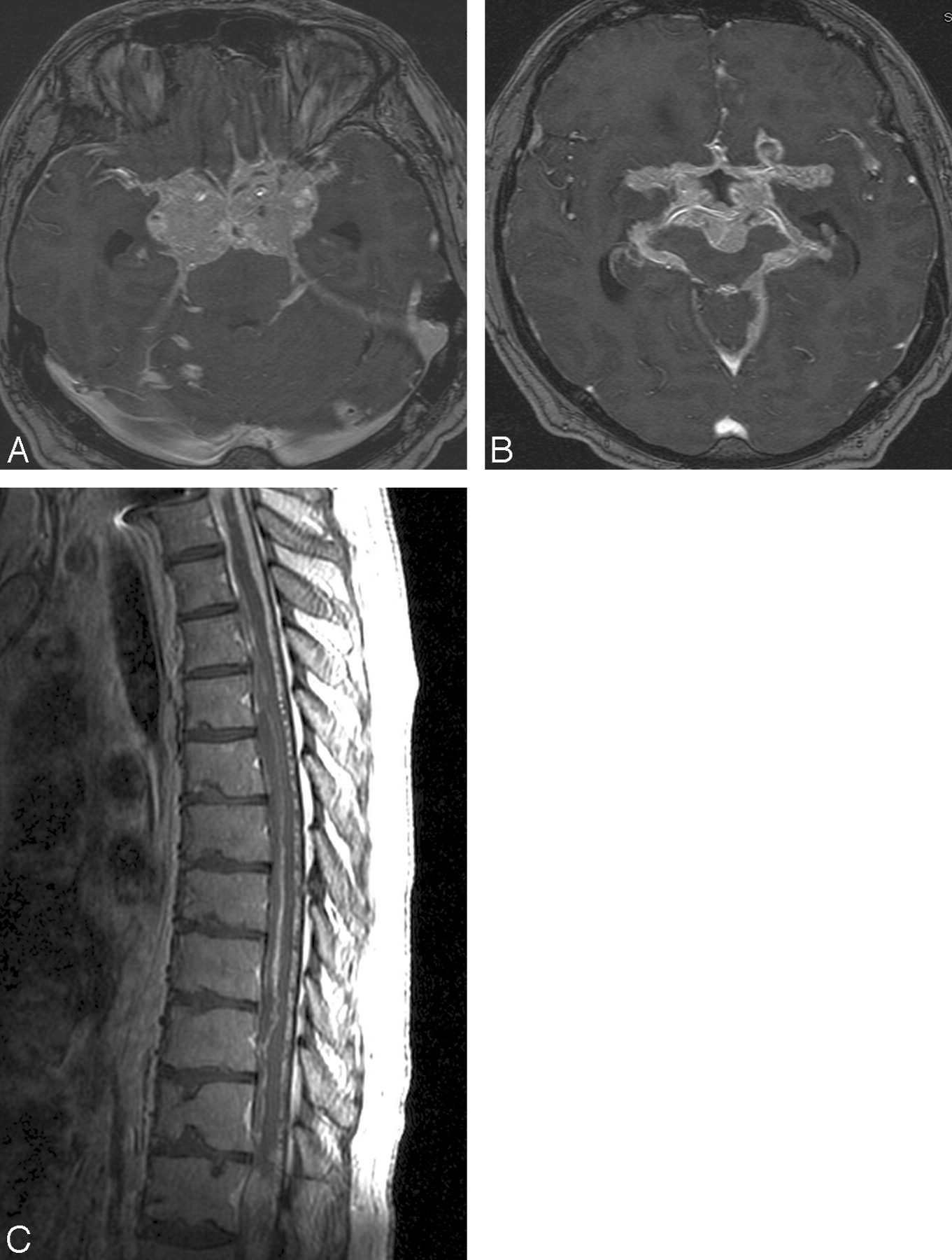
The tumor of the cervical cord was removed surgically, and immediately after recovery, the tumor of the pons was treated with radiation. On follow-up MR imaging scans, the tumor in the pons and the small cerebellar tumors appeared stable without evidence of growth. Approximately 2 years after completion of radiation therapy, an infiltration of the leptomeninges in the suprasellar cistern and in the spinal canal by HB had developed. Within the next 2 years, this process became gradually worse, with infiltration of the leptomeninges surrounding the cerebellar hemispheres, the brain stem, and the leptomeninges in the Sylvian fissures and the quadrigeminal cistern. There was tumor invasion of the hypothalamus, the temporal lobes midbrain, and brain stem. The optic chiasm, the arteries of the circle of Willis, and the basilar arteries were encased (Fig 1A, B). Extensive tumor invasion also progressed into the spinal canal, with prominent infiltration of the leptomeninges around the entire cord and the cauda equina (Fig 1C). From a clinical standpoint, the patient complained of increasing-intensity headaches, memory loss, and confusion. Multiple cranial nerve palsies primarily involving the III, IV, V, VII, and VIII nerves also developed. Shortly thereafter, the patient died of CNS decline and respiratory insufficiency.

  Fig 1.
  • Download figure
  • Open in new tab
  • Download powerpoint
Fig 1.

A and B, MR axial, postgadolinium images of the suprasellar region, obtained with use of the SPGR technique, demonstrate numerous hemangioblastomatous tumors surrounding the extraorbital parts of the optic nerves, the optic chiasm, the sella, and the arteries of the circle of Willis. C, Sagittal, postcontrast T1-weighted image of the thoracic spine shows diffuse leptomeningeal infiltration by hemangioblastomas.

Autopsy confirmed the presence of extensive tumor infiltration of the leptomeninges intracranially and in the spinal canal. On histologic examination, there was abundance of characteristic stromal cells with unusually large nuclei, small amounts of cytoplasm, and extensive epithelioid growth. There was also very prominent tumor vasculature with innumerable capillaries intervening between small nests and individual cells. Immunopexidase studies revealed positive neuron-specific enolase and negative epithelial membrane antigen, confirming the diagnosis of HB.

Discussion

In patients with VHL disease, HBs are multiple and usually involve the retina, the cerebellum, and the spinal cord.1–3 It has been reported that approximately 40% of patients with HBs in the CNS meet the criteria of VHL disease.1,3 It is well known that HBs of the CNS, regardless of location, have the same histologic characteristics supporting Lindau's hypothesis that the disease is an “angiomatosis of the central nervous system.”1,3,9 The individual angiomatous tumors in patients with VHL disease occur as a result of a separate gene defect in the cells of each tumor.3

HBs are known to be benign tumors and, once removed, do not usually recur at the surgical site unless a new mutation develops in the cells of the surgical bed. It has been reported that late recurrence occurs after surgery in only 15% to 25% of cases.4 The relative benign nature of these tumors has also been documented by the finding of Reyns et al,6 who reported that the recurrence rate is approximately 19 years in patients with VHL disease and 11 years in patients without disease.

On imaging studies, HBs in the brain or spinal cord usually present as solid nodules that enhance intensely on postcontrast scans.1 Edema is commonly encountered in the brain or the cord parenchyma surrounding an enhancing HB, caused by extravasation of fluid from the highly permeable capillaries of the tumor. When the amount of extravasated fluid exceeds the capability of the tissues to absorb it, a fluid-filled cyst develops in the periphery of the enhancing tumor. The extravasated fluid may also dissect the parenchyma in the cord and reach the central canal leading to syringomyelia.5

Diffuse and extensive leptomeningeal infiltration by HBs similar to that in our patient is an exceedingly rare phenomenon. The term leptomeningeal hemangioblastomatosis was first introduced by Bakshi et al5 to describe this unusual presentation. In a recent review of the literature, a total of 15 cases were reported with spinal infiltration but without supratentorial infiltration on imaging; 6 cases were associated with VHL disease and 9 caused by spontaneous mutations.7 In the case of our patient, hemangiomatous lesions were also located supratentorially. The involvement of the leptomeninges first appeared 2 years after surgical resection of the HB in the cervical cord and 2 years after completion of radiation therapy of the tumor of the pons. Infiltration was widespread and included the spine, the infratentorial brain, and the supratentorial brain. Soon after the development of the leptomeningeal disease, the growth of the tumor progressed relentlessly and led to the patient's death 2 years later.

The exact cause of this unusual biologic behavior observed in leptomeningeal hemangioblastomatosis has been debated but remains problematic. Bakshi et al5 suggested that this phenomenon occurs as a result of release by tumor cell factors that promote aggressive angiogenesis or increase vascular permeability. Mohan et al10 suggested that 1 or several other gene mutations, in addition to the VHL gene, may have induced this type of rapid growth and dissemination.10 Other investigators suggested that the underlying cause is uncontrolled angiogenesis from overexpression of vascular endothelial growth factors.3 Mechanical dissemination of tumor cells into the subarachnoid space after surgery or malignant transformation to angiosarcoma has also been speculated.7 Regardless of the cause(s) of leptomeningeal hemangioblastomatosis, this complication represents an ominous sign for which there is no specific treatment.

In conclusion, we present an aggressive case of disseminated meningeal hemangioblastomatosis involving both the spine and the brain that led to rapid clinical decline and eventual death. The exact causes of this rare complication of CNS HBs are not yet identified.

Footnotes

  • Author Contributions: Guarantors of integrity of entire study: N.A.C., N.J.P.; study concepts: N.A.C., N.J.P.; definition of intellectual content: N.A.C., N.J.P., P.K.P.; literature research: N.A.C.; manuscript preparation and review: N.A.C., N.J.P., P.K.P.; manuscript editing: N.A.C., N.J.P., P.K.P.

References

  1. ↵
    Choyke PL, Glenn GM, Walther McCM, et al. von Hippel-Lindau disease: genetic, clinical, and imaging features [published erratum appears in Radiology 1995;196:582]. Radiology 1995;194:629–42
    PubMed
  2. ↵
    Couch V, Lindor NM, Karnes PS, et al. von Hippel-Lindau disease. Mayo Clin Proc 2000;75:265–72
    CrossRefPubMed
  3. ↵
    Wanebo JE, Lonser RR, Glenn GM, et al. The natural history of hemangioblastomas of the central nervous system in patients with von Hippel-Lindau disease. J Neurosurg 2003;98:82–94
    CrossRefPubMed
  4. ↵
    Kato M, Ohe N, Okumura A, et al. Hemangioblastomatosis of the central nervous system without von Hippel-Lindau disease: a case report. J Neurooncol 2005;72:267–70
    CrossRefPubMed
  5. ↵
    Bakshi R, Mechtler LL, Patel MJ, et al. Spinal leptomeningeal hemangioblastomatosis in von Hippel-Lindau disease: magnetic resonance and pathologic findings. J Neuroimag 1997;7:242–44
    PubMed
  6. ↵
    Reyns N, Assaker R, Louis E, et al. Leptomeningeal hemangioblastomatosis in a case of von Hippel-Lindau disease: case report. Neurosurgery 2003;52:1212–16
    PubMed
  7. ↵
    Ramachandran R, Lee HS, Mathews B, et al. Intradural extramedullary leptomeningeal hemangioblastomatosis and paraneoplastic limbic encephalitis diagnosed at autopsy. Arch Pathol Lab Med 2008;132:104–08
    PubMed
  8. ↵
    Hanse MC, Vincent A, van den Bent MJ. Hemangioblastomatosis in a patient with von Hippel-Lindau disease. J Neurooncol 2007;82:163–64
    CrossRefPubMed
  9. ↵
    Lindau A. Zur Frage der Angiomatosis Retinae und Ihrer Hirncomplikation. Acta Ophthal 1927;4:193–226
  10. ↵
    Mohan J, Brownell B, Oppenheimer DR. Malignant spread of hemangioblastoma: report of two cases. J Neurol Neurosurg Psychiatry 1976;39:515–25
    Abstract/FREE Full Text
  • Received August 14, 2008.
  • Accepted after revision September 10, 2008.
  • Copyright © American Society of Neuroradiology
View Abstract
PreviousNext
Back to top

In this issue

American Journal of Neuroradiology: 30 (4)
American Journal of Neuroradiology
Vol. 30, Issue 4
April 2009
  • Table of Contents
  • Index by author
Advertisement
Print
Download PDF
Email Article

Thank you for your interest in spreading the word on American Journal of Neuroradiology.

NOTE: We only request your email address so that the person you are recommending the page to knows that you wanted them to see it, and that it is not junk mail. We do not capture any email address.

Enter multiple addresses on separate lines or separate them with commas.
Aggressive Leptomeningeal Hemangioblastomatosis of the Central Nervous System in a Patient with von Hippel-Lindau Disease
(Your Name) has sent you a message from American Journal of Neuroradiology
(Your Name) thought you would like to see the American Journal of Neuroradiology web site.
CAPTCHA
This question is for testing whether or not you are a human visitor and to prevent automated spam submissions.
Cite this article
N.A. Courcoutsakis, P.K. Prassopoulos, N.J. Patronas
Aggressive Leptomeningeal Hemangioblastomatosis of the Central Nervous System in a Patient with von Hippel-Lindau Disease
American Journal of Neuroradiology Apr 2009, 30 (4) 758-760; DOI: 10.3174/ajnr.A1360

Citation Manager Formats

  • BibTeX
  • Bookends
  • EasyBib
  • EndNote (tagged)
  • EndNote 8 (xml)
  • Medlars
  • Mendeley
  • Papers
  • RefWorks Tagged
  • Ref Manager
  • RIS
  • Zotero
0 Responses
Respond to this article
Share
Bookmark this article
Aggressive Leptomeningeal Hemangioblastomatosis of the Central Nervous System in a Patient with von Hippel-Lindau Disease
N.A. Courcoutsakis, P.K. Prassopoulos, N.J. Patronas
American Journal of Neuroradiology Apr 2009, 30 (4) 758-760; DOI: 10.3174/ajnr.A1360
del.icio.us logo Twitter logo Facebook logo Mendeley logo
  • Tweet Widget
  • Facebook Like
  • Google Plus One
Purchase

Jump to section

  • Article
    • Abstract
    • Case Report
    • Discussion
    • Footnotes
    • References
  • Figures & Data
  • Info & Metrics
  • Responses
  • References
  • PDF

Related Articles

  • No related articles found.
  • PubMed
  • Google Scholar

Cited By...

  • Spinal leptomeningeal hemangioblastomatosis occurring without craniospinal surgery in von Hippel-Lindau disease
  • Genetics of Von Hippel-Lindau Disease
  • Crossref (22)
  • Google Scholar

This article has been cited by the following articles in journals that are participating in Crossref Cited-by Linking.

  • Supratentorial hemangioblastoma: clinical features, prognosis, and predictive value of location for von Hippel-Lindau disease
    S. A. Mills, M. C. Oh, M. J. Rutkowski, M. E. Sughrue, I. J. Barani, A. T. Parsa
    Neuro-Oncology 2012 14 8
  • Posterior Fossa Tumors in Adult Patients
    Robert Y. Shih, James G. Smirniotopoulos
    Neuroimaging Clinics of North America 2016 26 4
  • Extraneuraxial Hemangioblastoma: Clinicopathologic Features and Review of the Literature
    Michele Bisceglia, Lucia A. Muscarella, Carlos A. Galliani, Nina Zidar, David Ben-Dor, Gianandrea Pasquinelli, Annamaria la Torre, Angelo Sparaneo, Julie C. Fanburg-Smith, Janez Lamovec, Michal Michal, Carlos E. Bacchi
    Advances in Anatomic Pathology 2018 25 3
  • Hemangioblastomas with leptomeningeal dissemination: case series and review of the literature
    Hae-Won Koo, Ji Eun Park, Jihoon Cha, Dong Joon Kim, S. G. Kang, S. C. Lim, Dae Chul Suh
    Acta Neurochirurgica 2016 158 6
  • Von Hippel–Lindau disease manifesting disseminated leptomeningeal hemangioblastomatosis: surgery or medication?
    Qiao zhang, Lu Ma, Wen–yan Li, Jing Chen, Yan Ju, Xu–hui Hui
    Acta Neurochirurgica 2011 153 1
  • CNS hemangioblastomatosis in a patient without von Hippel–Lindau disease
    Arie Franco, Peter Pytel, Rimas V Lukas, Rupa Chennamaneni, John M Collins
    CNS Oncology 2017 6 2
  • Genetics of Von Hippel-Lindau Disease
    D.C. Dwyer, R.K. Tu
    American Journal of Neuroradiology 2017 38 3
  • Masked malignant phenotype with a benign appearance: beat-up copy number profile may be the key for hemangioblastoma dissemination
    Soichi Oya, Shunsaku Takayanagi, Hirokazu Takami, Masahiro Indo, Takahisa Yamashita, Nobuhito Saito, Toru Matsui
    Brain Tumor Pathology 2021 38 1
  • Sporadic supratentorial hemangioblastoma with meningeal affection: A case report and literature review
    Juan Francisco Sánchez-Ortega, Marta Claramonte, Mónica Martín, Juan Calatayud-Pérez
    Surgical Neurology International 2021 12
  • Aggressive dissemination of central nervous system hemangioblastoma without association with von Hippel–Lindau disease: A case report and literature review
    François Dantas, Jair Leopoldo Raso, Patrícia Salomé Gouvea Braga, Ricardo Vieira Botelho, Fernando Luiz Rolemberg Dantas
    Surgical Neurology International 2022 13

More in this TOC Section

  • Progression of Microstructural Damage in Spinocerebellar Ataxia Type 2: A Longitudinal DTI Study
  • Usefulness of Quantitative Susceptibility Mapping for the Diagnosis of Parkinson Disease
  • Evaluating the Effects of White Matter Multiple Sclerosis Lesions on the Volume Estimation of 6 Brain Tissue Segmentation Methods
Show more Brain

Similar Articles

Advertisement

Indexed Content

  • Current Issue
  • Accepted Manuscripts
  • Article Preview
  • Past Issues
  • Editorials
  • Editors Choice
  • Fellow Journal Club
  • Letters to the Editor

Cases

  • Case Collection
  • Archive - Case of the Week
  • Archive - Case of the Month
  • Archive - Classic Case

Special Collections

  • Special Collections

Resources

  • News and Updates
  • Turn around Times
  • Submit a Manuscript
  • Author Policies
  • Manuscript Submission Guidelines
  • Evidence-Based Medicine Level Guide
  • Publishing Checklists
  • Graphical Abstract Preparation
  • Imaging Protocol Submission
  • Submit a Case
  • Become a Reviewer/Academy of Reviewers
  • Get Peer Review Credit from Publons

Multimedia

  • AJNR Podcast
  • AJNR SCANtastic
  • Video Articles

About Us

  • About AJNR
  • Editorial Board
  • Not an AJNR Subscriber? Join Now
  • Alerts
  • Feedback
  • Advertise with us
  • Librarian Resources
  • Permissions
  • Terms and Conditions

American Society of Neuroradiology

  • Not an ASNR Member? Join Now

© 2025 by the American Society of Neuroradiology All rights, including for text and data mining, AI training, and similar technologies, are reserved.
Print ISSN: 0195-6108 Online ISSN: 1936-959X

Powered by HighWire