Advertisement

Main menu

  • Home
  • Content
    • Current Issue
    • Accepted Manuscripts
    • Article Preview
    • Past Issue Archive
    • AJNR Case Collection
    • Case of the Week Archive
    • Classic Case Archive
    • Case of the Month Archive
  • Special Collections
    • Spinal CSF Leak Articles (Jan 2020-June 2024)
    • 2024 AJNR Journal Awards
    • Most Impactful AJNR Articles
  • Multimedia
    • AJNR Podcast
    • AJNR Scantastics
    • Video Articles
  • For Authors
    • Submit a Manuscript
    • Author Policies
    • Fast publishing of Accepted Manuscripts
    • Graphical Abstract Preparation
    • Manuscript Submission Guidelines
    • Imaging Protocol Submission
    • Submit a Case for the Case Collection
  • About Us
    • About AJNR
    • Editorial Board
  • More
    • Become a Reviewer/Academy of Reviewers
    • Subscribers
    • Permissions
    • Alerts
    • Feedback
    • Advertisers
    • ASNR Home
  • Other Publications
    • ajnr

User menu

  • Alerts
  • Log in

Search

  • Advanced search
American Journal of Neuroradiology

American Journal of Neuroradiology

ASHNR American Society of Functional Neuroradiology ASHNR American Society of Pediatric Neuroradiology ASSR
  • Alerts
  • Log in

Advanced Search

  • Home
  • Content
    • Current Issue
    • Accepted Manuscripts
    • Article Preview
    • Past Issue Archive
    • AJNR Case Collection
    • Case of the Week Archive
    • Classic Case Archive
    • Case of the Month Archive
  • Special Collections
    • Spinal CSF Leak Articles (Jan 2020-June 2024)
    • 2024 AJNR Journal Awards
    • Most Impactful AJNR Articles
  • Multimedia
    • AJNR Podcast
    • AJNR Scantastics
    • Video Articles
  • For Authors
    • Submit a Manuscript
    • Author Policies
    • Fast publishing of Accepted Manuscripts
    • Graphical Abstract Preparation
    • Manuscript Submission Guidelines
    • Imaging Protocol Submission
    • Submit a Case for the Case Collection
  • About Us
    • About AJNR
    • Editorial Board
  • More
    • Become a Reviewer/Academy of Reviewers
    • Subscribers
    • Permissions
    • Alerts
    • Feedback
    • Advertisers
    • ASNR Home
  • Follow AJNR on Twitter
  • Visit AJNR on Facebook
  • Follow AJNR on Instagram
  • Join AJNR on LinkedIn
  • RSS Feeds

Welcome to the new AJNR, Updated Hall of Fame, and more. Read the full announcements.


AJNR is seeking candidates for the position of Associate Section Editor, AJNR Case Collection. Read the full announcement.


Research ArticleBrainC

Suspicious Neuroimaging Pattern of Thrombotic Microangiopathy

T.N. Ellchuk, L.M. Shah, R.H. Hewlett and A.G. Osborn
American Journal of Neuroradiology April 2011, 32 (4) 734-738; DOI: https://doi.org/10.3174/ajnr.A2351
T.N. Ellchuk
  • Find this author on Google Scholar
  • Find this author on PubMed
  • Search for this author on this site
L.M. Shah
  • Find this author on Google Scholar
  • Find this author on PubMed
  • Search for this author on this site
R.H. Hewlett
  • Find this author on Google Scholar
  • Find this author on PubMed
  • Search for this author on this site
A.G. Osborn
  • Find this author on Google Scholar
  • Find this author on PubMed
  • Search for this author on this site
  • Article
  • Figures & Data
  • Info & Metrics
  • Responses
  • References
  • PDF
Loading

Article Figures & Data

Figures

  • Tables
  • Fig 1.
    • Download figure
    • Open in new tab
    • Download powerpoint
    Fig 1.

    Case 6. A, Axial noncontrast CT scan demonstrates nonspecific peripheral hypoattenuation (arrow). B, Axial T2 MR image demonstrates cortical and subcortical hyperintensity (arrow) without appreciable hemorrhage. C, B = 1000 DWI MR image reveals multifocal cortical and subcortical hyperintensities (arrows). D, The corresponding apparent diffusion coefficient map demonstrates hypointensities in a similar distribution (arrows), consistent with infarction. E and F, GRE MR image reveals subtle blooming from petechial hemorrhage (E, arrow), better demonstrated on the SWI MR image (F, arrow).

  • Fig 2.
    • Download figure
    • Open in new tab
    • Download powerpoint
    Fig 2.

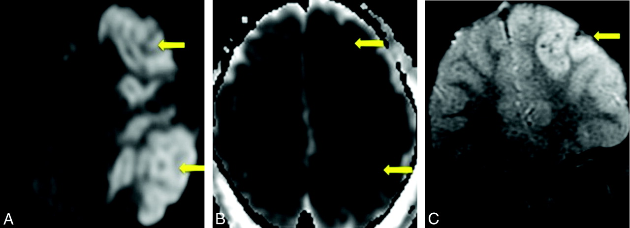
    Case 12. A, B = 1000 DWI MR image reveals multifocal cortical and subcortical hyperintensities (arrows). B, The corresponding apparent diffusion coefficient map demonstrates hypointensities in a similar distribution (arrows), consistent with infarction. C, GRE MR image reveals petechial hemorrhage (arrow) in a similar distribution.

  • Fig 3.
    • Download figure
    • Open in new tab
    • Download powerpoint
    Fig 3.

    Cases 4 and 8. Axial noncontrast CT scans demonstrate peripheral hemorrhagic infarctions (arrows) in 2 patients with DIC.

Tables

  • Figures
  • Patient demographics and clinical information

    No.SexAge (yr)DiagnosisImagingUnderlying Disease
    1M47DICCTMetastatic colon cancer
    MRISplenic infarct
    2F70DICCTStaphylococcus aureus bacterial endocarditis
    CTVaRenal, splenic infarcts
    MRI
    3M22DICCTStaphylococcus aureus bacterial endocarditis
    CTVaMeningitis
    MRI
    4M64DICCTGroup B streptococcal sepsis
    MRI
    MRVa
    5M60DICCTStreptococcus pneumoniae bacterial endocarditis
    MRICLL in remission
    6F44TTPCTAML
    MRIBreast cancer
    7M67DICCTMitral valve vegetation
    MRI
    MRVa
    8M7DICCTNone
    9M24mHTNCTRenal transplant
    MRI
    10M18mHTNCTCocaine use
    11F66mHTNMRIRenal failure
    12M7DICCTDown syndrome
    MRI
    • a Venography findings were negative.

PreviousNext
Back to top

In this issue

American Journal of Neuroradiology: 32 (4)
American Journal of Neuroradiology
Vol. 32, Issue 4
1 Apr 2011
  • Table of Contents
  • Index by author
Advertisement
Print
Download PDF
Email Article

Thank you for your interest in spreading the word on American Journal of Neuroradiology.

NOTE: We only request your email address so that the person you are recommending the page to knows that you wanted them to see it, and that it is not junk mail. We do not capture any email address.

Enter multiple addresses on separate lines or separate them with commas.
Suspicious Neuroimaging Pattern of Thrombotic Microangiopathy
(Your Name) has sent you a message from American Journal of Neuroradiology
(Your Name) thought you would like to see the American Journal of Neuroradiology web site.
CAPTCHA
This question is for testing whether or not you are a human visitor and to prevent automated spam submissions.
Cite this article
T.N. Ellchuk, L.M. Shah, R.H. Hewlett, A.G. Osborn
Suspicious Neuroimaging Pattern of Thrombotic Microangiopathy
American Journal of Neuroradiology Apr 2011, 32 (4) 734-738; DOI: 10.3174/ajnr.A2351

Citation Manager Formats

  • BibTeX
  • Bookends
  • EasyBib
  • EndNote (tagged)
  • EndNote 8 (xml)
  • Medlars
  • Mendeley
  • Papers
  • RefWorks Tagged
  • Ref Manager
  • RIS
  • Zotero
0 Responses
Respond to this article
Share
Bookmark this article
Suspicious Neuroimaging Pattern of Thrombotic Microangiopathy
T.N. Ellchuk, L.M. Shah, R.H. Hewlett, A.G. Osborn
American Journal of Neuroradiology Apr 2011, 32 (4) 734-738; DOI: 10.3174/ajnr.A2351
del.icio.us logo Twitter logo Facebook logo Mendeley logo
  • Tweet Widget
  • Facebook Like
  • Google Plus One
Purchase

Jump to section

  • Article
    • Abstract
    • Abbreviations
    • Case Series
    • Discussion
    • Conclusions
    • References
  • Figures & Data
  • Info & Metrics
  • Responses
  • References
  • PDF

Related Articles

  • No related articles found.
  • PubMed
  • Google Scholar

Cited By...

  • MRI Brain Findings in 126 Patients with COVID-19: Initial Observations from a Descriptive Literature Review
  • Cerebral Venous Thrombosis Associated with COVID-19
  • Retinal and intracranial hemorrhage in acute leukemic disseminated intravascular coagulation
  • Crossref (26)
  • Google Scholar

This article has been cited by the following articles in journals that are participating in Crossref Cited-by Linking.

  • Cerebrovascular disease in patients with COVID-19: neuroimaging, histological and clinical description
    Francisco Hernández-Fernández, Hernán Sandoval Valencia, Rosa Angélica Barbella-Aponte, Rosa Collado-Jiménez, Óscar Ayo-Martín, Cristina Barrena, Juan David Molina-Nuevo, Jorge García-García, Elena Lozano-Setién, Cristian Alcahut-Rodriguez, Álvaro Martínez-Martín, Antonio Sánchez-López, Tomás Segura
    Brain 2020 143 10
  • Cerebral Venous Thrombosis Associated with COVID-19
    D.D. Cavalcanti, E. Raz, M. Shapiro, S. Dehkharghani, S. Yaghi, K. Lillemoe, E. Nossek, J. Torres, R. Jain, H.A. Riina, A. Radmanesh, P.K. Nelson
    American Journal of Neuroradiology 2020 41 8
  • MRI Brain Findings in 126 Patients with COVID-19: Initial Observations from a Descriptive Literature Review
    E. Gulko, M.L. Oleksk, W. Gomes, S. Ali, H. Mehta, P. Overby, F. Al-Mufti, A. Rozenshtein
    American Journal of Neuroradiology 2020 41 12
  • The Spectrum of Neuroimaging Findings on CT and MRI in Adults With COVID-19
    Gul Moonis, Christopher G. Filippi, Claudia F. E. Kirsch, Suyash Mohan, Evan G. Stein, Joshua A. Hirsch, Amit Mahajan
    American Journal of Roentgenology 2021 217 4
  • Cerebral amyloid angiopathy: Review of clinico‐radiological features and mimics
    Rohit Sharma, Stephanie Dearaugo, Bernard Infeld, Richard O'Sullivan, Richard P Gerraty
    Journal of Medical Imaging and Radiation Oncology 2018 62 4
  • Cerebral MRI findings predict the risk of cognitive impairment in thrombotic thrombocytopenic purpura
    Ferras Alwan, Dina Mahdi, Sarrah Tayabali, Lisa Cipolotti, Grace Lakey, Harpreet Hyare, Marie Scully
    British Journal of Haematology 2020 191 5
  • Pediatric Neurology Part II
    Susanne Benseler, Daniela Pohl
    2013 112
  • Cerebral Magnetic Resonance Imaging Findings in Adults with Haemolytic Uraemic Syndrome Following an Infection with Escherichia coli, Subtype O104:H4
    U. Löbel, B. Eckert, O. Simova, M. Meier-Cillien, S. Kluge, C. Gerloff, J. Röther, T. Magnus, J. Fiehler
    Clinical Neuroradiology 2014 24 2
  • Cerebral Microhemorrhage: A Frequent Magnetic Resonance Imaging Finding in Pediatric Patients after Cardiopulmonary Bypass
    Paggie P C Kim, Benjamin W Nasman, Erica L Kinne, Udochukwu E Oyoyo, Daniel K Kido, J P Jacobson
    Journal of Clinical Imaging Science 2017 7
  • Childhood Primary Angiitis of the Central Nervous System With Metachronous Hemorrhagic Infarcts: A Postmortem Study With Clinicopathologic Correlation
    Christos D. Katsetos, Erica Poletto, Kathryn E. Kasmire, Diana Walleigh, Indira Kumar, Judy Mae Pascasio, Agustin Legido, Donald P. Goldsmith
    Seminars in Pediatric Neurology 2014 21 2

More in this TOC Section

  • Usefulness of Quantitative Susceptibility Mapping for the Diagnosis of Parkinson Disease
  • Evaluating the Effects of White Matter Multiple Sclerosis Lesions on the Volume Estimation of 6 Brain Tissue Segmentation Methods
  • White Matter Alterations in the Brains of Patients with Active, Remitted, and Cured Cushing Syndrome: A DTI Study
Show more Brain

Similar Articles

Advertisement

Indexed Content

  • Current Issue
  • Accepted Manuscripts
  • Article Preview
  • Past Issues
  • Editorials
  • Editors Choice
  • Fellow Journal Club
  • Letters to the Editor

Cases

  • Case Collection
  • Archive - Case of the Week
  • Archive - Case of the Month
  • Archive - Classic Case

Special Collections

  • Special Collections

Resources

  • News and Updates
  • Turn around Times
  • Submit a Manuscript
  • Author Policies
  • Manuscript Submission Guidelines
  • Evidence-Based Medicine Level Guide
  • Publishing Checklists
  • Graphical Abstract Preparation
  • Imaging Protocol Submission
  • Submit a Case
  • Become a Reviewer/Academy of Reviewers
  • Get Peer Review Credit from Publons

Multimedia

  • AJNR Podcast
  • AJNR SCANtastic
  • Video Articles

About Us

  • About AJNR
  • Editorial Board
  • Not an AJNR Subscriber? Join Now
  • Alerts
  • Feedback
  • Advertise with us
  • Librarian Resources
  • Permissions
  • Terms and Conditions

American Society of Neuroradiology

  • Not an ASNR Member? Join Now

© 2025 by the American Society of Neuroradiology All rights, including for text and data mining, AI training, and similar technologies, are reserved.
Print ISSN: 0195-6108 Online ISSN: 1936-959X

Powered by HighWire