
Article Figures & Data
Figures
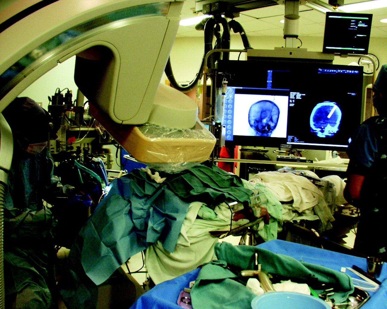
- Fig 1.
Ventricular drain placement. The surgeon is working adjacent to the C-arm during placement of the ventriculostomy catheter.
- Fig 2.
Axial images from a FPCT (left) and a standard noncontrast CT (right) demonstrate the tip of the ventricular catheter at the foramen of Monro.


In this issue


Jump to section
Related Articles
Cited By...
- How to iGuide: flat panel detector, CT-assisted, minimally invasive evacuation of intracranial hematomas
- Accuracy of image-guided percutaneous injection into a phantom spinal cord utilizing flat panel detector CT with MR fusion and integrated navigational software
- Accuracy of flat panel detector CT with integrated navigational software with and without MR fusion for single-pass needle placement
- Minimally invasive cone beam CT-guided evacuation of parenchymal and ventricular hemorrhage using the Apollo system: proof of concept in a cadaver model