
Article Figures & Data
Figures
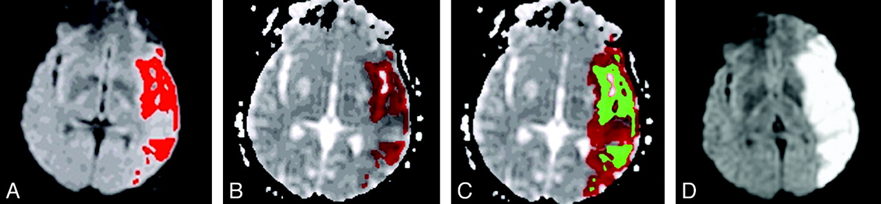
- Fig 1.
Prediction of MCA infarct growth in a hyperglycemic patient. The initial lesion was delineated on DWI images (A) and registered on the ADC map (B) in red; starting from the mask of the initial lesion obtained in B and represented in green (C), the region-growing model segmented the ADC map and iteratively added some voxels to the lesion mask, until a regional cutoff value was reached. (D) The observed final lesion visible as a bright area on the follow-up DWI.
- Fig 2.
Effect of SGL. Scatterplots of observed versus predicted infarct growths for hyperglycemic (open circles) and nonhyperglycemic patients (closed circles). The slope of the regression line for hyperglycemic patients (0.98 ± 0.16, P < .0001, R2 = 0.54) compared with the slope for nonhyperglycemic patients (0.38 ± 0.08, P < .0001, R2 = 0.27) was significantly higher (P = .0008).


Tables
- Table 1:
Clinical and radiologic characteristics in the hyperglycemic and nonhyperglycemic groups
Hyperglycemic Patients (n = 34) Nonhyperglycemic Patients (n = 60) P Value Age (yr) 60 62 .65 57–61 55–67 Thrombolytic treatment (%) 74 63 .38 Baseline NIHSS score 17 14 .003 15–20 11–16 Baseline SGL (mmol/L) 8.7 5.8 <.0001 7.8–10.9 5.4–6.2 Time to initial MRI (min) 132 150 .22 112–157 128–176 Time to follow-up MRI (hr) 32 28 .13 26.5–45.7 23–37.3 Recanalized patients (n, %) 23 44 .70 68% 72% Initial DWI volume (cm3) 29 18.3 .08 7.9, 47.5 16.6, 55.3 Mean ADC in initial DWI vol (× 10−6 mm2/s) 652 633 .56 609–694 580–674 Final DWI volume (cm3) 74.5 31.8 .004 34.8, 134.1 20.4, 57 Predicted final volume (cm3) 50.5 39.7 .25 21.3, 123.8 15.9, 82.2 Predicted infarct growth (cm3) 29.3 23.5 .47 (0.9, 53.3) (5.4, 44.5) Observed infarct growth (cm3) 40.2 9.2 .0003 (10.6, 85) (3.2, 30.8) Spared tissue-at-risk (cm3) −5.9 7 .0025 −55.1, 8.5 −3.6, 25.2 -
Note:Numbers are shown as the median and interquartile range.
-
- Table 2:
Coefficients of the multiple regression equations predicting observed infarct growth in the whole population and in the subgroups of nonrecanalized and recanalized patients
Population/Subgroup Variable Coefficient SE P Value Whole population (n = 94) PIG (cm3) 0.73 0.08 <.0001 Hyperglycemic status (yes = 1, no = 0) 29 6.9 .0001 MCA recanalization (yes = 0, no = 1) 24 7.4 .001 Constant −8.9 Recanalized patients (n = 67) PIG (cm3) 0.56 0.06 <.0001 Hyperglycemic status (yes = 1, no = 0) 18 5.5 .002 Constant 0.5 Nonrecanalized patients (n = 26) PIG (cm3) 1.07 0.22 .0001 Hyperglycemic status (yes = 1, no = 0) 51 18 .01 Constant −8
In this issue


Jump to section
Related Articles
Cited By...
- Impact of Glucose on Outcomes in Patients Treated With Mechanical Thrombectomy: A Post Hoc Analysis of the Solitaire Flow Restoration With the Intention for Thrombectomy Study
- Opposing Effects of Glucose on Stroke and Reperfusion Injury: Acidosis, Oxidative Stress, and Energy Metabolism
- Intensive Versus Subcutaneous Insulin in Patients With Hyperacute Stroke: Results From the Randomized INSULINFARCT Trial
- Glucose and Acute Stroke: Evidence for an Interlude