
Article Figures & Data
Figures
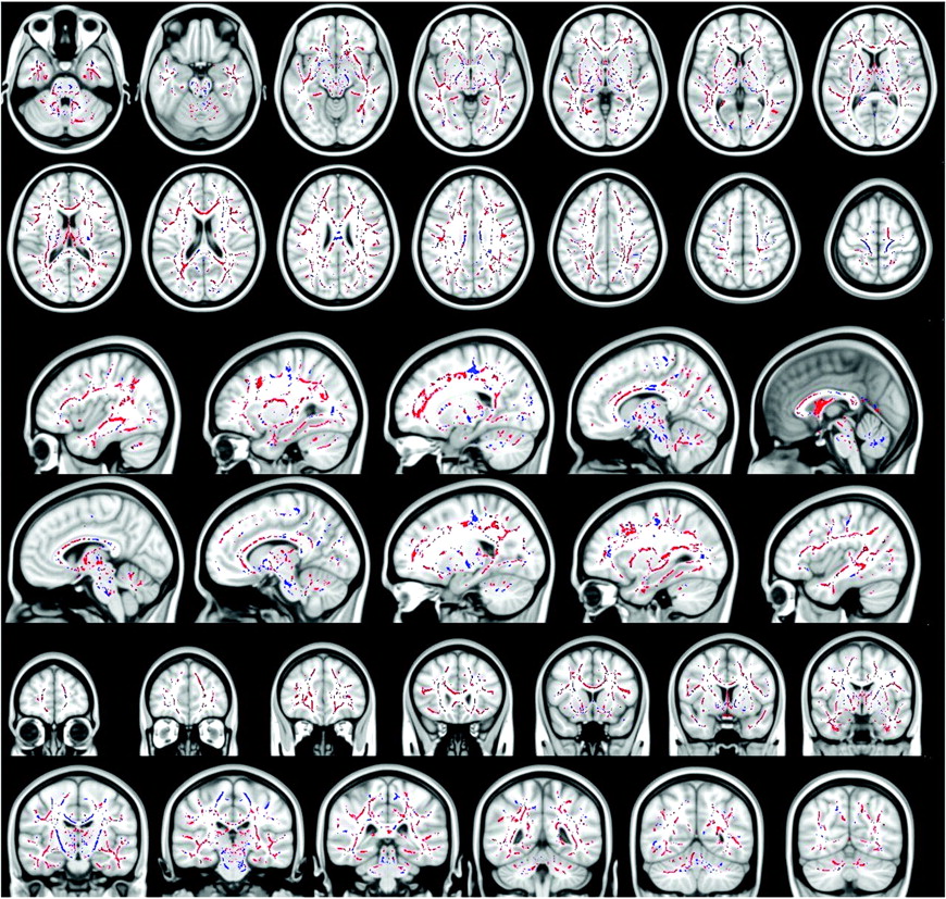
- Fig 1.
T1 MTC MR sagittal (A and B) and axial (C) images show a hyperintense signal intensity along the CST and in the posterior aspect of the CC (B).
- Fig 2.
Blue voxels show the regions within and outside the CST, where the FA is significantly reduced in patients with ALS with T1 MTC hyperintense signal intensity compared with patients with ALS without T1 MTC hyperintense signal intensity (all P values < .05, corrected at the cluster level). Red voxels show the regions where the FA is significantly increased in patients with ALS with T1 MTC hyperintense signal intensity compared with patients with ALS without T1 MTC hyperintense signal intensity (all P values < .05, corrected at cluster level). In the CC, patients with T1 MTC hyperintense signal intensity compared with patients without T1 MTC hyperintensity show a significantly lower FA only in the posterior aspect of the body of the CC, whereas they show higher FA in the entire CC, in the splenium, in the genu, and in the anterior body.


Tables
- Table 1:
Sensitivity and specificity of T1 MTC hyperintense signal in patients with ALS and controls
Patients with ALS Controls T1 MTC hyperintensity Present 16 0 Absent 27 43 Sensitivity (%) 37.2 Specificity (%) 100 Positive predictive value 1 Negative predictive value 0.61 - Table 2:
Mean FA of clusters with statistically significant differences between patients with ALS with T1 MTC hyperintense signal and those without it and controlsa
Patients with ALS Controls MT Hyperintensity No MT Hyperintensity Entire CST 0.403 (0.024) 0.432 (0.022) 0.454 (0.024) CST including internal capsule 0.373 (0.024) 0.411 (0.021) 0.431 (0.024) Middle cerebellar peduncles 0.435 (0.020) 0.464 (0.019) 0.480 (0.020) Pons 0.414 (0.014) 0.427 (0.014) 0.445 (0.015) Entire CC 0.580 (0.018) 0.569 (0.018) 0.619 (0.019) Genu CC 0.517 (0.022) 0.499 (0.022) 0.549 (0.023) Body CC 0.530 (0.014) 0.524 (0.014) 0.584 (0.015) Anterior body CC 0.532 (0.013) 0.514 (0.013) 0.571 (0.014) Posterior body CC 0.528 (0.015) 0.536 (0.015) 0.602 (0.016) Splenium CC 0.679 (0.015) 0.665 (0.015) 0.706 (0.015) Temporal white matter 0.268 (0.013) 0.245 (0.012) 0.281 (0.013) Parietal white matter 0.236 (0.016) 0.212 (0.015) 0.255 (0.016) Frontal white matter 0.243 (0.014) 0.234 (0.013) 0.259 (0.014) -
↵a All P values <.05. Numbers in parentheses are SDs.
-
In this issue


Jump to section
Related Articles
Cited By...
- A distinct imaging phenotype in amyotrophic lateral sclerosis confidently detected on T1 MTC
- Corticospinal Tract MR Signal-Intensity Pseudonormalization on Magnetization Transfer Contrast Imaging: A Potential Pitfall in the Interpretation of the Advanced Compromise of Upper Motor Neurons in Amyotrophic Lateral Sclerosis