
Article Figures & Data
Figures
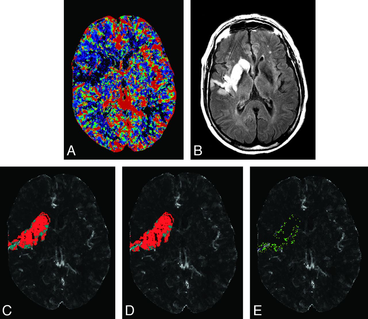
- Fig 1.
Admission CBF (A) and 7-day follow-up FLAIR (B) of a 74-year-old man with an ICA and M1 branch occlusion scanned 168 minutes after symptom onset and administered intravenous rtPA. The region of abnormality on follow-up FLAIR is coregistered to CBF and thresholded on the basis of without (C) and with covariate (D) thresholds from Table 1. Red voxels present predicted infarct, and cyan voxels present predicted noninfarct tissues on C and D. E, A subtraction of C and D representing voxels that were designated as infarcted tissue (green) and noninfarcted (pink, very few mainly located adjacent to blood vessels on E) only on the model with covariates.

Tables
- Table 1:
Model performance without and with baseline clinical covariates for distinguishing infarct from noninfarct regions for the best performing perfusion parametersa
In All Patients Cutoff Value Accuracy AIC P Value CBF-GM-Rel A) Without any covariate 0.64 0.88 2465.9 B) With covariates 0.78 0.91 2401.4 <.0001b CBF-WM-Rel A) Without any covariate 0.80 0.85 2693.6 B) With covariates 0.87 0.86 2593.6 <.0001b Tmax-GM-Abs A) Without any covariate 6.0 0.84 1377.8 B) With covariates 4.4 0.88 1358.5 .0113b Tmax-WM-Abs A) Without any covariate 5.9 0.79 1251.5 B) With covariates 3.7 0.80 1232.1 .0108b - Table 2:
Performance of selected nontransformed (model A) and covariate transformed (model B) perfusion thresholds for tissue fate prediction in derivation and validation data bases
Perfusion Parameter Derivation Data Base Validation Data Base Sens. Spec. Acc. Sens. Spec. Acc. CBF-GM-Rel A) Without any covariate 0.88 0.89 0.88 0.72 1 0.82 B) With covariates 0.89 0.93 0.91 0.78 1 0.86 CBF-WM-Rel A) Without any covariate 0.86 0.84 0.85 0.76 0.95 0.82 B) With covariates 0.90 0.80 0.86 0.79 0.91 0.83 Tmax-GM-Abs A) Without any covariate 0.80 0.92 0.84 0.70 0.93 0.78 B) With covariates 0.84 0.96 0.88 0.69 0.97 0.79 Tmax-WM-Abs A) Without any covariate 0.76 0.85 0.79 0.70 0.88 0.76 B) With covariates 0.80 0.80 0.80 0.64 0.94 0.76 Note:—Rel indicates relative to the contralateral side; Abs, absolute value; Sens, sensitivity; Spec, specificity; Acc, accuracy.
- Table 3:
Selected model performance without and with clinical covariates for good clinical outcome predictiona
In All Patients AIC G2 P Value CBF-GM-Rel Null model 291.5 – A) Without any covariate 291.7 1.796 .1803 B) With covariates 291.3 2.247 .1338 CBF-WM-Rel Null model 282.5 – A) Without any covariate 280.4 4.080 .0434 B) With covariates 268.9 15.613 <.0001b Tmax-GM-Abs Null model 291.5 – A) Without any covariate 291.3 2.255 .1332 B) With covariates 285.4 8.116 .0044b Tmax-WM-Abs Null model 282.5 – A) Without any covariate 280.9 3.643 .0563 B) With covariates 266.2 18.328 <.0001b Note:—Rel indicates relative to the contralateral side; Abs, absolute value; AIC, Akaike Information Criterion; G2, the difference between −2 L of the fitted model (transformed threshold) and the reference model (nontransformed threshold).
↵a Remaining parameters are presented in On-line Table 3.
↵b Significant.


Jump to section
Related Articles
Cited By...
- Reducing False-Positives in CT Perfusion Infarct Core Segmentation Using Contralateral Local Normalization
- A framework for testing different imputation methods for tabular datasets
- Acute Stroke Imaging Research Roadmap III Imaging Selection and Outcomes in Acute Stroke Reperfusion Clinical Trials: Consensus Recommendations and Further Research Priorities
- Effect of Collaterals on Clinical Presentation, Baseline Imaging, Complications, and Outcome in Acute Stroke
- Contribution and Additional Impact of Imaging to the SPAN-100 Score