
Article Figures & Data
Figures
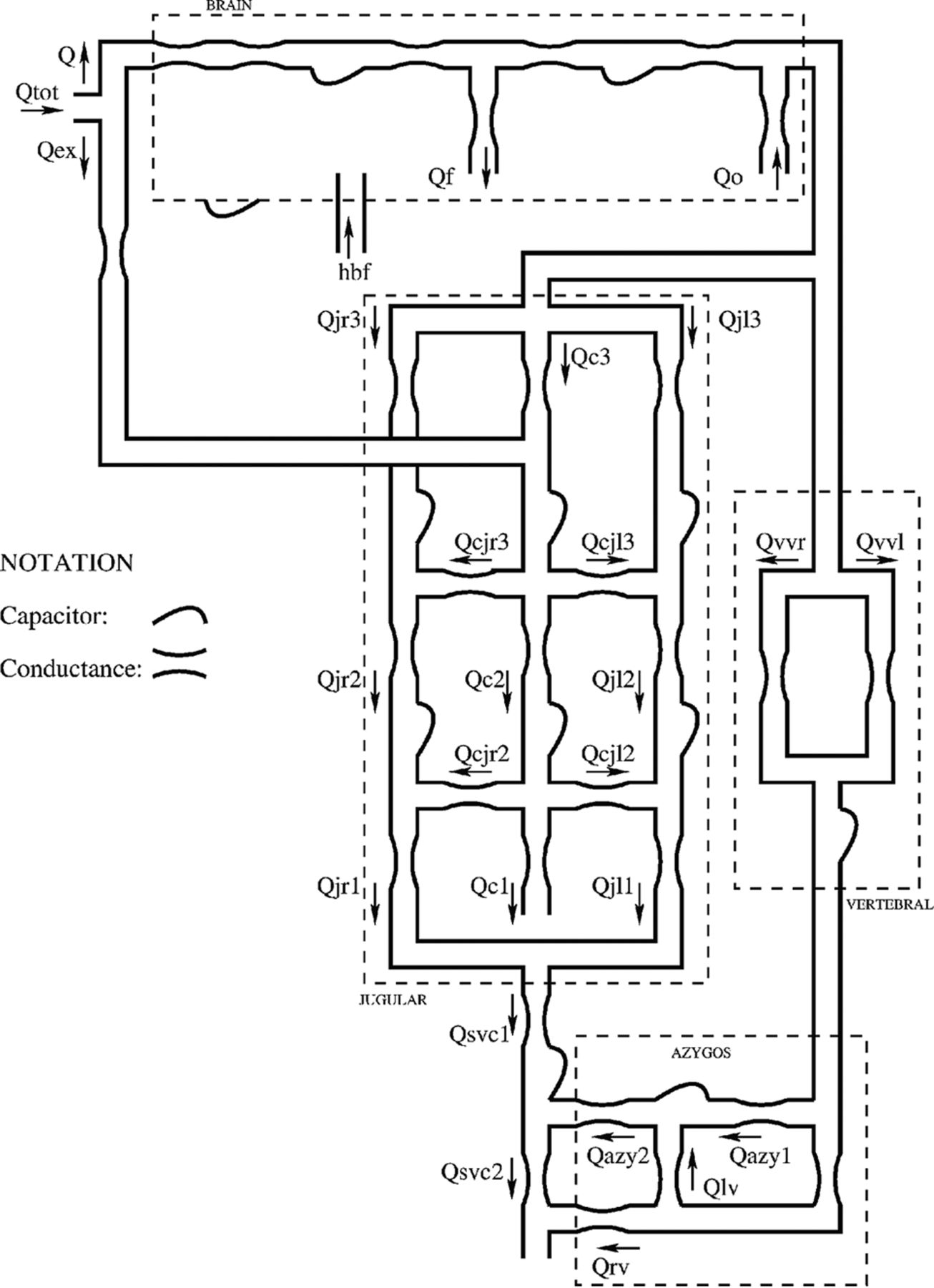
- Fig 1.
Scheme of the hemodynamic parameter model for the study of cerebral venous outflow.8
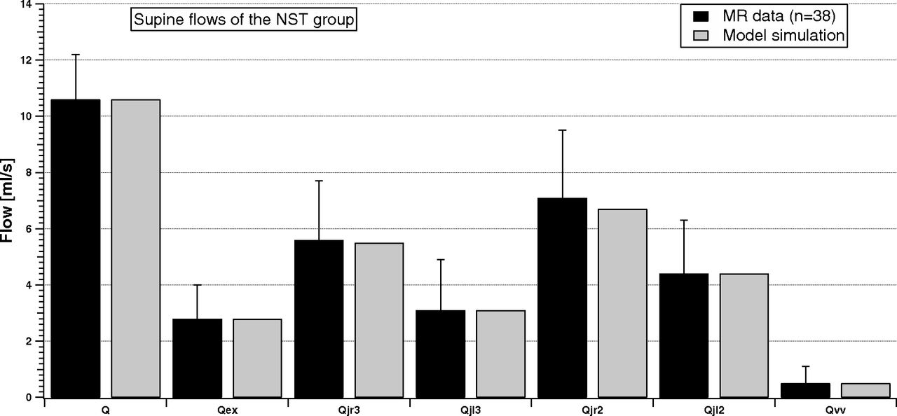
- Fig 2.
Comparison between supine MR imaging data (dark columns) and model simulation (light columns) of the NST group.
- Fig 3.
Comparison between supine MR imaging data (dark columns) and model simulation (light columns) of the LL-R ST group.
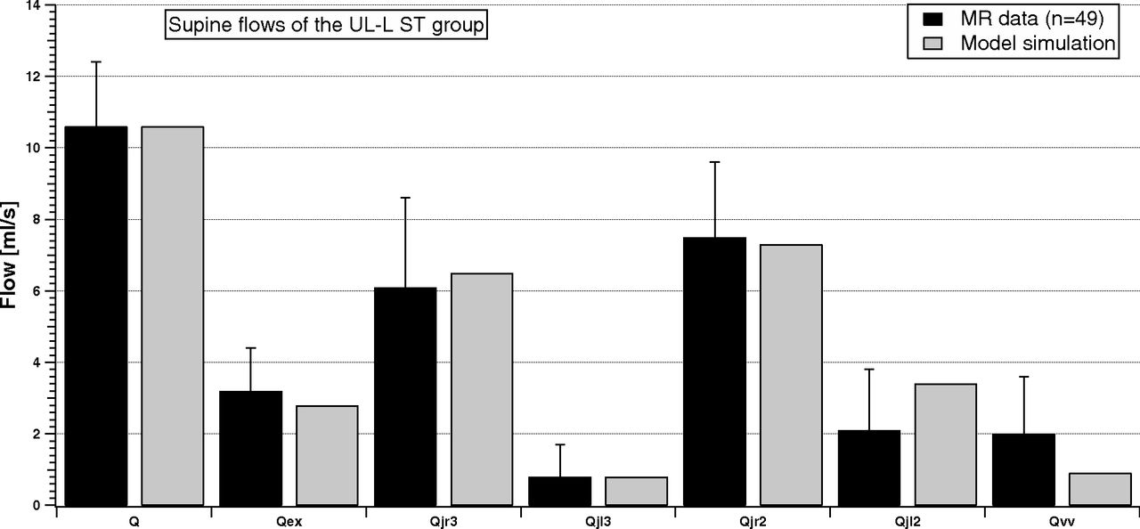
- Fig 4.
Comparison between supine MR imaging data (dark columns) and model simulation (light columns) of the UL-L ST group.
- Fig 5.
Comparison between supine and upright simulated total jugular (sum of left and right) and vertebral average flows of the NST group. The percentage changes are −32.6%, −40.5%, −8.1%, and +120%, which fit those in Table 3 very well.
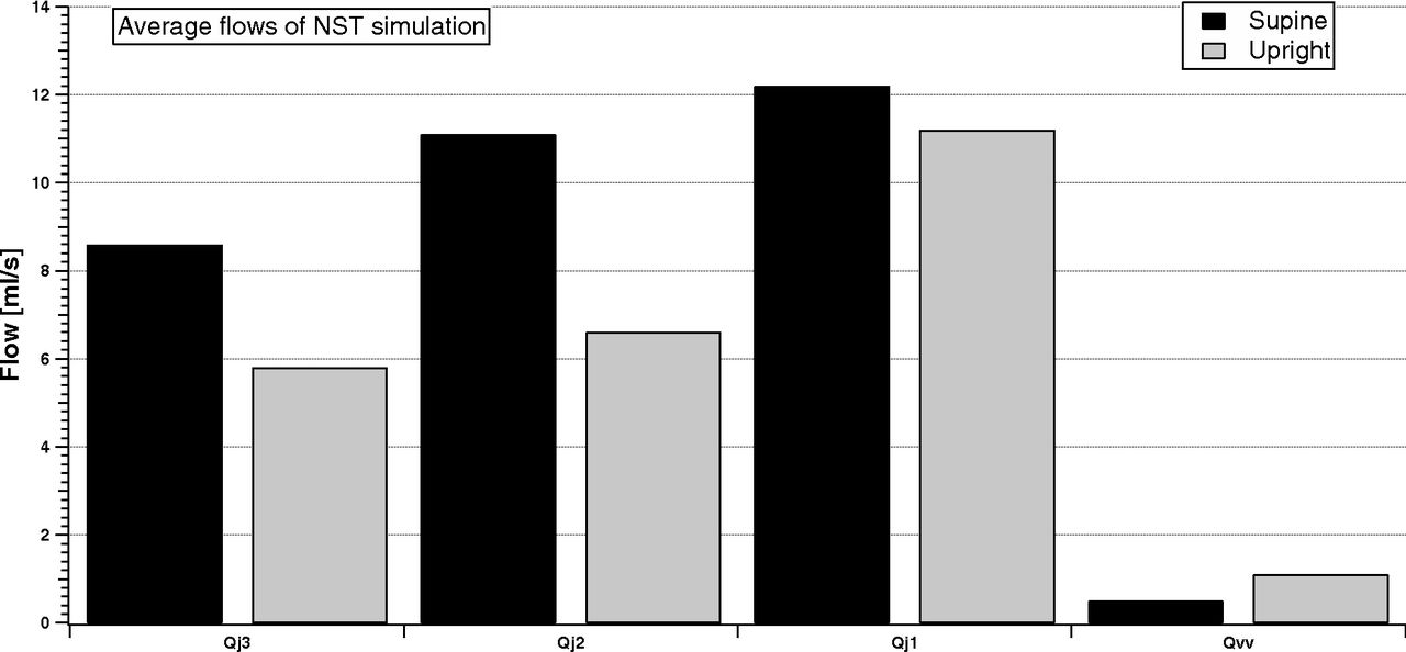
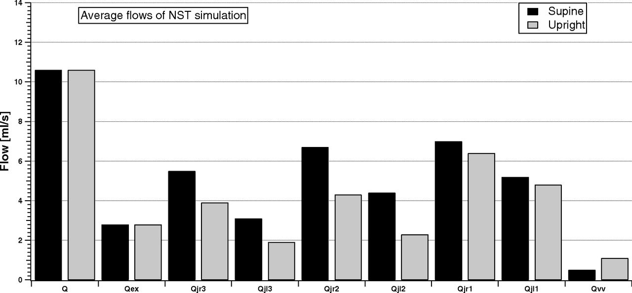
- Fig 6.
Model simulation of the cerebral, external, jugular (left and right), and vertebral average flows in supine and upright conditions for the NST group.
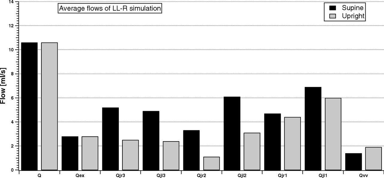
- Fig 7.
Model simulation of the cerebral, external, jugular (left and right), and vertebral average flows in the supine and upright condition for the LL-R ST group.
- Fig 8.
Model simulation of the cerebral, external, jugular (left and right), and vertebral average flows in the supine and upright condition for the UL-L ST group.
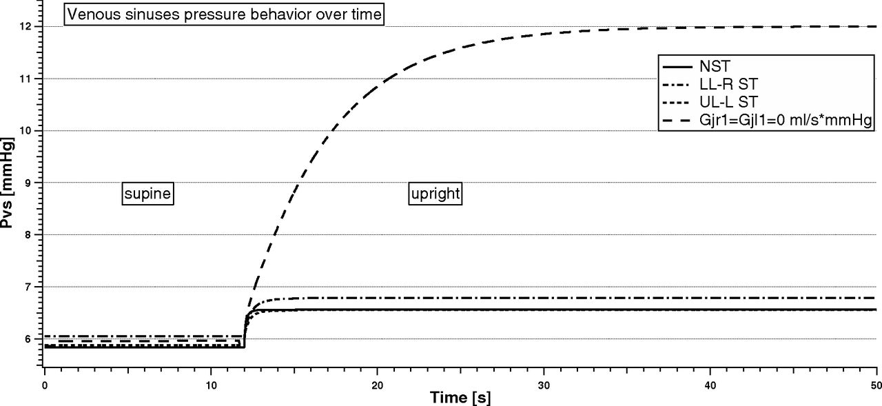
- Fig 9.
Venous sinuses pressure behavior with time in the supine and upright simulations for different stenotic patterns.


Tables
- Table 1:
List of the conductance values to reproduce the average flow data reported in Table 2 and the flow percentage variation reported in Table 3
G (mL/s × mm Hg) NST LL-R ST UL-L ST A) Gvv1 0.60 3.9 7.7 kjl3 6.00 6.00 0.86 kjr2 16.00 2.30 16.00 B) Gc2 11.00 Gc3 21.43 Gcjl2 6.67 Gcjl3 16.00 Gazy1 1.33 Gazy2 1.78 Gc1 1.18 Gcjr2 6.67 Gcjr3 21.00 Gex 0.03 Glv 0.89 Grv 0.41 Gsvc1 78.50 Gsvc2 81.17 Gvv2 0.83 kjl1 7.27 Kjl2 8.00 kjr1 7.27 Kjr3 13.00 Note:—G indicates conductance; Gvvl, conductance of the vertebral system (upper part); kjl3, parameter for the basal conductance of the upper segment of the left jugular vein; Gazy1, conductance of the distal azygos vein; Gazy2, conductance of the proximal azygos vein; Gc1, conductance of the lower segment of the collateral network; Gc2, conductance of the middle segment of the collateral network; Gc3, conductance of the upper segment of the collateral network; Gcjl2, conductance of the lower anastomotic connection (left side); Gcjl3, conductance of the upper anastomotic connection (left side); Gcjr2, conductance of the lower anastomotic connection (right side); Gcjr3, conductance of the upper anastomotic connection (right side); Gex, conductance of the external carotid arteries; Glv, conductance of the lumbar vein; Grv, conductance of the renal vein; Gsvc1, conductance of the upper segment of the superior vena cava (jugular confluence); Gsvc2, conductance of the lower segment of the superior vena cava; Gvv2, conductance of the vertebral system (lower part); kjl1, parameter for the basal conductance of the lower segment of the left jugular vein; kjl2, parameter for the basal conductance of the middle segment of the left jugular vein; kjr1, parameter for the basal conductance of the lower segment of the right jugular vein; kjr3, parameter for the basal conductance of the upper segment of the right jugular vein; kjr2, middle segment of the right jugular vein.
- Table 2:
MRI average data of flows related to cerebral, external, jugular, and vertebral circuits in the supine conditiona
MRI n Q Qex Qjr3 Qjl3 Qjr2 Qjl2 Qvv NST 38 10.6 ± 1.6 2.8 ± 1.2 5.6 ± 2.1 3.1 ± 1.8 7.1 ± 2.4 4.4 ± 1.9 0.5 ± 0.6 LL-R ST 20 10.7 ± 1.8 3.0 ± 1.1 3.5 ± 2.5 4.0 ± 2.1 3.6 ± 2.7 4.7 ± 2.6 1.8 ± 1.6 UL-L ST 49 10.6 ± 1.8 3.2 ± 1.2 6.1 ± 2.5 0.8 ± 0.9 7.5 ± 2.1 2.1 ± 1.7 2.0 ± 1.6 ↵a Flow values are reported in milliliters/second with SDs.
- Table 3:
ECD average data of flow related to cerebral, jugular, and vertebral circuits in the supine NST conditiona
ECD Q Qj3 Qj2 Qj1 Qvv Supine 10.6 ± 1.9 6.0 ± 2.6 8.9 ± 3.4 22.0 ± 10.3 1.1 ± 0.7 Upright 10.6 ± 1.9 4.1 ± 2.0 5.2 ± 3.3 20.4 ± 12.5 2.3 ± 1.2 Variation (%) 0 −32 −42 −7 +109 ↵a Flow values are reported in milliliters/second with SDs.


Jump to section
Related Articles
Cited By...
- No citing articles found.