Advertisement

Main menu

  • Home
  • Content
    • Current Issue
    • Accepted Manuscripts
    • Article Preview
    • Past Issue Archive
    • AJNR Case Collection
    • Case of the Week Archive
    • Classic Case Archive
    • Case of the Month Archive
  • Special Collections
    • Spinal CSF Leak Articles (Jan 2020-June 2024)
    • 2024 AJNR Journal Awards
    • Most Impactful AJNR Articles
  • Multimedia
    • AJNR Podcast
    • AJNR Scantastics
    • Video Articles
  • For Authors
    • Submit a Manuscript
    • Author Policies
    • Fast publishing of Accepted Manuscripts
    • Graphical Abstract Preparation
    • Manuscript Submission Guidelines
    • Imaging Protocol Submission
    • Submit a Case for the Case Collection
  • About Us
    • About AJNR
    • Editorial Board
  • More
    • Become a Reviewer/Academy of Reviewers
    • Subscribers
    • Permissions
    • Alerts
    • Feedback
    • Advertisers
    • ASNR Home
  • Other Publications
    • ajnr

User menu

  • Alerts
  • Log in

Search

  • Advanced search
American Journal of Neuroradiology

American Journal of Neuroradiology

ASHNR American Society of Functional Neuroradiology ASHNR American Society of Pediatric Neuroradiology ASSR
  • Alerts
  • Log in

Advanced Search

  • Home
  • Content
    • Current Issue
    • Accepted Manuscripts
    • Article Preview
    • Past Issue Archive
    • AJNR Case Collection
    • Case of the Week Archive
    • Classic Case Archive
    • Case of the Month Archive
  • Special Collections
    • Spinal CSF Leak Articles (Jan 2020-June 2024)
    • 2024 AJNR Journal Awards
    • Most Impactful AJNR Articles
  • Multimedia
    • AJNR Podcast
    • AJNR Scantastics
    • Video Articles
  • For Authors
    • Submit a Manuscript
    • Author Policies
    • Fast publishing of Accepted Manuscripts
    • Graphical Abstract Preparation
    • Manuscript Submission Guidelines
    • Imaging Protocol Submission
    • Submit a Case for the Case Collection
  • About Us
    • About AJNR
    • Editorial Board
  • More
    • Become a Reviewer/Academy of Reviewers
    • Subscribers
    • Permissions
    • Alerts
    • Feedback
    • Advertisers
    • ASNR Home
  • Follow AJNR on Twitter
  • Visit AJNR on Facebook
  • Follow AJNR on Instagram
  • Join AJNR on LinkedIn
  • RSS Feeds

Welcome to the new AJNR, Updated Hall of Fame, and more. Read the full announcements.


AJNR is seeking candidates for the position of Associate Section Editor, AJNR Case Collection. Read the full announcement.


Research ArticlePediatrics

Effectiveness of 3D T2-Weighted FLAIR FSE Sequences with Fat Suppression for Detection of Brain MR Imaging Signal Changes in Children

M. Mascalchi, A. Bianchi, M. Basile, P. Gulino, M.R. Trifan, D. Difeo, E. Bartolini, C. Defilippi and S. Diciotti
American Journal of Neuroradiology December 2016, 37 (12) 2376-2381; DOI: https://doi.org/10.3174/ajnr.A4915
M. Mascalchi
aFrom the “Mario Serio” Department of Experimental and Clinical Biomedical Sciences (M.M., A.B.), University of Florence, Florence, Italy
bStructural and Functional Neuroradiology Research Unit (M.M.)
  • Find this author on Google Scholar
  • Find this author on PubMed
  • Search for this author on this site
  • ORCID record for M. Mascalchi
A. Bianchi
aFrom the “Mario Serio” Department of Experimental and Clinical Biomedical Sciences (M.M., A.B.), University of Florence, Florence, Italy
  • Find this author on Google Scholar
  • Find this author on PubMed
  • Search for this author on this site
  • ORCID record for A. Bianchi
M. Basile
cDiagnostic Radiology (M.B., P.G., M.R.T., D.D., C.D.)
  • Find this author on Google Scholar
  • Find this author on PubMed
  • Search for this author on this site
  • ORCID record for M. Basile
P. Gulino
cDiagnostic Radiology (M.B., P.G., M.R.T., D.D., C.D.)
  • Find this author on Google Scholar
  • Find this author on PubMed
  • Search for this author on this site
  • ORCID record for P. Gulino
M.R. Trifan
cDiagnostic Radiology (M.B., P.G., M.R.T., D.D., C.D.)
  • Find this author on Google Scholar
  • Find this author on PubMed
  • Search for this author on this site
  • ORCID record for M.R. Trifan
D. Difeo
cDiagnostic Radiology (M.B., P.G., M.R.T., D.D., C.D.)
  • Find this author on Google Scholar
  • Find this author on PubMed
  • Search for this author on this site
  • ORCID record for D. Difeo
E. Bartolini
dNeurology Unit and Laboratories (E.B.), Meyer Children's Hospital, Florence, Italy
  • Find this author on Google Scholar
  • Find this author on PubMed
  • Search for this author on this site
  • ORCID record for E. Bartolini
C. Defilippi
cDiagnostic Radiology (M.B., P.G., M.R.T., D.D., C.D.)
  • Find this author on Google Scholar
  • Find this author on PubMed
  • Search for this author on this site
  • ORCID record for C. Defilippi
S. Diciotti
eDepartment of Electrical, Electronic, and Information Engineering “Guglielmo Marconi” (S.D.), University of Bologna, Cesena, Italy.
  • Find this author on Google Scholar
  • Find this author on PubMed
  • Search for this author on this site
  • ORCID record for S. Diciotti
  • Article
  • Figures & Data
  • Info & Metrics
  • Responses
  • References
  • PDF
Loading

Article Figures & Data

Figures

  • Tables
  • Fig 1.
    • Download figure
    • Open in new tab
    • Download powerpoint
    Fig 1.

    T2 hyperintensity of the WM adjacent to the occipital horns of the lateral ventricles (arrows) in a 9-year-old girl referred for MR imaging for episodes of hypersomnia. This is often observed as an isolated finding in children with otherwise normal MR imaging examination and was indicated in the training session to represent a borderline or normal finding to the 3 observers and not to be marked as abnormal T2 hyperintensity.
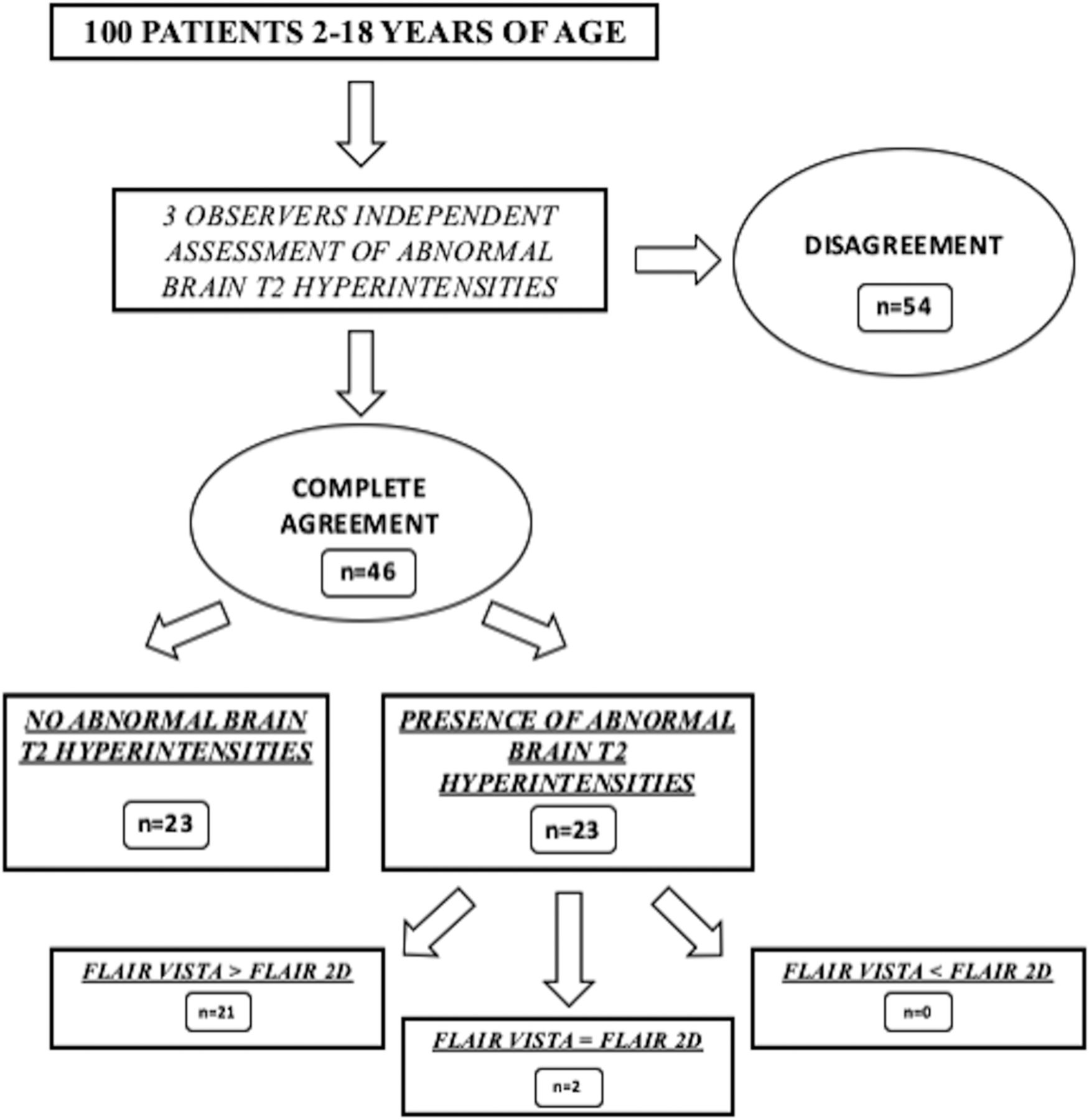
  • Fig 2.
    • Download figure
    • Open in new tab
    • Download powerpoint
    Fig 2.

    Results of the visual assessment by the 3 observers of T2 hyperintensities in 100 MR imaging examinations.

  • Fig 3.
    • Download figure
    • Open in new tab
    • Download powerpoint
    Fig 3.

    Abnormal focal T2 hyperintensity (arrows) consistent with focal cortical dysplasia type II in the right fusiform gyrus of a 10-year-old boy with partial epilepsy. All 3 observers judged that the abnormal T2 hyperintensity was more conspicuous in FLAIR-VISTA (B–D) than in axial 2D-FLAIR (A).
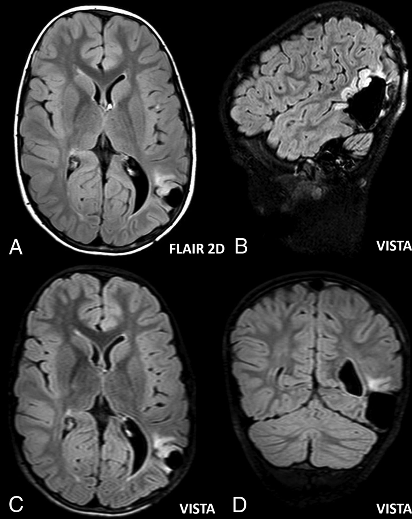
  • Fig 4.
    • Download figure
    • Open in new tab
    • Download powerpoint
    Fig 4.

    Abnormal focal T2 hyperintensity corresponding to pathologically verified recurrence of dysembryoplastic neuroepithelial tumor in the left parietal-occipital region of a 10-year-old boy. Two observers judged that the abnormal T2 hyperintensity was more conspicuous in FLAIR-VISTA (B–D) than in axial 2D-FLAIR (A), whereas 1 observer judged that the conspicuity was similar.
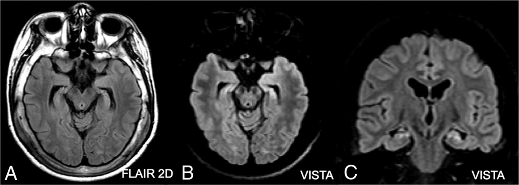
  • Fig 5.
    • Download figure
    • Open in new tab
    • Download powerpoint
    Fig 5.

    Abnormal focal T2 hyperintensity consistent with mesial temporal sclerosis in both hippocampi of a 13-year-old boy with partial epilepsy, which followed an acute encephalopathy of undefined etiology. The abnormal focal hyperintensity was judged to be present (and more conspicuous on FLAIR-VISTA) by 2 observers, whereas 1 observer did not report it. Note the marked diffuse T2 hyperintensity of the thickened skull in 2D-FLAIR (A), which is not present on axial (B) and coronal (C) reformatted images of FLAIR-VISTA with fat suppression.

Tables

  • Figures
    • View popup
    Table 1:

    Patient demographic characteristics, number of MRI examinations under sedation, and clinical indications in the training and test sets

    Training SetTest Set
    No.20100
    Age (mean) (range) (yr)9 ± 6.5, 2–179 ± 7, 2–18
    SexFemale, n = 10; male, n = 10Female, n = 45; male, n = 55
    No. of sedations834
    Clinical indications
        Epilepsy940
        Postsurgery for tumor or epilepsy313
        Headache–12
        Malformation–5
        Perinatal damage12
        Others728
    • View popup
    Table 2:

    Expert neuroradiologist's evaluation of the training set

    MRI DiagnosisNo.Presence of Abnormal T2 Hyperintensity
    None4–
    WM UBOs44
    Leukoencephalopathy22
    Focal cortical dysplasia11
    Neuronal migration disorders1–
    UBOs, neurofibromatosis type 111
    Tuber in tuberous sclerosis11
    Multiple sclerosis plaques11
    Brain infarct––
    Intra-axial tumor––
    Postsurgical tumor evaluation33
    Myelination delay11
    Brain malformations––
    Others11
    • Note:—UBOs indicates unidentified bright objects.

    • View popup
    Table 3:

    Expert neuroradiologist's evaluation of the test set

    MRI DiagnosisNo.Presence of Abnormal T2 Hyperintensity
    None28–
    WM UBOs2222
    Leukoencephalopathy99
    Focal cortical dysplasia44
    Neuronal migration disorders1–
    UBOs, neurofibromatosis type 122
    Tuber in tuberous sclerosis11
    Multiple sclerosis plaques––
    Brain infarct33
    Intra-axial tumor22
    Postsurgical tumor evaluation1111
    Myelination delay22
    Brain malformations5–
    Others1010
    • Note:—UBOs indicates unidentified bright objects.

    • View popup
    Table 4:

    Weighted κ statistic assessment of the agreement of the 3 observers

    Observers 1 and 2Observers 1 and 3Observers 2 and 3Mean Value
    Presence of abnormal T2 hyperintensities of the brain (n = 100)0.690.490.640.61
    Conspicuity of abnormal T2 hyperintensities in FLAIR-VISTA vs 2D-FLAIR (n = 23)0.270.380.230.29
PreviousNext
Back to top

In this issue

American Journal of Neuroradiology: 37 (12)
American Journal of Neuroradiology
Vol. 37, Issue 12
1 Dec 2016
  • Table of Contents
  • Index by author
  • Complete Issue (PDF)
Advertisement
Print
Download PDF
Email Article

Thank you for your interest in spreading the word on American Journal of Neuroradiology.

NOTE: We only request your email address so that the person you are recommending the page to knows that you wanted them to see it, and that it is not junk mail. We do not capture any email address.

Enter multiple addresses on separate lines or separate them with commas.
Effectiveness of 3D T2-Weighted FLAIR FSE Sequences with Fat Suppression for Detection of Brain MR Imaging Signal Changes in Children
(Your Name) has sent you a message from American Journal of Neuroradiology
(Your Name) thought you would like to see the American Journal of Neuroradiology web site.
CAPTCHA
This question is for testing whether or not you are a human visitor and to prevent automated spam submissions.
Cite this article
M. Mascalchi, A. Bianchi, M. Basile, P. Gulino, M.R. Trifan, D. Difeo, E. Bartolini, C. Defilippi, S. Diciotti
Effectiveness of 3D T2-Weighted FLAIR FSE Sequences with Fat Suppression for Detection of Brain MR Imaging Signal Changes in Children
American Journal of Neuroradiology Dec 2016, 37 (12) 2376-2381; DOI: 10.3174/ajnr.A4915

Citation Manager Formats

  • BibTeX
  • Bookends
  • EasyBib
  • EndNote (tagged)
  • EndNote 8 (xml)
  • Medlars
  • Mendeley
  • Papers
  • RefWorks Tagged
  • Ref Manager
  • RIS
  • Zotero
0 Responses
Respond to this article
Share
Bookmark this article
Effectiveness of 3D T2-Weighted FLAIR FSE Sequences with Fat Suppression for Detection of Brain MR Imaging Signal Changes in Children
M. Mascalchi, A. Bianchi, M. Basile, P. Gulino, M.R. Trifan, D. Difeo, E. Bartolini, C. Defilippi, S. Diciotti
American Journal of Neuroradiology Dec 2016, 37 (12) 2376-2381; DOI: 10.3174/ajnr.A4915
del.icio.us logo Twitter logo Facebook logo Mendeley logo
  • Tweet Widget
  • Facebook Like
  • Google Plus One
Purchase

Jump to section

  • Article
    • Abstract
    • ABBREVIATION:
    • Materials and Methods
    • Results
    • Discussion
    • Conclusions
    • References
  • Figures & Data
  • Info & Metrics
  • Responses
  • References
  • PDF

Related Articles

  • No related articles found.
  • PubMed
  • Google Scholar

Cited By...

  • No citing articles found.
  • Crossref (6)
  • Google Scholar

This article has been cited by the following articles in journals that are participating in Crossref Cited-by Linking.

  • Iterative denoising accelerated 3D SPACE FLAIR sequence for brain MR imaging at 3T
    Michael Eliezer, Alexis Vaussy, Solenn Toupin, Rémy Barbe, Stephan Kannengiesser, Alto Stemmer, Emmanuel Houdart
    Diagnostic and Interventional Imaging 2022 103 1
  • Small high-signal lesions posterior to the intracranial vertebral artery incidentally identified by 3D FLAIR: retrospective study of 127 patients
    Ryota Kogue, Masayuki Maeda, Maki Umino, Kazuhiro Tsuchiya, Hajime Sakuma
    Neuroradiology 2018 60 6
  • Cerebral hemodynamics associated with fluid-attenuated inversion recovery hyperintense vessels in patients with extracranial carotid artery stenosis
    Takuma Nishimoto, Hideyuki Ishihara, Fumiaki Oka, Mototsugu Shimokawa, Michiyasu Suzuki
    Neuroradiology 2020 62 6
  • Evaluation of cortical superficial siderosis in patients with cognitive dysfunction using 3D FLAIR and 3D DIR
    Maki Umino, Masayuki Maeda, Ryota Kogue, Satoshi Nakamura, Yuichiro Ii, Hidekazu Tomimoto, Hajime Sakuma
    European Radiology 2021 31 9
  • Evaluation of Intradural Ecchordosis Physaliphora With Three-Dimensional Fluid-Attenuated Inversion Recovery
    Ryota Kogue, Masayuki Maeda, Maki Umino, Seiya Kishi, Fumine Tanaka, Kazuhiro Tsuchiya, Hajime Sakuma
    Journal of Computer Assisted Tomography 2020 44 5
  • Fast Quantitative Magnetic Resonance Imaging Evaluation of Hydrocephalus Using 3-Dimensional Fluid-Attenuated Inversion Recovery: Initial Experience
    Hyun Woo Goo, Sang Hyub Park
    Journal of Computer Assisted Tomography 2024 48 2

More in this TOC Section

  • Comparison of Image Quality and Radiation Dose in Pediatric Temporal Bone CT Using Photon-Counting Detector CT and Energy-Integrating Detector CT
  • SyMRI & MR Fingerprinting in Brainstem Myelination
  • Axenfeld-Rieger Syndrome: Neuroimaging Findings
Show more Pediatrics

Similar Articles

Advertisement

Indexed Content

  • Current Issue
  • Accepted Manuscripts
  • Article Preview
  • Past Issues
  • Editorials
  • Editors Choice
  • Fellow Journal Club
  • Letters to the Editor

Cases

  • Case Collection
  • Archive - Case of the Week
  • Archive - Case of the Month
  • Archive - Classic Case

Special Collections

  • Special Collections

Resources

  • News and Updates
  • Turn around Times
  • Submit a Manuscript
  • Author Policies
  • Manuscript Submission Guidelines
  • Evidence-Based Medicine Level Guide
  • Publishing Checklists
  • Graphical Abstract Preparation
  • Imaging Protocol Submission
  • Submit a Case
  • Become a Reviewer/Academy of Reviewers
  • Get Peer Review Credit from Publons

Multimedia

  • AJNR Podcast
  • AJNR SCANtastic
  • Video Articles

About Us

  • About AJNR
  • Editorial Board
  • Not an AJNR Subscriber? Join Now
  • Alerts
  • Feedback
  • Advertise with us
  • Librarian Resources
  • Permissions
  • Terms and Conditions

American Society of Neuroradiology

  • Not an ASNR Member? Join Now

© 2025 by the American Society of Neuroradiology All rights, including for text and data mining, AI training, and similar technologies, are reserved.
Print ISSN: 0195-6108 Online ISSN: 1936-959X

Powered by HighWire