
Article Figures & Data
Figures
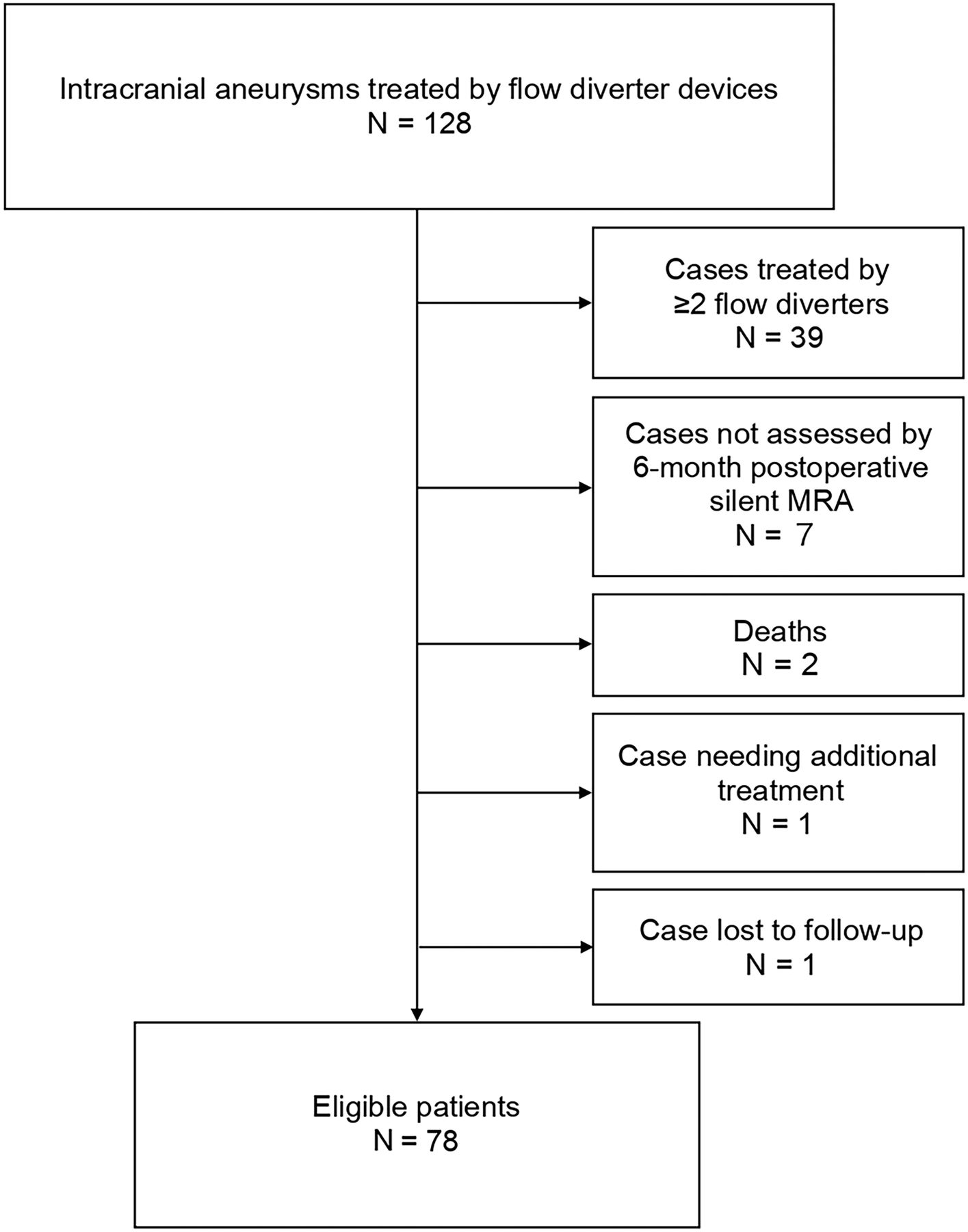
- Fig 1.
A flowchart of eligible patients assessed by Silent MR angiography after Pipeline Flex placement for intracranial aneurysms.
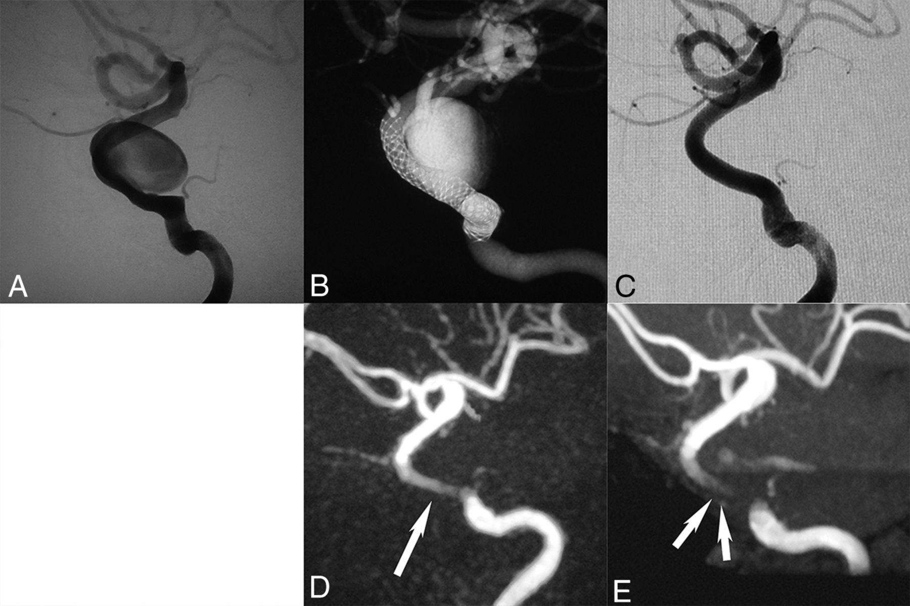
- Fig 2.
A, An 82-year-old woman. Flow-diverter placement in the left internal carotid artery cavernous segment aneurysm (20.1 × 6.3 mm) was performed with the Pipeline Flex (4.5 × 25 mm). B, Digital subtraction angiography shows the location of the Pipeline Flex by conebeam CT. C, The 6-month follow-up DSA shows complete occlusion of the aneurysm. D, This Silent MRA shows an excellent signal flow in the Pipeline Flex (arrow) but no intra-aneurysmal signal. E, This time-of-flight MRA almost lost the signal in the Pipeline Flex (double arrows) and has a faint signal in the aneurysm. The scores of the Silent MRA by 3 observers were 4, 3, and 4, and those of the TOF-MRA were 2, 2, and 2.
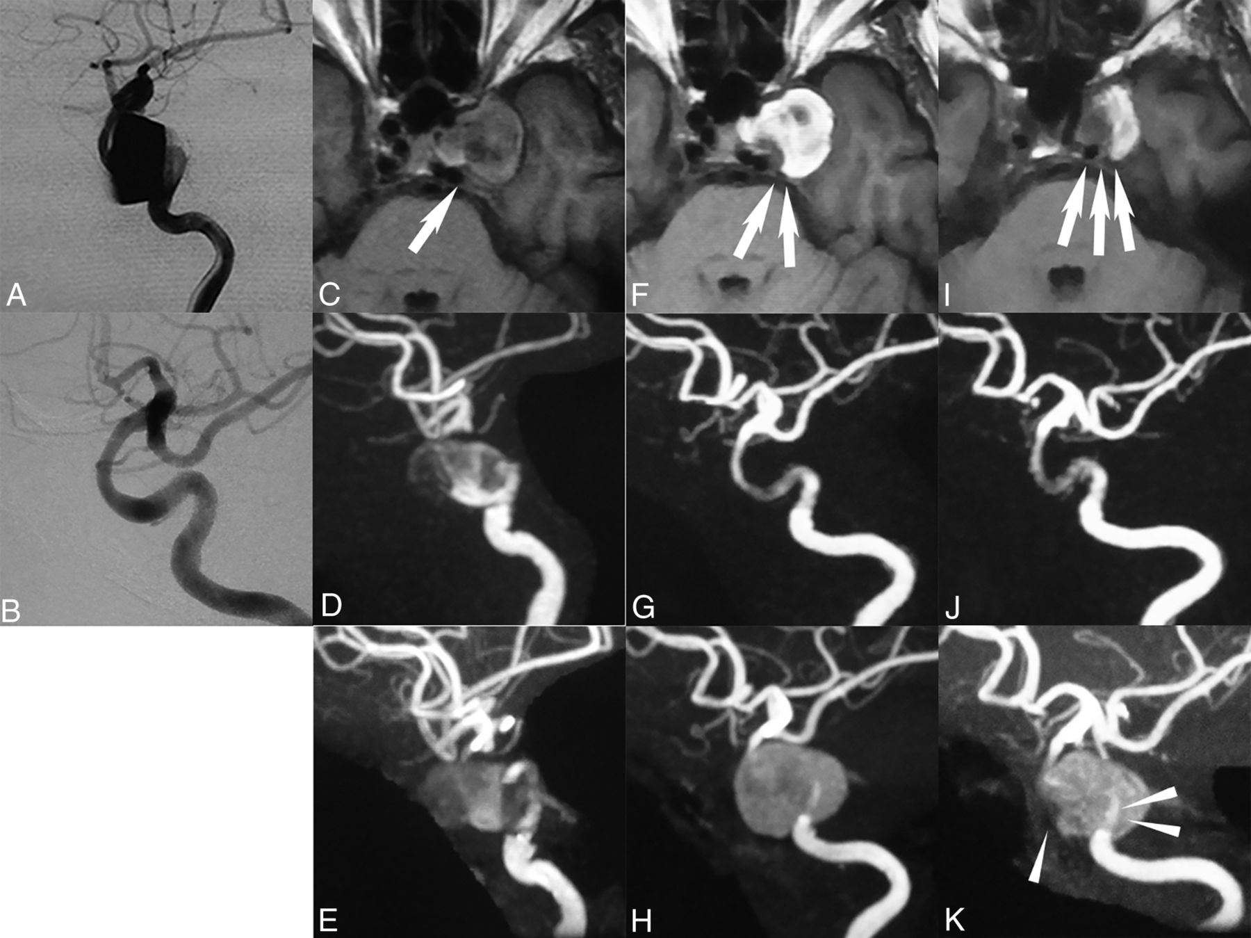
- Fig 3.
A 75-year-old woman. A, The aneurysm (26.2 × 6.1 mm) was located in the left internal carotid artery paraclinoid segment. B, A 6-month follow-up DSA shows almost complete occlusion of the aneurysm. C, The MRA T1-weighted image immediately after the operation shows an intra-aneurysmal isointensity signal (arrow). D and E, Both Silent MRA and TOF-MRA show an intra-aneurysmal signal that had a negative influence on the parent artery assessment. F, A 6-month follow-up T1-weighted image shows an intra-aneurysmal high-intensity signal, which means thrombosis (double arrows). G, A 6-month follow-up, Silent MRA shows excellent signal flow in the Pipeline Flex. H, A 6-month follow-up TOF-MRA shows that the image could not be assessed by the intra-aneurysmal thrombosed signal. I, The 1-year follow-up T1-weighted MR image shows a lower intra-aneurysmal high-intensity signal decrease than at the 6-month follow-up (triple arrows). J, The 1-year follow-up findings on the Silent MRA signal flow were the same as at the 6-month follow-up. K, The 1-year follow-up on TOF-MRA shows a better signal flow than at the 6-month follow-up TOF-MRA (arrowhead) by improvement of the thrombosed aneurysm (double arrowheads), but the signal flow on the Silent MRA is better than that on the TOF-MRA. The scores of the 6-month follow-up Silent MRA by 3 independent observers were 4, 4, and 4. The scores of the TOF-MRA were 1, 1, and 1.
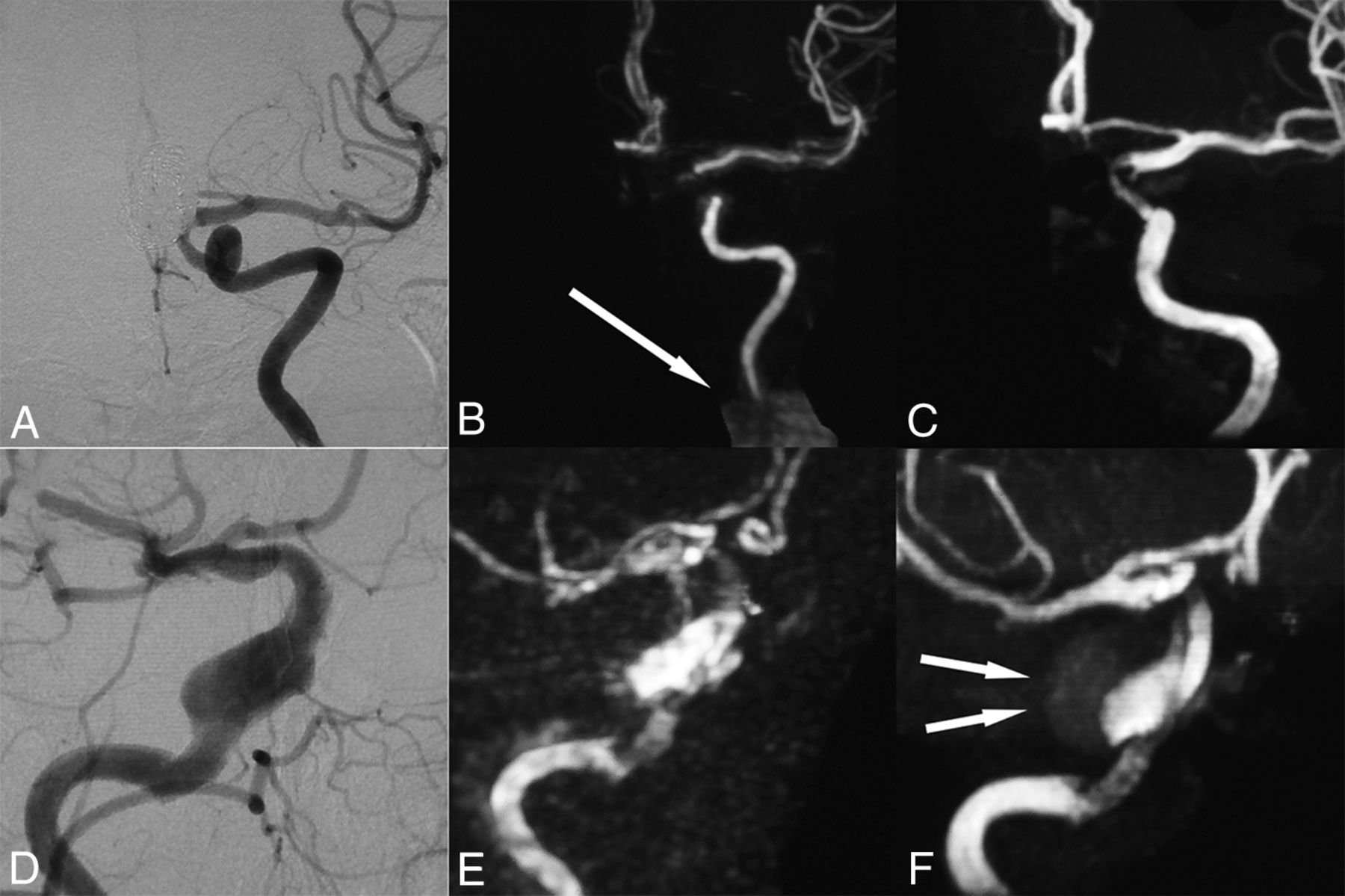
- Fig 4.
A–C, A 62-year-old woman. A, The 6-month follow-up DSA shows complete occlusion and good patency in the Pipeline Flex. B and C, The 6-month follow-up Silent MRA shows a worse signal flow than the TOF-MRA due to artifacts caused by the left denture (arrow). The Silent MRA scores by 3 independent observers were 2, 1, and 2, while the TOF-MRA scores were 3, 2, and 3. D–F, A 73-year-old woman. D, The 6-month follow-up DSA shows reduction of intra-aneurysmal flow. E and F, The 6-month follow-up Silent MRA shows a poor signal caused by motion artifacts because the patient was claustrophobic and moved during the procedure, and TOF-MRA also shows a poor signal flow caused by an intra-aneurysmal thrombosis after the Pipeline Flex placement (double arrows). The scores of Silent MRA by 3 independent observers were 3, 3, and 3. The scores of TOF-MRA were 2, 3, and 4.


Tables
- Table 1:
Characteristics of the patients assessed by DSA, Silent MRA, and TOF-MRA after placement of flow-diverter devices
Parameters Data Age (mean) (range) (yr) 61.4 ± 13.3 (19–82) Sex (M/F) 6:72 Side (R/L) 36:42 Location (cavernous segment/paraclinoid segment) 52:26 Aneurysmal size (mean) (range) (mm) 14.8 ± 5.5 (10.0–32.5) Aneurysmal neck diameter (mean) (range) (mm) 6.5 ± 2.2 (2.8–15.3) Symptomatic cases (No.) (%) 23 (29.5%) Additional coiling (No.) (%) 39 (50%) Pipeline diameter (mean) (range) (mm) 4.2 ± 0.6 (3–5) Pipeline length (mean) (range) (mm) 21.8 ± 6.3 (16–35) In-stent percutaneous transluminal angioplasty (No.) (%) 52 (66.7%) In-stent stenosis (No.) (%) None (0%) 28 (38.9%) Mild (0–25%) 41 (52.6%) Moderate (≥25%) 9 (11.5%) Duration of follow-up (mean) (range) (mo) 6.1 ± 0.6 (4–8) Note:—R indicates right; L, left.
- Table 2:
Comparison of correlations between Silent MRA and TOF-MRA from the viewpoint of all parameters for Silent MRA (3.18 ± 0.72)
High Score (>2, n = 66) Low Score (≤2, n = 12) P Value Aneurysmal size (mean) (mm) 14.8 ± 5.4 15.0 ± 6.6 .91 Aneurysmal neck diameter (mean) (mm) 6.5 ± 2.2 6.4 ± 2.3 .79 Pipeline diameter (mean) (mm) 4.3 ± 0.6 4.1 ± 0.6 .27 Pipeline length (mean) (mm) 21.7 ± 6.3 22.0 ± 6.8 .90 In-stent stenosis (No.) (%) 42/66 (63.6%) 8/12 (66.7%) .84 Location (paraclinoid/cavernous) 44:22 8:4 1.00 Additional coiling (No.) (%) 33/66 (50.0%) 7/12 (58.3%) .60 - Table 3:
Comparison of correlations between Silent MRA and TOF-MRA from the viewpoint of all parameters for TOF-MRA (2.31 ± 0.86)
High Score (>2, n = 44) Low Score (≤2, n = 34) P Value Aneurysmal size (mean) (mm) 13.5 ± 4.3 16.6 ± 6.5 <.05 Aneurysmal neck diameter (mean) (mm) 6.0 ± 1.6 7.2 ± 2.7 <.05 Pipeline diameter (mean) (mm) 4.2 ± 0.6 4.3 ± 0.5 .61 Pipeline length (mean) (mm) 20.8 ± 5.8 23.0 ± 6.9 .13 In-stent stenosis (No.) (%) 28/44 (63.6%) 22/34 (65.9%) .92 Location (paraclinoid/cavernous) 35:9 17:17 <.01 Additional coiling (No.) (%) 26/44 (59.1%) 14/34 (41.2%) .12 - Table 4:
Comparison of correlation between Silent MRA and TOF-MRA from the viewpoint of intra-aneurysmal thrombosis after flow-diverter device placement
Intra-Aneurysmal Thrombosis (+) (n = 13) Intra-Aneurysmal Thrombosis (−) (n = 65) P Value Location (paraclinoid segment/cavernous segment) 2:11 63:2 <.01 Aneurysmal size (mean) (range) (mm) 21.7 ± 5.3 (12.6–32.5) 13.4 ± 4.4 (8.1–28.7) <.01 Aneurysmal neck diameter (mean) (range) (mm) 7.8 ± 3.1 (2.8–15.3) 6.3 ± 1.9 (3.3–13.9) <.05 Additional coiling (No.) (%) 2 (15.4%) 37 (56.9%) <.01 Pipeline diameter (mean) (range) (mm) 4.21 ± 0.5 (3–5) 4.23 ± 0.6 (3–5) .91 Pipeline length (mean) (range) (mm) 27.5 ± 7.2 (18–35) 20.6 ± 5.4 (16–35) <.01 Silent MRA score (mean) 3.5 ± 0.5 3.1 ± 0.7 .10 TOF-MRA score (mean) 2.0 ± 0.8 2.3 ± 0.8 .20 Note:—+ indicates present; −, absent.
- Table 5:
Comparison of embolization assessment among DSA, Silent MRA, and TOF-MRA after flow-diverter device placement
DSA CO (n = 55) IO (n = 23) Silent MRA CO (n = 58) 53 5 IO (n = 20) 2 18 TOF-MRA CO (n = 52) 47 7 IO (n = 26) 8 16 Note:—CO indicates complete occlusion; IO, incomplete occlusion.


Jump to section
Related Articles
Cited By...
- Visualization of Intracranial Aneurysms Treated with Woven EndoBridge Devices Using Ultrashort TE MR Imaging
- Non-contrast enhanced silent MR angiography to evaluate hemodynamics and morphology of unruptured intracranial aneurysms: a comparative computational fluid dynamics study
- Diagnostic performance of silent magnetic resonance angiography for endovascularly-treated intracranial aneurysm follow-up: a prospective study
- Diagnostic Performance of High-Resolution Vessel Wall MR Imaging Combined with TOF-MRA in the Follow-up of Intracranial Vertebrobasilar Dissecting Aneurysms after Reconstructive Endovascular Treatment
- Differential Subsampling with Cartesian Ordering-MRA for Classifying Residual Treated Aneurysms
- Diagnostic performance of silent magnetic resonance angiography for endovascularly-treated intracranial aneurysm follow-up: a prospective study
- Follow-up of Intracranial Aneurysms Treated by Flow Diverters: Evaluation of Parent Artery Patency Using 3D-T1 Gradient Recalled-Echo Imaging with 2-Point Dixon in Combination with 3D-TOF-MRA with Compressed Sensing