Advertisement

Main menu

  • Home
  • Content
    • Current Issue
    • Accepted Manuscripts
    • Article Preview
    • Past Issue Archive
    • AJNR Case Collection
    • Case of the Week Archive
    • Classic Case Archive
    • Case of the Month Archive
  • Special Collections
    • Spinal CSF Leak Articles (Jan 2020-June 2024)
    • 2024 AJNR Journal Awards
    • Most Impactful AJNR Articles
  • Multimedia
    • AJNR Podcast
    • AJNR Scantastics
    • Video Articles
  • For Authors
    • Submit a Manuscript
    • Author Policies
    • Fast publishing of Accepted Manuscripts
    • Graphical Abstract Preparation
    • Manuscript Submission Guidelines
    • Imaging Protocol Submission
    • Submit a Case for the Case Collection
  • About Us
    • About AJNR
    • Editorial Board
  • More
    • Become a Reviewer/Academy of Reviewers
    • Subscribers
    • Permissions
    • Alerts
    • Feedback
    • Advertisers
    • ASNR Home
  • Other Publications
    • ajnr

User menu

  • Alerts
  • Log in

Search

  • Advanced search
American Journal of Neuroradiology

American Journal of Neuroradiology

ASHNR American Society of Functional Neuroradiology ASHNR American Society of Pediatric Neuroradiology ASSR
  • Alerts
  • Log in

Advanced Search

  • Home
  • Content
    • Current Issue
    • Accepted Manuscripts
    • Article Preview
    • Past Issue Archive
    • AJNR Case Collection
    • Case of the Week Archive
    • Classic Case Archive
    • Case of the Month Archive
  • Special Collections
    • Spinal CSF Leak Articles (Jan 2020-June 2024)
    • 2024 AJNR Journal Awards
    • Most Impactful AJNR Articles
  • Multimedia
    • AJNR Podcast
    • AJNR Scantastics
    • Video Articles
  • For Authors
    • Submit a Manuscript
    • Author Policies
    • Fast publishing of Accepted Manuscripts
    • Graphical Abstract Preparation
    • Manuscript Submission Guidelines
    • Imaging Protocol Submission
    • Submit a Case for the Case Collection
  • About Us
    • About AJNR
    • Editorial Board
  • More
    • Become a Reviewer/Academy of Reviewers
    • Subscribers
    • Permissions
    • Alerts
    • Feedback
    • Advertisers
    • ASNR Home
  • Follow AJNR on Twitter
  • Visit AJNR on Facebook
  • Follow AJNR on Instagram
  • Join AJNR on LinkedIn
  • RSS Feeds

Welcome to the new AJNR, Updated Hall of Fame, and more. Read the full announcements.


AJNR is seeking candidates for the position of Associate Section Editor, AJNR Case Collection. Read the full announcement.


Research ArticleInterventional

Pointwise Encoding Time Reduction with Radial Acquisition with Subtraction-Based MRA during the Follow-Up of Stent-Assisted Coil Embolization of Anterior Circulation Aneurysms

Y.J. Heo, H.W. Jeong, J.W. Baek, S.T. Kim, Y.G. Jeong, J.Y. Lee and S.-C. Jin
American Journal of Neuroradiology May 2019, 40 (5) 815-819; DOI: https://doi.org/10.3174/ajnr.A6035
Y.J. Heo
aFrom the Departments of Radiology (Y.J.H., H.W.J., J.W.B.)
  • Find this author on Google Scholar
  • Find this author on PubMed
  • Search for this author on this site
  • ORCID record for Y.J. Heo
H.W. Jeong
aFrom the Departments of Radiology (Y.J.H., H.W.J., J.W.B.)
  • Find this author on Google Scholar
  • Find this author on PubMed
  • Search for this author on this site
  • ORCID record for H.W. Jeong
J.W. Baek
aFrom the Departments of Radiology (Y.J.H., H.W.J., J.W.B.)
  • Find this author on Google Scholar
  • Find this author on PubMed
  • Search for this author on this site
  • ORCID record for J.W. Baek
S.T. Kim
bNeurosurgery (S.T.K., Y.G.J.)
  • Find this author on Google Scholar
  • Find this author on PubMed
  • Search for this author on this site
  • ORCID record for S.T. Kim
Y.G. Jeong
bNeurosurgery (S.T.K., Y.G.J.)
  • Find this author on Google Scholar
  • Find this author on PubMed
  • Search for this author on this site
  • ORCID record for Y.G. Jeong
J.Y. Lee
cInternal Medicine (J.Y.L.), Inje University Busan Paik Hospital, Busan, Korea
  • Find this author on Google Scholar
  • Find this author on PubMed
  • Search for this author on this site
  • ORCID record for J.Y. Lee
S.-C. Jin
dDepartment of Neurosurgery (S.-C.J.), Inje University Haeundae Paik Hospital, Busan, Korea.
  • Find this author on Google Scholar
  • Find this author on PubMed
  • Search for this author on this site
  • ORCID record for S.-C. Jin
  • Article
  • Figures & Data
  • Info & Metrics
  • Responses
  • References
  • PDF
Loading

Abstract

BACKGROUND AND PURPOSE: Time-of-flight MR angiography, though widely used after coil embolization, is associated with limitations owing to magnetic susceptibility and radiofrequency shielding following stent-assisted coil embolization. We evaluated the pointwise encoding time reduction with radial acquisition (PETRA) sequence in subtraction-based MRA (qMRA) using an ultrashort TE relative to TOF-MRA during the follow-up of stent-assisted coil embolization for anterior circulation aneurysms.

MATERIALS AND METHODS: Twenty-five patients (3 men and 22 women; mean age, 59.1 ± 14.0 years) underwent stent-assisted coil embolization for anterior circulation aneurysms and were retrospectively evaluated using TOF-MRA and PETRA qMRA data from the same follow-up session. Two neuroradiologists independently reviewed both MRA findings and subjectively graded flow within the stents (relative to the latest DSA findings) and occlusion status (complete occlusion or neck/aneurysm remnant). Interobserver and intermodality agreement for TOF-MRA and PETRA qMRA were evaluated.

RESULTS: The mean score for flow visualization within the stents was significantly higher in PETRA qMRA than in TOF-MRA (P < .001 for both observers), and good interobserver agreement was reported (κ = 0.63). The aneurysm occlusion status of PETRA qMRA (observer 1, 92.0%; observer 2, 88.0%) was more consistent with DSA than with TOF-MRA (observer 1, 76.0%; observer 2, 80.0%), and there was a better intermodality agreement between DSA and PETRA qMRA than between DSA and TOF-MRA.

CONCLUSIONS: These findings indicate that PETRA qMRA is a useful follow-up technique for patients who have undergone stent-assisted coil embolization for anterior circulation aneurysms.

ABBREVIATIONS:

PETRA
pointwise encoding time reduction with radial acquisition
qMRA
subtraction-based MRA

Stent-assisted coil embolization has been widely used for cases in which conventional coil embolization is considered difficult, such as in wide-neck aneurysms. Considering the possibility of recanalization, follow-up imaging is important even after complete occlusion has been achieved. DSA is considered the criterion standard for follow-up to evaluate intra-aneurysmal flow or in-stent restenosis; however, this technique has risks associated with contrast media, procedural invasiveness, and radiation exposure. Contrast-enhanced MRA provides visualization of the remnant neck and stented segments that is similar to that of DSA.1⇓–3 However, contrast-enhanced MRA has contrast-associated risks, including nephrogenic systemic fibrosis, anaphylaxis, gadolinium deposition, and the potential for misdiagnosing perianeurysmal enhancement as recanalization.

Nonenhanced TOF-MRA is another established technique used for follow-up after coil embolization. Specifically, it is useful for the evaluation of the residual lumen of aneurysms.4 However, this approach is difficult in cases with stent-assisted coil embolization because visualizing blood flow within intracranial stents and the parent artery is complicated by radiofrequency shielding and susceptibility artifacts.2,5 These can obscure the neck remnant and adjacent structures as a result of signal loss. Several trials have attempted to overcome these problems when using MRA after stent-assisted coil embolization.6⇓⇓⇓–10 For example, ultrashort-TE MRA is known to decrease the susceptibility artifacts caused by metallic devices and to increase the signal intensity within the stent.11,12 Recently, a few studies7⇓–9 have highlighted the efficacy of Silent MRA (GE Healthcare, Milwaukee, Wisconsin) using an ultrashort TE after stent-assisted coil embolization. The present study evaluated another nonenhanced MRA technique using ultrashort TE, known as pointwise encoding time reduction with radial acquisition (PETRA). Our results indicate that PETRA showed a higher SNR compared with the conventional ultrashort TE. This was due to a shorter encoding time for whole k-space, which was required before the signal decayed.13 To the best of our knowledge, PETRA with subtraction-based MRA (qMRA) has not been studied following stent-assisted coil embolization. Therefore, we aimed to evaluate its utility relative to TOF-MRA during follow-up after stent-assisted coil embolization for anterior circulation aneurysms.

Materials and Methods

Patients

This retrospective study evaluated 25 patients (22 women and 3 men; mean age, 59.1 ± 14.0 years) who underwent stent-assisted coil embolization for unruptured aneurysms in the anterior circulation (24 cases in the paraclinoid segment of the ICA, 1 case in the anterior communicating artery) between September 2017 and October 2018. All subjects underwent TOF-MRA and PETRA qMRA during the same follow-up session. The retrospective protocol of the study was approved by the Inje University Busan Paik Hospital institutional review board.

Eighteen patients were treated using a Neuroform stent (Stryker Neurovascular, Kalamazoo, Michigan), 6 patients were treated with an Enterprise stent (Codman & Shurtleff, Raynham, Massachusetts), and 1 patient was treated using both stents. Endovascular therapy and DSA were performed using a biplane angiographic unit (Integris V, Philips Healthcare, Best, the Netherlands; or Artis zee biplane system, Siemens, Erlangen, Germany). Complete occlusion was identified in 15 cases, and a residual neck was observed in 10 cases, based on the latest DSA findings.

Imaging Acquisition

Intracranial TOF-MRA and PETRA qMRA were performed during the same follow-up session using 3T MR imaging (Magnetom Skyra; Siemens) with a 64-channel head-neck coil. The PETRA qMRA technique has been described previously14 and is commercially available. Briefly, labeled and control data were obtained; the labeled data were applied to the slice-selective saturation pulse for the carotid artery (ie, arterial inflow appears dark). The saturation pulse of control data was applied to the 10 mm above the vertex of the head, and the subtracted MRA showed dark background tissue and suppressed venous flow. The detailed scan parameters for PETRA qMRA: TR/TE, 3.52/0.07 ms; flip angle, 6°; FOV, 230 × 230 mm; matrix, 320 × 320; radial sampling, 20,000 radial spokes; voxel size, 0.72 × 0.72 × 0.72 mm3; section thickness, 0.72 mm; NEX, 1; bandwidth, 363 Hz/pixel; slice-selective saturation pulse applied once per 20 TRs; and acquisition time, 2 minutes 10 seconds. The detailed scan parameters for TOF-MRA: TR/TE, 24/3.99 ms; flip angle, 20°; FOV, 220 × 177 mm; matrix, 576 × 302; voxel size, 0.38 × 0.38 × 0.38 mm3; section thickness, 0.6 mm; NEX, 1; bandwidth, 185 Hz/pixel; acquisition time, 6 minutes 40 seconds; number of slab, 6. The maximum intensity projections for both sequences were reconstructed in the same manner as the DSA images.

Image Analysis

Two neuroradiologists independently reviewed both MRA findings at 2-week intervals and subjectively graded the visualization of flow within each stent (relative to the latest DSA findings) using the following scale: 1, no signal within the stent; 2, slightly visible but not of diagnostic quality due to image blurring; 3, good quality with minimal blurring; or 4, excellent quality. The occlusion status of the coiled aneurysm was also evaluated. The locations of the aneurysms were provided to the observers, but they were blinded to the DSA results. However, observers were not blinded to the type of MR images because the 2 MR images are distinctively different in appearance. One experienced interventional neuroradiologist also assessed the occlusion status of the coiled aneurysm using the Montreal Neurological Institute scale.15 Specifically, patients were rated as having complete occlusion, subtotal occlusion (residual neck, residual aneurysm), or incomplete occlusion. The latest DSA images were used as references, and the average interval between the latest DSA and the MRA follow-up was 16 months, with a range of 1 day to 127 months.

Statistical Analyses

Continuous variables are presented as mean ± SD. We compared the mean scores for each MRA technique and type of intracranial stent using the Wilcoxon signed rank test and the Mann-Whitney test. Weighted κ coefficients were used to evaluate the interobserver and intermodality agreement for TOF-MRA and PETRA qMRA. The results were classified as poor (<0.20), fair (0.21–0.40), moderate (0.41–0.60), good (0.61–0.80), or excellent (0.81–1.00).16 All statistical analyses were performed using SPSS software (Version 24.0; IBM, Armonk, New York) and MedCalc for Windows (Version 18.11.3; MedCalc Software, Mariakerke, Belgium). P values < .05 were considered statistically significant.

Results

The mean score for assessing blood flow within the intracranial stent was significantly higher in the PETRA qMRA than in the TOF-MRA (observer one: 3.40 ± 0.87 for PETRA qMRA and 2.20 ± 0.87 for TOF-MRA, P < .001; observer two: 3.60 ± 0.76 for PETRA qMRA and 2.44 ± 0.77 for TOF-MRA, P < .001) with good interobserver agreement for both MRA techniques (κ = 0.63 for PETRA qMRA, 95% CI, 0.35–0.90; κ = 0.63 for TOF-MRA, 95% CI, 0.43–0.83). The Neuroform stents (open-cell design) were associated with higher scores than the Enterprise stents (closed-cell design) for both PETRA qMRA and TOF-MRA; however, no significant differences were detected between the 2 intracranial stents (Table 1). On the basis of the latest DSA findings, complete occlusion was achieved in 15 cases (60.0%) and a residual neck was observed in 10 cases (40.0%). PETRA qMRA findings were consistent with DSA findings in 23 cases (92.0%) according to observer 1 and 22 cases (88.0%) according to observer 2. Therefore, the intermodality agreement between PETRA qMRA and DSA was excellent according to observer 1 (weighted κ = 0.83; 95% CI, 0.60–1.00) and good according to observer 2 (weighted κ = 0.73; 95% CI, 0.47–1.00) (Table 2). However, only 19 cases (76.0%) of TOF-MRA findings were consistent with DSA according to observer 1 and 20 cases (80.0%) according to observer 2. Thus, the intermodality agreement between TOF-MRA and DSA was moderate in both observer 1 (weighted κ= 0.44; 95% CI, 0.12–0.77) and observer 2 (weighted κ = 0.55; 95% CI, 0.23–0.86) (Figs 1 and 2).

View this table:
  • View inline
  • View popup
Table 1:

Comparison of PETRA qMRA and TOF-MRA according to stent designa

View this table:
  • View inline
  • View popup
Table 2:

Comparison of occlusion status of coiled aneurysms between PETRA qMRA and TOF-MRAa

Fig 1.
  • Download figure
  • Open in new tab
  • Download powerpoint
Fig 1.

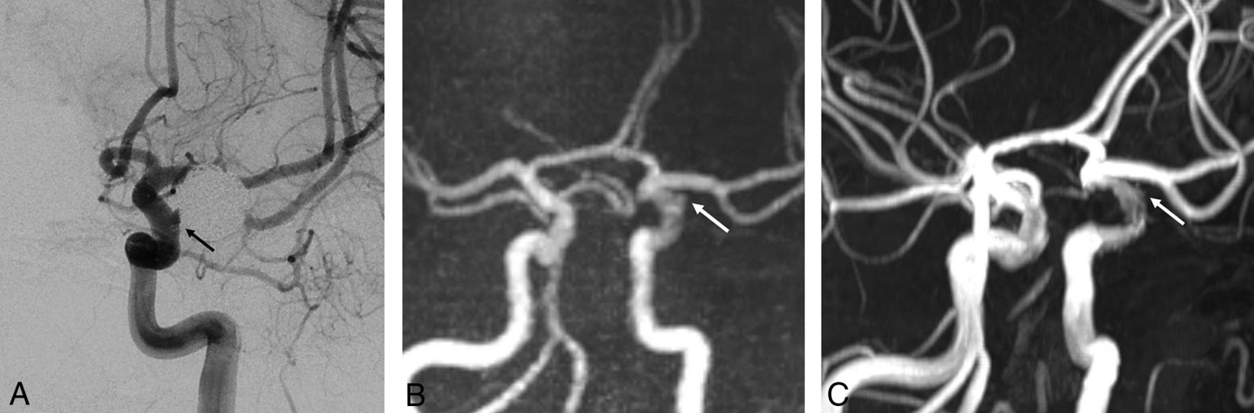
A 54-year-old man with a left paraclinoid internal carotid artery aneurysm who underwent stent-assisted coil embolization using a Neuroform stent. A, The follow-up DSA reveals the neck remnant (arrow). B, The PETRA qMRA reveals minimal signal loss at the stent insertion site, and the neck remnant is clearly visible (arrow). C, The TOF-MRA reveals mild signal loss at the stent insertion site and a less prominent neck remnant relative to the PETRA qMRA.

Fig 2.
  • Download figure
  • Open in new tab
  • Download powerpoint
Fig 2.

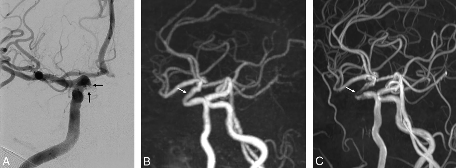
A 70-year-old woman with a right paraclinoid internal carotid artery aneurysm who underwent stent-assisted coil embolization using an Enterprise stent. A, The follow-up DSA reveals the neck remnant (arrows). B, The PETRA qMRA reveals minimal signal loss at the stent-insertion-site remnant (arrow), and the neck remnant is clearly visible. C, The TOF-MRA reveals marked signal loss at the stent-insertion-site remnant (arrow) and poor differentiation of the prominent neck remnant relative to PETRA qMRA.

Discussion

The present study revealed that PETRA qMRA provided better visualization of intracranial stent flow than TOF-MRA, regardless of stent type. Furthermore, PETRA qMRA showed higher intermodality agreement with DSA than TOF-MRA. In the 2 cases in which PETRA qMRA failed to detect the residual neck, the intervals between the last DSA and the MRA were 31 and 15 months. Santillan et al17 reported cumulative occlusion after the first treatment, and our cases may have involved progressive occlusion within the aneurysms. In cases in which PETRA qMRA identified the residual neck, its configuration was more similar to DSA findings than to TOF-MRA findings. These results suggest that PETRA qMRA could be a useful imaging technique following stent-assisted coil embolization.

The increased use of intracranial stent placement has also increased the difficulty of using follow-up imaging to assess the recanalization status or stability of the stent. Although DSA is an established technique for follow-up, it also has risks related to contrast media (eg, nephrogenic systemic fibrosis), neurologic complications (eg, embolic infarction),18 and radiation exposure. Contrast-enhanced MRA provides better delineation of the parent artery and is correlated with DSA findings; however, it can also cause contrast-related complications. Furthermore, contrast-enhanced MRA can produce a false-positive result that indicates a residual neck, which is caused by peripheral enhancement of the thrombus or vasa vasorum within the aneurysmal wall.19,20 In contrast, TOF-MRA is a noninvasive imaging technique that is used following intracranial stent insertion. However, TOF-MRA is prone to susceptibility artifacts, which can generate false-positive or false-negative results at the stent insertion site. Various trials have attempted to overcome these problems.4,10,21 Additionally, some reports have described the value of the ultrashort-TE technique in this setting,4,6⇓⇓–9 which can significantly reduce susceptibility artifacts and decrease the phase dispersion of labeled flow.22,23

Irie et al6 reported that Silent MRA with an ultrashort TE is useful for visualizing flow within an intracranial stent and the occlusion status of aneurysms. Furthermore, there is a strong positive correlation between the results obtained using Silent MRA and DSA. PETRA qMRA also uses an ultrashort TE, and our findings indicate that the results from this technique are strongly correlated with those from Silent MRA. Similar to Silent MRA, PETRA qMRA uses subtraction of scanned images from before and after labeling. This makes it difficult to assess static tissue and may prevent detection of in-stent thrombus, which is visualized as a high-intensity signal during TOF-MRA. Although in-stent thrombosis can occasionally occur after stent insertion for atherosclerotic stenosis, it is uncommon and usually asymptomatic or it spontaneously regresses in patients who undergo stent-assisted coil embolization.17,24 PETRA qMRA is associated with poorer imaging quality than TOF-MRA, which is another disadvantage of PETRA qMRA, like Silent MRA. Although, Silent MRA is associated with better background suppression, it has been reported that it showed more blurring and a lower SNR than TOF-MRA because of its longer readout time.25 Conversely, PETRA qMRA had a shorter acquisition time (approximately 2 minutes) than Silent MRA and TOF-MRA (approximately 6–7 minutes) using a highly undersampled outer k-space.13

The degree of artifacts associated with an intracranial stent can vary according to the material, cell design, and strut thickness. For example, previous reports have indicated that stents made using stainless steel or cobalt are associated with more artifacts than nitinol stents.10,26,27 The present study included only patients who were treated with nitinol stents, albeit with different cell designs (open cell versus closed cell) and strut thicknesses. Although the Neuroform stent has an open-cell design, thinner struts, and better intrastent flow than the Enterprise stent (a closed-cell design with thicker struts), there were no significant differences between the 2 intracranial stents according to the analyses of the 2 observers. We believe that this was due to the small sample size of this study. Similarly, previous studies5,21,28 have demonstrated that the Neuroform stent generated fewer artifacts than the Enterprise stent because the thinner struts generated less radiofrequency shielding.5,7,11

The present study has several limitations. First, the retrospective design is prone to selection bias. Second, there was a long interval between the latest DSA and MRA, which may indicate that the patients were evaluated with MRA when they were at a slightly different status from that in their last DSA. However, we do not regularly perform DSA for patients who do not show evidence of recanalization during follow-up MRA due to the invasiveness of the procedure and the possibility of neurologic complications. Therefore, shorter intervals between the latest DSA and MRA could not be achieved. Additionally, we did not compare our results with those of contrast-enhanced MRA, which is considered a useful follow-up technique for stent-assisted coil embolization.21,29 Thus, future studies comparing PETRA qMRA with contrast-enhanced MRA are necessary. Furthermore, we evaluated cases that involved nitinol stents, which have fewer artifacts than stents made using other materials. Further studies should be conducted to determine whether our findings are consistent with the use of stainless steel, platinum, and cobalt stents. Finally, this study had a small sample size; future studies should include a large sample size to confirm the effectiveness of PETRA qMRA.

Conclusions

Our results show that PETRA qMRA provides clear visualization of intracranial stent flow in the anterior circulation. Additionally, PETRA qMRA was more similar to the occlusion status of DSA than TOF-MRA. Overall, it appears that PETRA qMRA could be a useful follow-up technique for patients who have undergone stent-assisted coil embolization for anterior circulation aneurysms.

Acknowledgments

The authors thank Kang Soo Kim (Siemens Healthcare, Korea) for technical advice.

Footnotes

  • Disclosures: Young Jin Heo—RELATED: Grant: 2018 Inje University research grant.

  • This work was supported by the 2018 Inje University research grant.

REFERENCES

  1. 1.↵
    1. Agid R,
    2. Willinsky R,
    3. Lee SK, et al
    . Characterization of aneurysm remnants after endovascular treatment: contrast-enhanced MR angiography versus catheter digital subtraction angiography. AJNR Am J Neuroradiol 2008;29:1570–74 doi:10.3174/ajnr.A1124 pmid:18499789
    Abstract/FREE Full Text
  2. 2.↵
    1. Lubicz B,
    2. Levivier M,
    3. Sadeghi N, et al
    . Immediate intracranial aneurysm occlusion after embolization with detachable coils: a comparison between MR angiography and intra-arterial digital subtraction angiography. J Neuroradiol 2007;34:190–97 doi:10.1016/j.neurad.2007.05.002 pmid:17582496
    CrossRefPubMed
  3. 3.↵
    1. Kaufmann TJ,
    2. Huston J 3rd.,
    3. Cloft HJ, et al
    . A prospective trial of 3T and 1.5T time-of-flight and contrast-enhanced MR angiography in the follow-up of coiled intracranial aneurysms. AJNR Am J Neuroradiol 2010;31:912–18 doi:10.3174/ajnr.A1932 pmid:20019107
    Abstract/FREE Full Text
  4. 4.↵
    1. Gönner F,
    2. Heid O,
    3. Remonda L, et al
    . MR angiography with ultrashort echo time in cerebral aneurysms treated with Guglielmi detachable coils. AJNR Am J Neuroradiol 1998;19:1324–28 pmid:9726477
    Abstract/FREE Full Text
  5. 5.↵
    1. Cho WS,
    2. Kim SS,
    3. Lee SJ, et al
    . The effectiveness of 3T time-of-flight magnetic resonance angiography for follow-up evaluations after the stent-assisted coil embolization of cerebral aneurysms. Acta Radiol 2014;55:604–13 doi:10.1177/0284185113502335 pmid:24003259
    CrossRefPubMed
  6. 6.↵
    1. Irie R,
    2. Suzuki M,
    3. Yamamoto M, et al
    . Assessing blood flow in an intracranial stent: a feasibility study of MR angiography using a silent scan after stent-assisted coil embolization for anterior circulation aneurysms. AJNR Am J Neuroradiol 2015;36:967–70 doi:10.3174/ajnr.A4199 pmid:25523588
    Abstract/FREE Full Text
  7. 7.↵
    1. Takano N,
    2. Suzuki M,
    3. Irie R, et al
    . Usefulness of non–contrast-enhanced MR angiography using a silent scan for follow-up after Y-configuration stent-assisted coil embolization for basilar tip aneurysms. AJNR Am J Neuroradiol 2017;38:577–81 doi:10.3174/ajnr.A5033 pmid:28007767
    Abstract/FREE Full Text
  8. 8.↵
    1. Takano N,
    2. Suzuki M,
    3. Irie R, et al
    . Non-contrast-enhanced silent scan MR angiography of intracranial anterior circulation aneurysms treated with a low-profile visualized intraluminal support device. AJNR Am J Neuroradiol 2017;38:1610–16 doi:10.3174/ajnr.A5223 pmid:28522664
    Abstract/FREE Full Text
  9. 9.↵
    1. Ye J,
    2. Luo X,
    3. Qu J, et al
    . Follow-up assessment of coiled intracranial aneurysms using zTE MRA as compared with TOF MRA: a preliminary image quality study. Eur Radiol 2017;27:4271–80 doi:10.1007/s00330-017-4794-z pmid:28382536
    CrossRefPubMed
  10. 10.↵
    1. Meyer JM,
    2. Buecker A,
    3. Spuentrup E, et al
    . Improved in-stent magnetic resonance angiography with high flip angle excitation. Invest Radiol 2001;36:677–81 doi:10.1097/00004424-200111000-00007 pmid:11606845
    CrossRefPubMed
  11. 11.↵
    1. Choi JW,
    2. Roh HG,
    3. Moon WJ, et al
    . Optimization of MR parameters of 3D TOF-MRA for various intracranial stents at 3.0T MRI. Neurointervention 2011;6:71–77 doi:10.5469/neuroint.2011.6.2.71 pmid:22125752
    CrossRefPubMed
  12. 12.↵
    1. Ikushima Y,
    2. Hashido T,
    3. Watanabe Y, et al
    . Effects of imaging parameters on the quality of contrast-enhanced MR angiography of cerebral aneurysms treated using stent-assisted coiling: a phantom study. Magn Reson Med Sci 2017;16:146–51 doi:10.2463/mrms.mp.2016-0042 pmid:27599584
    CrossRefPubMed
  13. 13.↵
    1. Grodzki DM,
    2. Jakob PM,
    3. Heismann B
    . Ultrashort echo time imaging using pointwise encoding time reduction with radial acquisition (PETRA). Magn Reson Med 2012;67:510–18 doi:10.1002/mrm.23017 pmid:21721039
    CrossRefPubMed
  14. 14.↵
    1. Yutaka Natsuaki RG,
    2. Grimm R,
    3. Grodzki D, et al
    . PETRA quiet MRA: improved robustness with 3D elastic cross-registration. In: Proceedings of the Annual Meeting and Exhibition of the International Society for Magnetic Resonance in Medicine, Singapore. May 7–8, 2016
  15. 15.↵
    1. Raymond J,
    2. Guilbert F,
    3. Weill A, et al
    . Long-term angiographic recurrences after selective endovascular treatment of aneurysms with detachable coils. Stroke 2003;34:1398–403 doi:10.1161/01.STR.0000073841.88563.E9 pmid:12775880
    Abstract/FREE Full Text
  16. 16.↵
    1. Landis JR,
    2. Koch GG
    . The measurement of observer agreement for categorical data. Biometrics 1977;33:159–74 pmid:843571
    CrossRefPubMed
  17. 17.↵
    1. Santillan A,
    2. Greenberg E,
    3. Patsalides A, et al
    . Long-term clinical and angiographic results of Neuroform stent-assisted coil embolization in wide-necked intracranial aneurysms. Neurosurgery 2012;70:1232–37; discussion 1237 doi:10.1227/NEU.0b013e3182422a68 pmid:22095221
    CrossRefPubMed
  18. 18.↵
    1. Willinsky RA,
    2. Taylor SM,
    3. terBrugge K, et al
    . Neurologic complications of cerebral angiography: prospective analysis of 2,899 procedures and review of the literature. Radiology 2003;227:522–28 doi:10.1148/radiol.2272012071 pmid:12637677
    CrossRefPubMed
  19. 19.↵
    1. Gauvrit JY,
    2. Leclerc X,
    3. Pernodet M, et al
    . Intracranial aneurysms treated with Guglielmi detachable coils: usefulness of 6-month imaging follow-up with contrast-enhanced MR angiography. AJNR Am J Neuroradiol 2005;26:515–21 pmid:15760858
    Abstract/FREE Full Text
  20. 20.↵
    1. Gauvrit JY,
    2. Leclerc X,
    3. Caron S, et al
    . Intracranial aneurysms treated with Guglielmi detachable coils: imaging follow-up with contrast-enhanced MR angiography. Stroke 2006;37:1033–37 doi:10.1161/01.STR.0000209236.06451.3b pmid:16528000
    Abstract/FREE Full Text
  21. 21.↵
    1. Choi JW,
    2. Roh HG,
    3. Moon WJ, et al
    . Time-resolved 3D contrast-enhanced MRA on 3.0T: a non-invasive follow-up technique after stent-assisted coil embolization of the intracranial aneurysm. Korean J Radiol 2011;12:662–70 doi:10.3348/kjr.2011.12.6.662 pmid:22043147
    CrossRefPubMed
  22. 22.↵
    1. Schmalbrock P,
    2. Yuan C,
    3. Chakeres DW, et al
    . Volume MR angiography: methods to achieve very short echo times. Radiology 1990;175:861–65 doi:10.1148/radiology.175.3.2343137 pmid:2343137
    CrossRefPubMed
  23. 23.↵
    1. Robson MD,
    2. Gatehouse PD,
    3. Bydder M, et al
    . Magnetic resonance: an introduction to ultrashort TE (UTE) imaging. J Comput Assist Tomogr 2003;27:825–46 doi:10.1097/00004728-200311000-00001 pmid:14600447
    CrossRefPubMed
  24. 24.↵
    1. Fiorella D,
    2. Albuquerque FC,
    3. Woo H, et al
    . Neuroform in-stent stenosis: incidence, natural history and treatment strategies. Neurosurgery 2006;59:34–42 doi:10.1227/01.NEU.0000219853.56553.71
    CrossRefPubMed
  25. 25.↵
    1. Holdsworth SJ,
    2. Macpherson SJ,
    3. Yeom KW, et al
    . Clinical evaluation of Silent T1-weighted MRI and Silent MR angiography of the brain. AJR Am J Roentgenol 2018;210:404–11 doi:10.2214/AJR.17.18247 pmid:29112472
    CrossRefPubMed
  26. 26.↵
    1. Bartels LW,
    2. Bakker CJ,
    3. Viergever MA
    . Improved lumen visualization in metallic vascular implants by reducing RF artifacts. Magn Reson Med 2002;47:171–80 doi:10.1002/mrm.10004 pmid:11754456
    CrossRefPubMed
  27. 27.↵
    1. Bartels LW,
    2. Smits HF,
    3. Bakker CJ, et al
    . MR imaging of vascular stents: effects of susceptibility, flow, and radiofrequency eddy currents. J Vasc Interv Radiol 2001;12:365–71 doi:10.1016/S1051-0443(07)61918-6 pmid:11287516
    CrossRefPubMed
  28. 28.↵
    1. Lövblad KO,
    2. Yilmaz H,
    3. Chouiter A, et al
    . Intracranial aneurysm stenting: follow-up with MR angiography. J Magn Reson Imaging 2006;24:418–22 doi:10.1002/jmri.20642 pmid:16795090
    CrossRefPubMed
  29. 29.↵
    1. Agid R,
    2. Schaaf M,
    3. Farb R
    . CE-MRA for follow-up of aneurysms post stent-assisted coiling. Interv Neuroradiol 2012;18:275–83 doi:10.1177/159101991201800305 pmid:22958765
    CrossRefPubMed
  • Received November 23, 2018.
  • Accepted after revision March 11, 2019.
  • © 2019 by American Journal of Neuroradiology
View Abstract
PreviousNext
Back to top

In this issue

American Journal of Neuroradiology: 40 (5)
American Journal of Neuroradiology
Vol. 40, Issue 5
1 May 2019
  • Table of Contents
  • Index by author
  • Complete Issue (PDF)
Advertisement
Print
Download PDF
Email Article

Thank you for your interest in spreading the word on American Journal of Neuroradiology.

NOTE: We only request your email address so that the person you are recommending the page to knows that you wanted them to see it, and that it is not junk mail. We do not capture any email address.

Enter multiple addresses on separate lines or separate them with commas.
Pointwise Encoding Time Reduction with Radial Acquisition with Subtraction-Based MRA during the Follow-Up of Stent-Assisted Coil Embolization of Anterior Circulation Aneurysms
(Your Name) has sent you a message from American Journal of Neuroradiology
(Your Name) thought you would like to see the American Journal of Neuroradiology web site.
CAPTCHA
This question is for testing whether or not you are a human visitor and to prevent automated spam submissions.
Cite this article
Y.J. Heo, H.W. Jeong, J.W. Baek, S.T. Kim, Y.G. Jeong, J.Y. Lee, S.-C. Jin
Pointwise Encoding Time Reduction with Radial Acquisition with Subtraction-Based MRA during the Follow-Up of Stent-Assisted Coil Embolization of Anterior Circulation Aneurysms
American Journal of Neuroradiology May 2019, 40 (5) 815-819; DOI: 10.3174/ajnr.A6035

Citation Manager Formats

  • BibTeX
  • Bookends
  • EasyBib
  • EndNote (tagged)
  • EndNote 8 (xml)
  • Medlars
  • Mendeley
  • Papers
  • RefWorks Tagged
  • Ref Manager
  • RIS
  • Zotero
0 Responses
Respond to this article
Share
Bookmark this article
Pointwise Encoding Time Reduction with Radial Acquisition with Subtraction-Based MRA during the Follow-Up of Stent-Assisted Coil Embolization of Anterior Circulation Aneurysms
Y.J. Heo, H.W. Jeong, J.W. Baek, S.T. Kim, Y.G. Jeong, J.Y. Lee, S.-C. Jin
American Journal of Neuroradiology May 2019, 40 (5) 815-819; DOI: 10.3174/ajnr.A6035
del.icio.us logo Twitter logo Facebook logo Mendeley logo
  • Tweet Widget
  • Facebook Like
  • Google Plus One
Purchase

Jump to section

  • Article
    • Abstract
    • ABBREVIATIONS:
    • Materials and Methods
    • Results
    • Discussion
    • Conclusions
    • Acknowledgments
    • Footnotes
    • REFERENCES
  • Figures & Data
  • Info & Metrics
  • Responses
  • References
  • PDF

Related Articles

  • No related articles found.
  • PubMed
  • Google Scholar

Cited By...

  • Non-contrast enhanced silent MR angiography to evaluate hemodynamics and morphology of unruptured intracranial aneurysms: a comparative computational fluid dynamics study
  • Crossref (22)
  • Google Scholar

This article has been cited by the following articles in journals that are participating in Crossref Cited-by Linking.

  • Silent zero TE MR neuroimaging: Current state-of-the-art and future directions
    Emil Ljungberg, Nikou L. Damestani, Tobias C. Wood, David J. Lythgoe, Fernando Zelaya, Steven C.R. Williams, Ana Beatriz Solana, Gareth J. Barker, Florian Wiesinger
    Progress in Nuclear Magnetic Resonance Spectroscopy 2021 123
  • Usefulness of Noncontrast-Enhanced Silent Magnetic Resonance Angiography (MRA) for Treated Intracranial Aneurysm Follow-up in Comparison with Time-of-Flight MRA
    Kyeong Hwa Ryu, Hye Jin Baek, Jin Il Moon, Bo Hwa Choi, Sung Eun Park, Ji Young Ha, Hyun Park, Seung Soo Kim, Jung-Soo Kim, Soo Buem Cho, Michael Carl
    Neurosurgery 2020 87 2
  • Usefulness of Silent MRA for Evaluation of Aneurysm after Stent-Assisted Coil Embolization
    You Na Kim, Jin Wook Choi, Yong Cheol Lim, Jihye Song, Ji Hyun Park, Woo Sang Jung
    Korean Journal of Radiology 2022 23 2
  • Pointwise encoding time reduction with radial acquisition in subtraction-based magnetic resonance angiography to assess saccular unruptured intracranial aneurysms at 3 Tesla
    Qing Fu, Ding-Xi Liu, Xiao-Yong Zhang, Xian-Bo Deng, Chuan-Sheng Zheng
    Neuroradiology 2021 63 2
  • Use of PETRA-MRA to assess intracranial arterial stenosis: Comparison with TOF-MRA, CTA, and DSA
    Junxia Niu, Yuncai Ran, Rui Chen, Feifei Zhang, Xiaowen Lei, Xiao Wang, Tengfei Li, Jinxia Zhu, Yong Zhang, Jingliang Cheng, Yan Zhang, Chengcheng Zhu
    Frontiers in Neurology 2023 13
  • Clinical evaluation of subtracted pointwise encoding time reduction with radial acquisition-based magnetic resonance angiography compared to 3D time-of-flight magnetic resonance angiography for improved flow dephasing at 3 Tesla
    Qing Fu, Xiao-yong Zhang, Xian-bo Deng, Ding-xi Liu
    Magnetic Resonance Imaging 2020 73
  • The Use of Pointwise Encoding Time Reduction With Radial Acquisition MRA to Assess Middle Cerebral Artery Stenosis Pre- and Post-stent Angioplasty: Comparison With 3D Time-of-Flight MRA and DSA
    Feifei Zhang, Yuncai Ran, Ming Zhu, Xiaowen Lei, Junxia Niu, Xiao Wang, Yong Zhang, Shujian Li, Jinxia Zhu, Xuemei Gao, Mahmud Mossa-Basha, Jingliang Cheng, Chengcheng Zhu
    Frontiers in Cardiovascular Medicine 2021 8
  • Follow-up imaging of clipped intracranial aneurysms with 3-T MRI: comparison between 3D time-of-flight MR angiography and pointwise encoding time reduction with radial acquisition subtraction-based MR angiography
    Jae Ho Kim, Sung Jun Ahn, Mina Park, Yong Bae Kim, Bio Joo, Woosung Lee, Sang Hyun Suh
    Journal of Neurosurgery 2022 136 5
  • Imaging follow-up strategy after endovascular treatment of Intracranial aneurysms: A literature review and guideline recommendations
    Yong-Hwan Cho, Jaehyung Choi, Chae-Wook Huh, Chang Hyeun Kim, Chul Hoon Chang, Soon Chan KWON, Young Woo Kim, Seung Hun Sheen, Sukh Que Park, Jun Kyeung Ko, Sung-kon Ha, Hae Woong Jeong, Hyen Seung Kang
    Journal of Cerebrovascular and Endovascular Neurosurgery 2024 26 1
  • Noncontrast Magnetic Resonance Angiography in the Era of Nephrogenic Systemic Fibrosis and Gadolinium Deposition
    Ruth P. Lim, Elizabeth M. Hecht, Patricia M. Desmond
    Journal of Computer Assisted Tomography 2021 45 1

More in this TOC Section

  • SAVE vs. Solumbra Techniques for Thrombectomy
  • CT Perfusion&Reperfusion in Acute Ischemic Stroke
  • Delayed Reperfusion Post-Thrombectomy&Thrombolysis
Show more Interventional

Similar Articles

Advertisement

Indexed Content

  • Current Issue
  • Accepted Manuscripts
  • Article Preview
  • Past Issues
  • Editorials
  • Editors Choice
  • Fellow Journal Club
  • Letters to the Editor

Cases

  • Case Collection
  • Archive - Case of the Week
  • Archive - Case of the Month
  • Archive - Classic Case

Special Collections

  • Special Collections

Resources

  • News and Updates
  • Turn around Times
  • Submit a Manuscript
  • Author Policies
  • Manuscript Submission Guidelines
  • Evidence-Based Medicine Level Guide
  • Publishing Checklists
  • Graphical Abstract Preparation
  • Imaging Protocol Submission
  • Submit a Case
  • Become a Reviewer/Academy of Reviewers
  • Get Peer Review Credit from Publons

Multimedia

  • AJNR Podcast
  • AJNR SCANtastic
  • Video Articles

About Us

  • About AJNR
  • Editorial Board
  • Not an AJNR Subscriber? Join Now
  • Alerts
  • Feedback
  • Advertise with us
  • Librarian Resources
  • Permissions
  • Terms and Conditions

American Society of Neuroradiology

  • Not an ASNR Member? Join Now

© 2025 by the American Society of Neuroradiology All rights, including for text and data mining, AI training, and similar technologies, are reserved.
Print ISSN: 0195-6108 Online ISSN: 1936-959X

Powered by HighWire