
Article Figures & Data
Figures
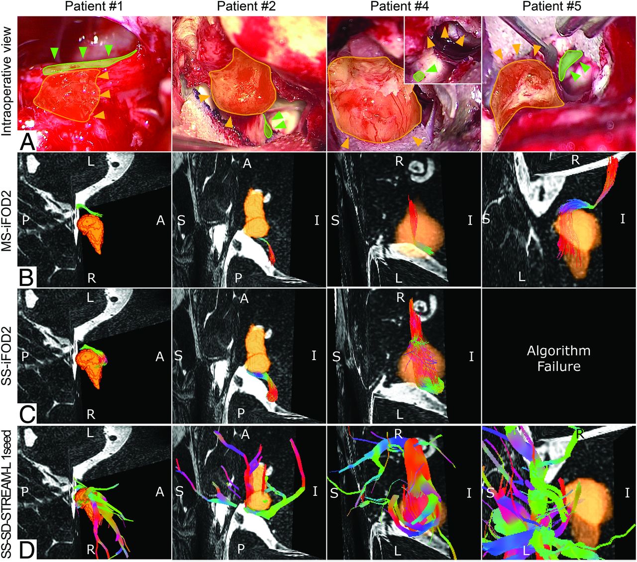
- Fig 1.
Each column shows a specific patient, from top to bottom. A, The intraoperative view of the VS (yellow area) with the position of the VS highlighted (orange arrows) and the position (green arrows) of the FN (green area). B, C, and D, MS-iFOD2, the SS-iFOD2, and the SS-SD-STREAM-L with 1 anatomic seed tractography reconstruction of the FN with a 3D reconstruction of the VS (orange) displayed in the same orientation as in the intraoperative view, respectively. A, For patient 4, two images of the VS, before and after its debulking, highlight the residual tumor capsule (orange arrows) and the FN (green arrow).
- Fig 2.
Results for patient 3 (see text for details). A, The intraoperative view of the VS (yellow area) highlighting the position of the VS (orange arrows) and the position of the FN (green arrows). Also shown is the stimulation site suggesting that the FN was frayed and spread on the tumor surface. Note that the FN is posterior compared with the VS position. B, C, and D, The MS-iFOD2, the SS-iFOD2, and the SS-SD-STREAM-L with 1 anatomic seed tractography reconstruction of the FN with a 3D reconstruction of the VS (orange) displayed in the same orientation as in the intraoperative view.


Tables
Patient No. 1 2 3 4 5 Sex F F F M M Age (yr) 52 57 64 45 53 Symptoms Deafness, right tinnitus, dizziness Right earache, hearing loss Tinnitus, right hearing loss Tinnitus, right hearing loss Tinnitus, left hearing loss Tumor side Right Right Right Right Left Tumor volume (mm3) 347.4 370.1 1941 1153 460.6 Tumor size (mm) 15.5 × 7 15 × 6.7 22 × 17 16.2 × 14.6 15.5 × 9.8 Extrameatal tumor dimension (mm) 8.36 × 5 4.3 × 6.7 17 × 12 14.6 × 10.5 9.8 × 6.6 Preoperative HB scaleb I I I I I Postoperative HB scale I I VI III–IV I Koos classificationc II II III III II Note:—HB indicates House and Brackmann.
↵a Both preoperative and postoperative FN functions are reported according to the scale of House and Brackmann.8
↵b House and Brackmann scale: I (normal), normal facial function in all areas; III (moderate dysfunction), gross: obvious but not disfiguring difference between 2 sides; noticeable-but-not severe synkinesis, contracture, and/or hemifacial spasm. At rest, normal symmetry and tone. Motion forehead: slight-to-moderate movement. Eye: complete closure with effort. Mouth: slightly weak with maximum effort; IV (moderately severe dysfunction), gross: obvious weakness and/or disfiguring asymmetry. At rest: normal symmetry and tone. Motion forehead: none. Eye: incomplete closure. Mouth: asymmetric with maximum effort; VI (total paralysis), no movement.
↵c Classification of Koos et al:9 grade II, small tumor with protrusion into the cerebellopontine angle; no contact with the brain stem; grade III, tumor occupying the cerebellopontine cistern with no brain stem displacement.
- Table 2:
Intraoperative location of FN relative to the VS, assessed at the origin of the nerve and in the cisternal passage tract according to the classification of Sampath et al20
Patient Intraoperative Findings FN Origin FN Passage 1 AI AM 2 AI AM 3 AM P 4 AM AS 5 AM AS Note:—AI indicates anterior-inferior; AM, anterior-medial; AS, anterior-superior; P, posterior.
- Table 3:
Results of the fiber-tracking reconstructions obtained with the 4 adopted approaches for each patient
Patient MS-iFOD2 SS-iFOD2 SS-SD-STREAM SS-SD-STREAM-L 1 Anatomic Seed 2 Anatomic Seeds 1 Accurate Different nerve reconstruction Algorithm failure Accurate, presence of FP Accurate 2 Accurate Accurate, presence of PVE Accurate Accurate, presence of FP and PVE Accurate, presence of FP 3 Partially accurate Partially accurate, presence of FP Algorithm failure Partially accurate, presence of FP and PVE Inconsistent reconstruction 4 Accurate Accurate, presence of PVE Algorithm failure Inconsistent reconstruction Algorithm failure 5 Accurate Algorithm failure Algorithm failure Inconsistent reconstruction Algorithm failure Note:—FP indicates false-positives.


